Angioma is one of the common diseases in adults. It is a benign neoplasm that occurs due to abnormal development of blood and lymphatic vessels. The formation looks like an ordinary mole and does not cause trouble until a certain point. A similar pathology can appear on the skin, mucous membranes, and internal organs. Angioma can be single or multiple. When numerous foci are found on the surface of the skin and internal organs, they speak of angiomatosis. Conservative treatment will not get rid of this disease. Most often, removal of the tumor using various methods is indicated.
Reasons for development
The most common cause of the development of pathology is congenital. Even during intrauterine development, the formation of the vascular system in the fetus is disrupted. There can be many factors that contribute to this. Bad habits and stress that a pregnant woman experiences have a detrimental effect on the development of the fetus.
Another reason for the development of angioma is considered to be traumatic damage to the skin and internal organs.
There are cases when the number of external tumors increases sharply. This indicates a serious disruption of the functioning of internal organs. With such a rapid development, you must immediately consult a doctor. He will determine the etiology of the disease and prescribe the correct treatment.
Types of neoplasms
Depending on which vessel the angioma is formed from, they are of two types: hemangioma - from blood vessels and lymphangioma (from lymphatic vessels).
Hemangiomas also have several subtypes:
- Experts attribute more than 90% of all cases of angiomas to simple or capillary angiomas. It is formed from capillaries and is localized on the surface of the skin. Its color is purple. If such a tumor is pressed a little, it turns pale.
- Cavernous hemangiomas are formed from spongy cavities filled with blood. Externally, such a tumor looks like a tuberous node. Most often it forms under the skin. A fairly common place for the development of pathology is the brain.
- A branched neoplasm is usually located on the head, face or limbs.
- The largest are arterial and venous tumors. They are rare, have a red-bluish color and occupy large areas.
- Intraosseous pathologies are localized mainly on the bones of the skull. The danger is due to the increased risk of bleeding.
- Tumors that combine the characteristics of simple and cavernous subtypes are called combined.
Lymphangiomas are less common . They also have several subspecies:
- Simple lymphangioma is a painless, flat thickening. If you press on it, it disappears. Most often it develops in the muscles of the lips and tongue.
- A yellowish-red swelling that does not have clear boundaries is called cavernous lymphangioma. Most often localized on the face and lips.
- Cystic neoplasms develop in the intestinal mesentery, groin and neck. They are multiple elastic cysts.
In order to determine the type of pathology, specialists prescribe different research methods .
The meaning of the word Angioma according to the Brockhaus and Efron dictionary:
Angioma (Greek, vascular tumor
) - a tumor consisting almost exclusively of newly formed blood vessels (
true
A.), or lymphatic spaces (
lymphangioma
).
True A. occurs in two forms: 1) in the form of a simple angioma
(
telantiectasia
) and 2) in the form of
a cavernous vascular tumor.
Simple A. is a flat or slightly elevated tumor of dark cherry to steel blue color, consisting of dilated, tortuous and curled hair vessels like a corkscrew. These are mostly congenital tumors, known as birthmarks. They are observed mainly on the forehead and cheeks and can reach the size of a palm. Cavernous A. are tense, pulsating tumors of a dark purple color, similar to typical swelling tissues of the body. Their structure is such that blood is brought into their wide cavernous cavities by very narrow arteries and removed from them by wide veins. They are found mainly in the liver of elderly people, as well as in the fatty tissue of the orbit and in the bones. A. can give rise to severe bleeding, and therefore it is better to remove them by artificial means. For this purpose, you can use anointing with caustic substances, pulling with ligatures, annealing with a galvano-caustic loop or peeling with a knife.
Symptoms of pathology development
Depending on where the angioma has formed, the following symptoms may occur.
- With pathology in the brain area, the patient is bothered by headaches, nausea, and tinnitus. In severe cases, speech and thinking disturbances and paralysis of certain areas of the face occur. In severe cases, it can lead to stroke, epileptic seizures, and visual impairment.
- The appearance of tumors on the mucous membranes of the larynx and pharynx can lead to problems with swallowing and breathing.
- On the skin, at the site of the angioma, hair begins to grow rapidly. This is especially true for angioma on the face.
- Localization of the tumor in the lower extremities can lead to swelling, fever, and pain at the site of the lesion.
- If the tumor develops in the liver, no symptoms are observed at the initial stage. Later, the patient feels heaviness in the side and nausea. Over time, the patient's general condition worsens.
As the tumor grows, it can cause the development of thrombosis and phlebitis.
How to treat
Not all angiomas require treatment. If the neoplasms are small and do not affect the functioning of vital organs, then dynamic monitoring of them is possible.
Sometimes pinpoint angiomas cause people purely cosmetic discomfort. This problem is especially relevant for people who have them on the face and open areas of the body.
You can get rid of such a tumor using low-traumatic methods. Electrocoagulation, laser cauterization, and cryodestruction come to the rescue. It is important that the tumors are located at a shallow depth.
If angiomas are small, but lie in the deep structures of the dermis, then they can be eliminated using sclerotherapy. In this case, ethyl alcohol with a concentration of 70% is injected into the tumor area. As a result, it becomes inflamed, but without infection. In the future, its tissues simply die and scar.
On this topic
- Circulatory system
10 common causes of leukopenia
- Olga Vladimirovna Khazova
- February 22, 2020
If the tumor is located on internal organs and it is not possible to get rid of it using minimally traumatic methods, then surgical excision is indicated. In this case, there are two options. The doctor either ties off the vessels that feed the angioma, which leads to its death, or completely excises it. Healthy tissues are not affected.
There are certain indications that require immediate treatment of the tumor. These include:
- large size angioma;
- its localization in the area of vital organs with disruption of their functioning;
- open bleeding;
- rapid growth ;
- its suppuration and inflammation.
If the angioma does not cause any inconvenience, then you can adhere to a wait-and-see approach, remembering to regularly see a doctor.
Diagnosis of the disease
If an angioma begins to bother a person, it is necessary to consult a doctor. Diagnosing superficial skin pathology is quite simple. It has a characteristic cherry color and shrinks slightly in size when squeezed.
To diagnose deep and internal tumors, there are several types of studies: radiography and angiography of the suspected site of pathology, ultrasound, which will determine the depth of the tumor.
In some cases, an angioma is punctured to determine the degree of danger and type of pathology .
Common types of pathology
There are several variants of angiomas , which are the most common:
- Cherry hemangioma most often affects older people. It spreads to the chest area and consists of multiple small red pustules. This form of the disease is benign. However, in case of extensive lesions, it is recommended to consult an oncologist.
- Point angiomas are considered the safest. Experts do not recommend removing them, since they do not pose a threat, and removal can provoke the growth of tumors.
- Angiomas on the skin of the face and other surfaces of the body are not dangerous. They should be removed if they are in the way or are in places where they are constantly exposed to injury.
- Neoplasms in the uterus are often found in middle-aged women. These are benign tumors that do not cause discomfort and are not dangerous. At the first signs of degeneration of the tumor into malignant, surgical intervention is prescribed. It is also indicated for the rapid growth of pathology.
At the first signs indicating a change in the status of the angioma , you should immediately contact a specialist.
Angioma - clinical picture (symptoms)
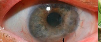
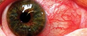
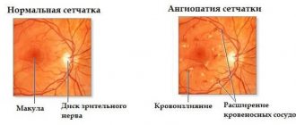
There are different types of angiomas. The most common are capillary and cavernous hemangiomas. Angiomas can be located in different parts of the body (face, neck, limbs, mucous membranes, internal organs) and be single or multiple. Angiomas are observed on the skin of the eyelids, conjunctiva, choroid, retina (see Retinal angiomatosis) and in the orbit. Most often, angiomas are detected in childhood and account for 70-80% of all neoplasms in children.
Capillary angioma is located in the thickness of the skin of the eyelid, has a bluish tint, grows along a plane, and does not spread in depth. Nodular hemangioma protrudes above the surface of the skin. Superficial cavernous hemangioma of the eyelids is usually limited, soft, and protrudes from under the skin of the eyelid. The palpebral fissure is narrowed, the skin of the eyelid is somewhat pasty, bluish in color, dilated blood vessels are visible. Deep cavernous hemangioma of the eyelids spreads deep into the tissue and surrounding tissues. A combination of hemangioma of the eyelids and orbit is observed, with the upper eyelid most often affected.
Hemangiomas of the conjunctiva of the eyeball are observed in the form of capillary, nodular and cavernous forms. Often conjunctival hemangioma is combined with lymphangioma. In the choroid, a cavernous angioma is most often found, which is a tumor located near the optic nerve head, grayish-white or greenish-gray in color, sometimes with a bluish tint, with an uneven spotty surface. There are no signs of inflammation, tumor growth is slow.
Visual functions are impaired. Scotomas appear in the field of vision: visual acuity decreases due to changes in the macula.
With orbital hemangiomas, exophthalmos is often observed with or without displacement of the eyeball, depending on the location of the tumor. Congestive optic disc develops early. Localization of the tumor in the lower part of the orbit is often accompanied by a feeling of fullness. Swelling of the eyelids as one of the early signs of a developing tumor is observed in 1/3 of patients. This symptom is more typical for hemangiomas located in the upper or lower part of the orbit. As the tumor grows over several years, exophthalmos and displacement of the eye appear. Exophthalmos with cavernous hemangioma occurs in 80% of patients. The degree of exophthalmos varies.
With orbital angiomas, 40% of patients experience a persistent decrease in visual acuity due to the development of a congestive optic disc or its atrophy. Often, decreased visual acuity is an early sign of a developing tumor and is observed even before the appearance of exophthalmos. Scotomas appear in the field of vision; sometimes a ceral scotoma may occur without any changes in the fundus.
The diagnosis of angiomas of the eyelids and conjunctiva is usually not difficult. For the diagnosis of choroidal angioma, the presence of vascular tumors on the face, as well as the light red or yellow-red color of the tumor with bluish or greenish reflexes, and slight or very slight pigmentation of the tumor are important. These features distinguish angioma from choroidal melanoblastoma.
Orbital angioma should be differentiated from hemangioendothelioma and angiosarcoma.
Orbital angioma is characterized by exophthalmos almost from birth, relatively slow growth, an increase in exophthalmos when the child cries, cries, or when the head is tilted. Often, orbital angioma is combined with angiomatous areas in the skin of the eyelids and face. Visual functions are preserved; radiographically, only thinning of the walls of the orbit can be noted; angiolitis is often observed - lime deposits in the thrombosed vessels of the angioma. The eyeball is easily reduced.
Rapidly increasing exophthalmos with displacement of the eyeball, corneal damage, decreased visual acuity, destructive changes in the bone walls of the orbit indicate the transition of angioma to a malignant tumor (hemangioendothelioma or angiosarcoma).
Treatment methods for angioma
Angiomas cannot be treated conservatively. Modern medicine offers different ways to painlessly get rid of the disease:
- Laser treatment is considered a popular method. Blood loss during the procedure is minimal, and the likelihood of adverse reactions is very low.
- Radiation therapy is used to treat hard-to-reach tumors.
- A painless method for removing simple types of pathology is cryotherapy.
- Electrocoagulation is the cauterization of a tumor with a high current discharge. The method is painful, so it is not used for large formations.
- Small and hard-to-reach angiomas are eliminated with sclerotherapy. The method involves injecting ethyl alcohol into the tumor.
- If the above methods are ineffective, surgical intervention is performed.
There are many traditional medicine methods for removing angiomas. Patients should be aware that independent intervention can lead to unpredictable consequences. In such a situation, self-medication is strictly prohibited and can cause irreparable harm to health.