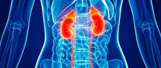
Causes
Most often, the cause of the appearance of cysts is congenital; it lies in an anomaly in the intrauterine formation of organs. The risk increases if alcohol, nicotine, or intrauterine infections occurred during pregnancy.
The disease can also be acquired due to the following reasons:
- consequences of urolithiasis;
- heart attack;
- glomerulonephritis;
- tuberculosis;
- pyelonephritis;
- harmful work factors;
- kidney tumor;
- organ injury;
- improper diet;
- disturbance of urine outflow.
The predisposing age for cyst formation begins after 45 years. If the situation is neglected, it may rupture and become infected with internal tissues. The formation can also cause renal failure and hydronephrosis. To recognize the disease at an early stage, you need to listen to your body and monitor the appearance of symptoms.
Classification and types
| Category | Character traits | Further tactics |
| 1 | A simple benign thin-walled cyst without septations, calcifications or solid component. The contents have liquid density and are not contrasted. | No further observation is required. The formations are benign. |
| 2 | Benign cyst. May contain several thin partitions. There may be small or slightly thickened calcifications in the walls and septa. The same group includes formations of homogeneous high density measuring {amp}lt;3 cm with clear contours that do not accumulate contrast. | |
| 3 | Cysts may have many thin septa. There may be minimal uniform thickening, as well as “apparent contrast” of the walls or partitions. They may contain calcifications, incl. wide and nodular, but no measurable accumulation of contrast occurs. The contours are usually clear. This category also includes high-density, non-contrast-accumulating formations with dimensions {amp}gt; or = 3 cm, completely inside the kidney. | Dynamic observation. A small part of the formations are malignant. |
| 4 | Cystic formations with uniformly or unevenly thickened walls or septa with measurable contrast uptake. | Surgery or follow-up. Over 50% of neoplasms are malignant. |
| 5 | Obviously malignant cystic formations that have all the characteristics of category 3 and, in addition, contain soft tissue components that accumulate contrast. | Surgery is recommended. Mostly malignant tumors. |
In medicine, tumors of the left and right kidneys, as well as both sides, are distinguished. Intrasinus and multilocular tumors are rare.
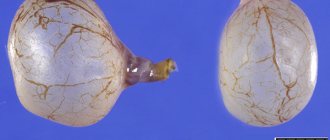
The cystic body contains several chambers, between which there are thick and durable walls.
Surgery involves the removal of affected tissues and cells. Repeated relapses of the disease often occur because some parts of the tumor remain after surgery.
Multilocular sinus cysts are congenital. Advanced forms of pathology affect the entire organ, which is completely removed during surgery.
In this case, the infection spreads to nearby healthy areas. Multiple formations affect the ureteric tract, renal canals at the entrance, as well as tissues located inside the organ.
Dangerous formations that spread over large areas. Doctors are forced to remove the cyst and the organ itself. Multiple tumors are easy to identify, especially if they have grown to medium size.
Right body
A simple sinus cyst in this case is rarely accompanied by indicating signs. In the early stage there are no symptoms. Over the course of many years, the tumor slowly grows and increases in size. Its presence may be indicated by disturbances in the functioning of the genitourinary system.
Multicystic right kidney causes chronic renal failure and serious disturbances in the functioning of the genitourinary system.
The neoplasm, compared to multiple sinus tumors, is firmly attached to the kidney. Pathological processes affect the organ's channels. The affected area enlarges. During surgery, it is difficult for doctors to separate the cyst from the organ tissue.
Sinus formations are oval or round in shape and resemble a capsule. It is located near the poles of the organ or at the entrance to the ureteric canals.
Symptoms
There are no symptoms when a sinus cyst appears; it appears after reaching a certain size. The formations are usually small, they can remain that way, then the person will not feel any discomfort at all. In this case, it can be detected during a routine examination.
But if it reaches more than 5 cm, then compression of neighboring organs occurs, and that’s when the first symptoms are felt:
Recommended topic:
Solitary renal cyst
- Aching, dull pain in the lower back, in the right or left hypochondrium, can radiate to the groin area. It appears due to impaired urine output and stagnation. The pain may intensify during movement. Stagnation provokes the formation of kidney stones.
- An increase in blood pressure occurs due to disruption of the renin-angiotensin-aldosterone hormonal system and compression of the renal vessels. Here additional symptoms may appear in the form of dizziness and tachycardia.
- A reduced portion of urine, the presence of blood in it, cloudiness, the appearance of sediment.
Depending on the concentration of pain on the right or left side, it is possible to determine accordingly whether a formation has appeared on the left kidney or on the right kidney. When inflammation of the cyst occurs, an increase in body temperature is noted.
The presence of blood in the urine is one of the symptoms of a kidney cyst.
Forecast
For a simple, benign cyst, the prognosis is 99.9% positive. And it doesn’t matter what kind of treatment tactics will be implemented. If the cyst does not cause dangerous complications (infection, cancer, changes in the kidneys, etc.), then treatment will lead to a complete recovery of the patient.
But even given such positive statistics, this disease should not be ignored. There is always a risk of cyst rupture. Then its contents will end up in the abdominal cavity. And this is fraught with peritonitis - severe and life-threatening inflammation.
Diagnostics
The most negative consequences occur after the cyst ruptures. To avoid this, you cannot ignore the symptoms; you must immediately consult a doctor to confirm the diagnosis. Diagnosis is not made based on symptoms, as they apply to many kidney diseases. Various examinations are needed here.
Congenital cysts in children are difficult to identify, since the formations are small. Therefore, examinations are usually carried out on an adult. Methods for diagnosing sinus cysts:
- collection of medical history and complaints;
- general urine test, or according to Nechiporenko;
- Ultrasound diagnostics is the most popular method for detecting kidney diseases. Using ultrasound, they not only determine the presence of a sinus cyst, but also its location, size down to a millimeter, and the amount of fluid in it.
After these methods, the clinical picture becomes clear, but there are exceptions. If the doctor is still in doubt, he may prescribe excretory urography of the kidneys, tomography and MRI. Examinations should be carried out by experienced diagnosticians, since pathologies can be confused with other diseases.
A sinus cyst can appear on both the right and left kidney. Due to physiological characteristics, damage to the right kidney is diagnosed more often, while damage to the second paired organ may not be noticed. If the cyst was congenital, then most likely it will be found on both kidneys. In case of acquisition, education is singular in nature.
Complications of untreated cysts
Infection of the cyst contents is one of the common complications. It is dangerous due to perforation of the wall of the neoplasm and leakage of pus into the pyelocaliceal system (PLS) of the kidney. Delayed treatment leads to organ abscess and toxic shock.
Possible complications of sinus cysts:
- hypertonic disease,
- kidney stones,
- hydronephrosis,
- renal failure,
- uremia.
A large sinus cyst of the right kidney damages the renal arteries and veins, which leads to hemorrhages into the pelvis or retroperitoneum. Excessive blood loss is dangerous due to a sharp decrease in blood pressure and brain hypoxia.
Treatment
A sinus cyst of simple formations, if there are no pronounced symptoms and its size is normal, then surgical intervention is not required. It is enough to follow the doctor’s instructions and monitor the situation once every six months. Pills or medication are not used.
Recommended topic:
Parapelvic cyst of the kidney
Surgery is indicated in the following cases:
- patient discomfort caused by pronounced symptoms;
- sinus cyst rupture;
- cancerous formations or the preconditions for their occurrence;
- intensive growth of the cyst;
- purulent infection;
- the appearance of diseases and complications (in this case, their treatment is additionally prescribed).
Laparoscopic surgery
Modern technologies make it possible to remove the formation without abdominal surgery, by puncture of a sclerosing substance through the skin. The solution promotes the resorption of the cyst, but this is only possible if it is small. The laparoscopy method is also used, when the formation is cut off using a small incision and laser. These are low-traumatic methods, rehabilitation after them takes about a week.
A major operation (resection) is resorted to only if the cyst is larger than 70 millimeters, or the entire organ needs to be removed. This happens if the kidney has atrophied, or the formation of the left kidney turns out to be malignant, or the vessels are located too close to the affected area. Full recovery requires several weeks in hospital.
Diet therapy
The diet is prescribed without fail when diagnosing the disease and after surgery, if any. This helps reduce the likelihood of progression of renal sinus cyst growth, reducing the load on the organs, allowing them to recover normally.
Basic principles of the diet:
- strictly exclude alcohol and smoking (hookah included);
- reduce salt intake as much as possible; if possible, it should be eliminated; it contributes to fluid retention and accumulation;
- limiting water consumption is required if kidney function is impaired, otherwise you need, on the contrary, to drink more so that harmful substances are eliminated faster in the urine;
- reduce the amount of protein foods - seafood and fish, meat, legumes;
- exclude coffee, canned and pickled foods, spicy, fried, smoked.
If you have a kidney cyst, it is recommended to completely avoid drinking alcohol.
You can use traditional methods, in particular, drink herbal teas and infusions that have diuretic properties. It is important to know that it is impossible to cure a cyst with diet; it only affects the functioning of the kidneys.
Complications and consequences
Lack of timely treatment of a renal sinus cyst of the left kidney can lead to the development of complicated pyelonephritis and renal failure.
With significant growth of the growth, the risk of its rupture increases with further release of pus onto adjacent organs and tissues. This can lead to intoxication of the body.
In addition, an increase in the size of the tumor leads to the displacement of organ tissue, as a result of which hydronephprosis develops, which threatens the loss of functionality of the affected kidney.
Any of these complications can cause serious damage to a person’s health, so only timely therapy will help avoid this.
If the patient ignores the doctor’s recommendations or is treated incorrectly, he will face unpleasant consequences. Complications may include:
- renal failure;
- complication of the general condition of the body;
- development of pyeloniphritis.
The most dangerous outcome of irresponsible behavior can be a purulent process in the cavity of the sinus cyst. The risk of rupture is high; surgical intervention in such a situation cannot be avoided.
Prevention
The consequences of the appearance of an intrasinus cyst are unpredictable, so it is better to adhere to measures to prevent it. The best prevention is a healthy lifestyle. Eat a balanced diet and do minimal physical activity.
It is important to consult a doctor at the first signs of a renal cyst. The cyst itself is not dangerous; you can live with it for several years only if it does not cause complications. If this happens, you can lose your kidneys.
Since it does not manifest itself for a long time, it is necessary to undergo an ultrasound examination at least once a year, and tests every six months. This will help bring cyst formation under control at an early stage. A sinus cyst requires qualified observation; self-medication is excluded here.
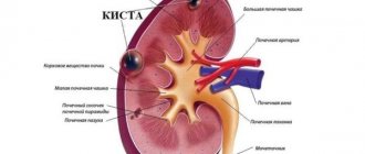
Structure of the renal sinuses
The sinus is an intrarenal cavity filled with blood vessels, a layer of fat and containing the pelvis. It opens outward through the renal gate, through which blood vessels, the ureter, and nerves pass. Through them, the sinuses are connected to the perinephric layer - the fatty capsule of the kidney. From the outside they border with:
- pyramids - seals formed by the medulla,
- cortical columns (renal columns) - an area of the cortex with blood and lymphatic vessels.
Inside the sinus there are:
- pelvis,
- nerves,
- vessels,
- adipose and fibrous tissue.