3367
Obstructive jaundice is a pathological condition of the biliary system, in which the lumen of the gallbladder or ducts is closed by some obstacle that disrupts the normal outflow of secretions. Clinically, this pathology is manifested by rapid discoloration of the skin and visible mucous membranes in yellow shades, painful sensations in the right side, darkening of urine and an increase in some specific indicators in a blood test.
Treatment of obstructive jaundice can only be surgical, since this condition is characterized by severe disturbances in the body. In fact, this pathology is a blockage of the lumen of the bile ducts, and it is impossible to eliminate the obstacle with medications.
If timely assistance is not provided in time, a serious complication may develop and death may occur.
Description of the syndrome
Obstructive jaundice is a pathological condition that develops due to a violation of the passage of bile through the bile ducts. The result is liver failure, which can be fatal if left untreated. The disease most often occurs in adults over 50 years of age, most of them are women. In elderly people over 70 years of age, the overall prevalence of diseases of the gallbladder and its ducts is 40%.
Since this stops the release of bilirubin with bile into the intestinal lumen, this substance accumulates in the tissues of the body. The normal content of total bilirubin (TB) in the blood is 5.1-21.5 µmol/l. This indicator increases tenfold in obstructive jaundice. When the level of bilirubin increases above 27–34 µmol/l, it begins to bind to the fibers of the skin and sclera of the eyes, which manifests itself in the form of their icteric coloration.
The syndrome is classified according to several criteria:
- For the reasons for its occurrence: benign (narrowing of the bile ducts due to scar changes, blockage with stones and others);
- malignant (cancer tumor localized in the head of the pancreas, major duodenal papilla, bile ducts).
- ABOUT
- full;
- low block - violation of the passage of bile at the level of the terminal (final) part of the common bile duct;
The duration of jaundice in adults before the onset of severe liver failure ranges from 10 days to six months. The severity of the patient’s condition no longer depends on the level of bilirubin in the blood, but on the duration of the syndrome and the complications caused. Obstructive jaundice must be treated as quickly as possible from the moment of its occurrence, since it is accompanied by a high risk of developing inflammation of the bile ducts and liver failure.
The mechanism of pathological processes in the human body with obstructive jaundice is as follows:
- impaired excretion of bile leads to its stagnation;
- pressure in the bile ducts increases;
- bile capillaries dilate and rupture;
- a large amount of bilirubin enters the blood;
- hypercholesterolemia occurs - an abnormal increase in cholesterol levels in the blood due to the circulation of bile acids in the bloodstream;
- the digestion process is disrupted, due to the lack of bile entering the intestines, bile pigment is not released, the stool becomes discolored;
- the activation of lipase, an enzyme that breaks down fats, decreases; therefore, 70% of lipids are not digested, but are excreted along with feces;
- intestinal dysbiosis develops, since there is no bile, which has a bactericidal effect;
- bile acids, excreted through the kidneys, color the urine dark (the color of “beer”);
- prolonged increase in pressure in the bile ducts leads to impaired liver function;
- the permeability of cell membranes increases, an inflammatory process develops in the liver;
- Necrosis of liver cells occurs, the hepatic form of jaundice occurs, and acute pancreatitis develops.
Due to the fact that bile acids stop entering the intestines, the absorption of vitamins A, E, and K is also impaired. The patient develops vitamin deficiency, blood clotting decreases, and increased bleeding occurs. Functional disorders in the liver lead to the accumulation of toxic substances that have an adverse effect on all organs and systems, in particular, renal and respiratory failure occurs.
What types of jaundice exist?
There are three types of jaundice:
- Prehepatic jaundice - an increase in bilirubin levels is caused by a disruption in the destruction of red blood cells. Due to high levels of bilirubin, the liver cannot cope with its binding and removal from the body.
- Hepatic jaundice is associated with a direct disorder of the liver, such as cirrhosis. Due to pathology, the organ cannot cope with the transformation and excretion of toxic bilirubin.
- Obstructive, also known as obstructive jaundice, occurs due to a violation of the outflow of bile. In this case, there may be no problems at the stage of bilirubin conversion, but the body cannot remove it with bile due to kidney and gallbladder diseases.
Symptoms
Characteristic signs of the syndrome are:
- Pain (70-85% of patients). Its character can be different: when the obstruction of the ducts is caused by a tumor located in the head of the pancreas, pain occurs in the right hypochondrium. If the tumor formation is located in the tail or body of the gland, then the patient feels pain in the left hypochondrium. When the tumor becomes large and compresses the solar plexus, the pain becomes unbearable and diffuse. It can radiate to the spine or to the area between the shoulder blades.
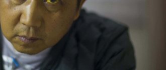
- Yellow coloration of the skin and conjunctiva of the eyes, severe skin itching as a result of irritation of skin receptors by bile acids. Itching is most common with malignant tumors, and the skin color may acquire an earthy tint, and with a long course of the disease, bronze. Jaundice usually appears after a painful symptom and develops gradually.
- Feverish state, increased body temperature.
- Darkening of urine, foaming.
- Palpation reveals tension in the abdominal muscles in the area of the right hypochondrium.
- When bile acids enter the bloodstream, a decrease in heart rate and blood pressure is observed.
The following changes occur in the digestive tract:
- Deterioration of appetite (more than half of patients), the appearance of aversion to fatty, meat foods.
- Loss of body weight due to intoxication due to tumor and disruption of the digestive process. It is often the first sign of the disease.
- Discoloration of feces, increase in their volume. The stool takes on a gray-clay color and a fetid odor, leaving greasy marks on the walls of the toilet.
- Nausea, vomiting, feeling of heaviness after eating, heartburn.
- Flatulence.
- Impaired intestinal motility, weakening of its tone, the appearance of constipation, which is replaced by diarrhea due to increased putrefactive and fermentative processes.
- Undigested muscle fibers appear in the stool, which is associated with impaired penetration of enzymes into the food bolus.
Yellow coloration of the skin and sclera
Not only the skin and mucous membranes, but also all tissues of the human body, fluids that accumulate as a result of edema, turn yellow. The natural color remains in tears, saliva and gastric juice. In patients with dark skin, jaundice may not be detected immediately.
Bile acids also have a toxic effect on the nervous system. This manifests itself in the form of the following signs:
- increase in general weakness, increased fatigue;
- irritability;
- depression;
- drowsiness during the day and insomnia at night;
- headache.
Types of jaundice and their treatment
Article verified 08/04/2015 . The article was checked by a specialist: Trandofilov Mikhail Mikhailovich
, hepatologist, oncologist surgeon, MD.
Have you noticed that your skin has recently taken on an unusual yellowish or reddish tint? Have the whites of your eyes, which just recently shone white, become dull or bright yellow? Does your liver ache, and is your general condition causing concern? Drop everything and immediately go to the doctor - it is quite possible that you have jaundice.
But what is jaundice? Strictly speaking, this is not a disease, it is a condition in which there is an excessive accumulation of bilirubin in the blood, which is deposited in the tissues and stains the mucous membranes, sclera of the eyes and skin yellow.
Bilirubin is a special bile pigment found in red blood cells (erythrocytes).
As a rule, after living a certain number of days, red blood cells are destroyed, and the bile pigment enters the liver, where, after processing, it is sent back into the blood in the form of new blood cells.
But sometimes this process is disrupted and bilirubin begins to accumulate in large quantities: due to too rapid destruction of red blood cells or due to any problems with the liver, when it is not able to process the amount of bilirubin that enters it. In some cases, the development of jaundice occurs due to blockage of the hepatic ducts through which bilirubin must leave the liver.
Therefore, we can say that jaundice in adults is a consequence of pathological processes developing in the liver and bile ducts. And, depending on the cause of this process, three types of jaundice can be distinguished:
- suprahepatic or hemolytic jaundice (due to too rapid breakdown of bilirubin)
- hepatic (due to liver disease)
- subhepatic or obstructive jaundice (due to closed hepatic ducts)
Differential diagnosis of jaundice
To prescribe the correct treatment, it is necessary to accurately determine the cause of the disease.
To do this, a differential diagnosis of jaundice is carried out, in which, first of all, an anamnesis is collected, taking into account many factors: the possible presence of contacts with infectious patients, taking certain medications, the possibility of poisoning with hemolytic poisons, parenteral manipulations (blood transfusion, tattooing, piercing ears), finding out other reasons that can cause problems with the destruction or removal of bilirubin.
After collecting an anamnesis, laboratory and instrumental studies are required for the differential diagnosis of jaundice.
Prehepatic jaundice develops against the background of excessive formation and rapid breakdown of bilirubin. As a result of such increased destruction, the liver is not able to remove all bilirubin, which leads to its return to the blood and, as a consequence, to the development of the disease.
Causes of development of suprahepatic jaundice:
- hemolytic anemia (congenital or acquired - microspherocytosis hereditary anemia, ovalocytosis, hemoglobinopathy, sickle cell anemia, thalassemia, acute post-transfusion anemia);
- diseases of ineffective erythropoiesis (primary shunt hyperbilirubinemia, erythropoietic uroporphyria, B12-deficiency anemia);
- the result of extensive infarctions of internal organs (usually the lungs) or hematomas;
- medicinal and toxic damage (arsenic, hydrogen sulfide, phosphorus, trinitrotoluene, sulfonamides).
Also, this type of jaundice can be a symptom of other diseases: lobar pneumonia, Addison-Birmer disease, malaria, or manifest itself in the presence of malignant tumors and certain liver lesions.
The main symptoms of prehepatic jaundice: lemon-yellow tint of the skin, mucous membrane and sclera of the eyes, pale skin due to anemia, slight enlargement of the liver.
Treatment of prehepatic jaundice
The choice of treatment for prehepatic jaundice depends on the cause of excess formation of bile pigment - a disease that causes the accumulation of bilirubin in the blood, and usually consists of drug therapy aimed at normalizing liver function.
Hepatic (parenchymal jaundice) occurs against the background of massive damage to liver cells (their structures, functions), which causes a disruption in the consumption of bilirubin by liver cells and problems with its excretion. Parenchymal jaundice is one of the most common liver diseases.
In the differential diagnosis of parenchymal jaundice, it is first necessary to exclude the possibility of an infectious disease.
Reasons for the development of parenchymal jaundice:
- acute viral hepatitis
- Infectious mononucleosis
- leptospirosis
- toxic drug or alcohol damage to the liver
- chronic aggressive hepatitis
- cirrhosis
- hepatocellular cancer
The main symptoms of parenchymal jaundice: reddish, ruby skin tone, enlarged liver, pain in the liver area, “liver palms” (spotted redness), spider veins, itchy skin.
Treatment of parenchymal jaundice:
The choice of treatment for this type of jaundice depends on the cause of the disease. But, in any case, we can distinguish between conservative (drug) treatment, which consists of getting rid of the underlying disease, and radical surgical intervention for advanced cases of jaundice (liver transplantation).
Conservative treatment includes the use of medications (antihistamines, steroids, etc.), plasmapheresis, phototherapy and diet. Therapy is aimed at curing the virus, suppressing the possibility of infecting others, and preventing the development of liver cirrhosis. When treating chronic autoimmune hepatitis, corticosteroid therapy is used.
Unfortunately, therapeutic treatment methods are not able to completely rid a person of the virus, but constant, systematic treatment reduces the inflammatory process in the body.
In the case of a far advanced pathological process, when it is not possible to achieve remission and various complications are already beginning to develop, we can talk about liver transplantation.
Subhepatic (obstructive) jaundice in adults develops due to a violation of the free flow of bile into the duodenum due to narrowing of the bile ducts.
Narrowing can develop for various reasons:
- presence of stones in the common bile duct
- the presence of tumors of the pancreas, liver, common bile duct, gallbladder
- parasites
- scarring (often occurs after surgery)
- atresia or hypoplasia of the biliary tract (developmental anomaly leading to tissue underdevelopment)
- cholangiocarcinoma (malignant tumor of the bile ducts)
- benign narrowing of the ducts
Benign narrowing of the ducts and cholangiocarcinoma (malignant tumor) can also cause duct damage in primary sclerosing cholangitis (chronic progressive damage to the bile ducts).
The main symptoms of obstructive jaundice: yellow coloring of the skin, mucous membrane and sclera of the eyes with a greenish tint, fever, itching.
Recently, there has been a progressive increase in benign and malignant pathology of the organs of the hepatopancreatoduodenal zone (duodenum, liver with bile ducts and gallbladder, pancreas).
This inevitably leads to an increase in the number of patients with obstructive jaundice, among whom elderly and senile people predominate with severe concomitant pathology, which directly affects the severity of the disease, leading to a fairly high mortality rate in obstructive jaundice.
Treatment of obstructive jaundice
With obstructive jaundice caused by benign processes, the main problem is the presence of stones in the common bile duct, which interfere with the normal outflow of bile. Stones can periodically cause blockage of the bile ducts, which is manifested by biliary colic, inflammation of the bile ducts (cholangitis), pancreatitis or obstructive jaundice.
Endoscopic papillosphincterotomy
Today, endoscopic papillosphincterotomy (EPST) has become widespread in solving the problem of restoring normal bile flow.
To perform EPST, it is necessary to have an accurate understanding of the nature of the obstacle (size, number and location of stones, length of narrowing of the bile duct). An important role is played by the ratio of the size of the stones and the width of the bile ducts below the level of blockage, since when the stones are large (above the diameter of the underlying duct), it is necessary to crush them (lithotripsy).
Endoscopic papillosphincterotomy can remove about 90% of stones located in the bile duct.
No less pressing is the problem of treating obstructive jaundice in malignant diseases of the hepatobiliary tract and pancreas. This complication occurs in almost all patients with malignant tumors in this area: due to compression or blockage of the lumen of the biliary tract.
Unfortunately, not many patients with malignant tumors of the hepatobiliary tract and pancreas can undergo radical treatment.
As a rule, patients with obstructive jaundice caused by a violation of the outflow of bile by malignant neoplasms undergo palliative interventions aimed at reducing the level of cholestasis (insufficient bile flow).
The following methods of treating obstructive jaundice are distinguished:
Open:
- cholecysto-, choledocho-, bihepaticojejunostomy
Minimally invasive:
- external-internal drainage
- stenting
- external drainage of bile ducts
Currently, more and more attention is being paid to antegrade (transcatheter) interventions for diseases of the biliary tract and pancreas.
Percutaneous transhepatic cholangiostomy
There are two methods for treating obstructive jaundice:
- two-stage (using a Lunderquist stylet catheter)
- one-stage (using a Chiba needle)
The technique of two-stage percutaneous transhepatic cholangiostomy using a Lunderquist stylet catheter involves performing a liver puncture from the right lateral approach.
Using an injected X-ray contrast agent and under the control of an electron-optical converter, puncture of the visualized bile duct is performed with a Lunderquist stylet catheter.
After removing the stylet and ensuring that bile is flowing from the sheath catheter, a special guidewire is inserted and guided to the point where the duct narrows.
An intrahepatic canal is formed, a permanent catheter is installed and, after X-ray control of the position of the catheter, it is fixed to the skin.
With the one-stage percutaneous transhepatic cholangiostomy technique, a fine-needle puncture of the intrahepatic bile duct is performed with a Chiba needle. The Lunderquist guide is passed through the lumen of the needle to the site of narrowing of the duct.
The needle is removed, an exchange catheter with a metal tube inserted into it is threaded onto the guidewire, which is passed to the wall of the punctured duct. Through a catheter, a conductor is inserted into the lumen of the duct to the point of narrowing and the intrahepatic canal is widened.
After the permanent catheter is installed, an X-ray examination is performed to monitor its position, and the catheter is fixed to the skin.
To improve the patient’s quality of life after relieving pressure in the ducts, the lumen of the bile ducts is restored (recanalization).
There are the following methods for recanalization of the bile ducts:
- catheter internal drainage
- endoprosthetics
- formation of compression ductal-intestinal anastomoses using magnetic elements
- formation of biliogastric anastomosis.
Catheter internal drainage of bile ducts in obstructive jaundice
There are:
- one-time
- deferred
Simultaneous internal drainage of the bile ducts is carried out only if, during external drainage, it was possible to insert the conductor beyond the level of the narrowing of the duct. Otherwise, delayed internal drainage is performed after insufficient bile flow and biliary hypertension are relieved by external drainage of the bile ducts.
Delayed internal drainage is usually performed 10 days after adequate external drainage.
Endoprosthetics of bile ducts
It is performed to restore the natural flow of bile in the gastrointestinal tract. Endoprosthesis replacement is possible only if the patient's life expectancy is no more than 6 months, due to the inevitable blockage of the endoprosthesis lumen after 6 months from the moment of stent installation.
Percutaneous transhepatic microcholecystostomy
The operation is carried out:
- to eliminate insufficient bile flow due to blockage of the distal parts of the common bile duct
- to reduce pressure inside the gallbladder in acute cholecystitis in elderly people and in patients with severe concomitant pathology that does not allow radical surgical treatment.
Microcholecystostomy using the Seldinger method
On the skin, under the control of ultrasound scanning, a place for optimal puncture of the gallbladder is selected, a skin incision is made and the gallbladder is punctured using a Chiba needle. Through the lumen of the needle, a conductor is inserted into the lumen of the bladder, through which the channel is expanded. After X-ray control of the position of the drainage, it is fixed to the skin.
Services Price Appointment
| Radiofrequency ablation of the liver | from 40,000 rub. |
| Biopsy of the liver and liver tumors | 17,000 rub. |
| Liver surgeries | from 15,000 rub. |
Source: https://www.klinikasoyuz.ru/patient/articles/vidy-zheltux-lechenie/
Causes
In medicine, there are several causes of this pathology:
- Congenital deviations of the normal development of the biliary system: absence or fusion of the biliary tract;
- underdevelopment of ducts;
- cysts in the main bile duct;
- diverticula (protrusions) of the duodenal wall near the major duodenal papilla (MDP).
- cholelithiasis (up to 35% of all cases);
- liver;
Main causes of obstructive jaundice
In patients under 30 years of age, the most common cause of this syndrome is cholelithiasis, and in those over 40 years of age, malignant neoplasms.
Diagnosis of obstructive jaundice
To make a correct diagnosis and prescribe appropriate therapy, the doctor needs to conduct a differential diagnosis to exclude parenchymal and hemolytic jaundice, as well as identify the cause of bile duct obstruction. For this purpose the following is carried out:
- detailed anamnesis and study of medical history;
- laboratory tests of blood and urine for bilirubin - with obstructive jaundice this indicator is always elevated, as is the activity of alkaline phosphatase;
- blood for cholesterol (indicators for obstructive jaundice are increased);
- ultrasound examination of the liver, gallbladder and ducts (the images show dilated areas of the ducts, stones in them, tumors, hydatid cysts and metastases);
- ERCP (endoscopic retrograde cholangiopancreatography) (with unclear or questionable ultrasound results, if a tumor of the major duodenal papilla is suspected);
- percutaneous transhepatic cholangiography under ultrasound or MRI control is used if there is a suspicion of blockage of the bile ducts in the area of the porta hepatis;
- splenportography and celiacography are used when malignant tumors of the liver and bile ducts are suspected (atypical clusters of blood vessels are visible on the images);
- if there is a suspicion of damage to the duodenal papilla (tumors, diverticula) and other pathologies of the duodenum, duodenography is used.
After establishing the full picture, the doctor prescribes a comprehensive treatment for obstructive jaundice.
Complications
Obstructive jaundice causes the following complications:
- liver and kidney failure (up to 70% of cases), accompanied by organic lesions of these organs;
- local infectious inflammation of the bile ducts (20-40% of patients) with a high risk of transition to a systemic inflammatory reaction and sepsis;
- liver abscesses with prolonged inflammation;
- acute pancreatitis.
The cause of these complications is intoxication of the body due to stagnation of bile and migration of pathogenic microflora from the intestines.
What it is?
Obstructive jaundice is a pathological condition of the liver. When it affects the flow of bile, the skin and mucous membranes turn yellow. This is due to the fact that bile pigments enter the blood. The disease is of a tumor nature in most cases.
Malignant tumors are usually more common in patients than benign tumors. In some cases, this jaundice is non-tumoral in nature.
The clinical picture of the disease usually develops gradually. An acute onset is observed quite rarely; most often, obstructive jaundice is a consequence of inflammation of the biliary tract.
Prevalence and significance
It occurs more often in the elderly and senile than in the young. Moreover, this disease is diagnosed in women much more often than in men. With choledocholithiasis, obstructive jaundice develops in approximately 70% of cases.
Risk factors
The risk of developing obstructive jaundice is caused by:
- obesity;
- sudden weight loss;
- abdominal injuries;
- liver and pancreas infections;
- surgical interventions on the liver and biliary tract.
Diagnostics
The following laboratory tests are performed in patients with jaundice:
- OAM - a dark color is detected, the absence of urobilin - a bile pigment that colors urine yellow, and the presence of other bile pigments.
- UAC – increase in ESR, leukocyte level; in case of intoxication – decrease in hemoglobin.
- Biochemical blood test - an increase in the content of bilirubin, cholesterol, enzymes of thyroid hormone, GGTP, with a long course of the disease - an increase in ALT, AST, a violation of the ratio between protein fractions, with the development of renal failure - an increase in creatinine and urea, with acute pancreatitis - an increase in the activity of C-reactive protein and lipase.
Before surgery, it is also necessary to take a blood test to determine the level of glucose, coagulation, group and Rh factor. Based on laboratory data, a preliminary diagnosis can be correctly established in 70-75% of patients.
Instrumental studies include:
- Ultrasound of the abdominal organs. It is the main diagnostic method. The following signs are detected: an enlargement of the main bile duct by more than 8 mm, intrahepatic bile duct by more than 4 mm, thickening of their walls, the presence of suspensions, enlargement and deformation of the gallbladder; in case of cholelithiasis, stones are visualized. Ultrasound examination is informative in 75% of patients.
- FGDS with biopsy according to indications. Conducted for morphological examination of tumor tissues.
- MRI with cholangiopancreatography, CT or percutaneous transhepatic cholangiography, endoscopic retrograde cholangiopancreatography (if indicated). The last 2 methods are the most accurate (diagnosis accuracy is close to 100%).
- Additionally, the doctor may prescribe an X-ray of the abdominal cavity, chest, laparoscopy, ultrasound, ECG.
External manifestations of jaundice are also characteristic of other diseases, including infectious ones. The answer to the question of whether the pathology is transmitted to other people depends on the cause that caused this condition. About 40% of patients with icteric discoloration of the skin upon initial treatment are referred to infectious diseases departments of hospitals to exclude infectious etiology. When diagnosing obstructive jaundice, differentiation is made with diseases such as:
- Viral and infectious hepatitis caused by pathogens of brucellosis, pleuropneumonia, leptospirosis, amoebiasis, typhoid, herpes, opisthorchiasis, malaria, syphilis and others.
- Hepatitis that develops when exposed to toxic substances (arsenic, pesticides, phosphorus, toluene, fungal toxins and others) and drugs (sulfonamide antibiotics, Levomycetin, thiazide diuretics, hormonal, anti-tuberculosis, cytotoxic drugs and others).
- Chronic hepatitis.
- Cirrhosis of the liver.
- Constitutional familial jaundice, Dubin-Johnson syndrome.
- Liver cancer.
Treatment
Obstructive jaundice is treated in two stages. At the first stage, various minimally invasive methods are used to eliminate bile stagnation, that is, cholestasis. Conservative therapy is also recommended.
Drugs
Conservative treatment includes vitamin therapy and drugs to improve liver function: Sirepar, Essentiale, Cocarboxylase. In addition, the doctor may prescribe Prednisolone, a metabolism stimulant Pentoxyl and amino acids in the form of Methionine. The patient is prescribed intravenous glucose, Glutamic acid, Vicasol, B vitamins, Trental. Antibiotics, detoxification procedure and blood purification - plasmapheresis are used if necessary.
Surgery
Direct surgical interventions are prescribed in the absence of the desired effect from minimally invasive methods. These include:
- lithoextraction;
- drainage of ducts;
- percutaneous cholangiostomy with external or internal drainage;
- laparoscopic drainage;
- reconstructive operations on the biliary tract and bilio-intestinal anastomoses;
- laparotomy with removal of the gallbladder.
At the second stage of treatment, intensive infusion therapy and forced diuresis will be used. The patient is also given intravenous drips of hemodesis, blood substitutes, glucose solutions with insulin, and saline solutions.
Treatment with folk remedies
If yellowing of the skin or sclera appears, you should contact a qualified specialist. Taking traditional medicine in combination with traditional medicines will create conditions for a quick recovery. Healing plants help strengthen the immune system and restore strength.
But therapy with medicinal plants should only be a complement to traditional medicine, and the folk remedies taken should be approved by the attending physician.
Traditional medicine for obstructive jaundice:
- decoction of immortelle;
- a decoction of peppermint, immortelle flowers, three-leaf watch, coriander fruits;
- tincture of wormwood;
- tincture of hazel leaf in white wine;
- infusion of horseradish root, but patients with gastritis, enterocolitis and nephritis should not take the remedy;
- sauerkraut juice;
- infusion of sage leaves;
- infusion of hop cones;
- infusion of corn silk.
Diet
The diet should consist of vegetables, fruits, and dairy products. Meals should be fractional. It is better to boil or wipe dishes. It is important to drink as much fluid as possible.
Therapy
At the initial stage of obstructive jaundice, due to the increased risk of complications during surgery, conservative treatment is used. If the pathological process increases or there is no tendency to improve within 2-3 days after the patient’s admission to the hospital, surgical intervention is performed. Conservative treatment is effective in only 23% of patients.
Conservative treatment
The doctor most often prescribes the following drugs:
- detoxification infusion therapy: parenteral administration of 1.5-2 liters of liquid per day (sodium chloride or bicarbonate, amino acid mixtures, Albumin, protein, Mannitol, Sorbitol, Hepatosteril, Vamin, Reopoliglyukin, Trometamol, glucose);
- antibacterial agents for the prevention of purulent complications: aminoglycosides, Ampicillin, Vancomycin, Clindamycin, Metronidazole, Cefoxitin, carbapenems, fluoroquinolones;
- vitamins and antioxidants: ATP, coenzyme A, vitamins B, D, E, ascorbic acid, Mexidol;
- hepatoprotective drugs: essential phospholipids, Hepa-Merz, Silymarin, Heptral, Hepatosan;
- reparative agents and medications that improve microcirculation in tissues: Trental, Pentoxifylline, Actovegin and others;
- for the prevention of erosions and ulcers in the gastrointestinal tract: proton pump inhibitors, antacids (Omeprazole, Losek, Omez, Kvamatel and others);
- enterosorbents for binding toxic substances in the intestines, reducing the load on the liver and quickly restoring liver tissue (Enterosgel and its analogues);
- for the prevention or elimination of bleeding: vitamin K, Vikasol, aminocaproic acid.
Surgical intervention
Elimination of bile stagnation is carried out in several ways:
- Open abdominal surgery (laparotomy).
- Laparoscopic surgery.
- Endoscopic and percutaneous transhepatic minimally invasive methods of surgery.
Traditionally, a two-stage surgical treatment scheme is used: at the first stage, drainage of the gallbladder is performed using minimally invasive techniques, and after eliminating hyperbilirubinemia, the cause of obstructive jaundice is finally eliminated (removal of stones, tumors, etc.). This approach makes it possible to improve the outcome for patients by 2.5 times, since in 50-80% of cases the severe condition of patients upon admission to the hospital does not allow radical surgery to be performed due to the high risks of complications and mortality.
Drainage helps eliminate jaundice, hepatic-renal failure, and intoxication of the body, which creates more favorable conditions for subsequent surgery. Minimally invasive endoscopic treatment methods are carried out using a duodenoscope and an electrosurgical unit. The probe is inserted through the oral cavity into the digestive tract, the BDS is found and a catheter is inserted through it. The following main endoscopic methods of bile drainage are used in surgery:
- Retrograde cholangiopancreaticography. A thin catheter (tube) is inserted into the mouth of the BDS, and fluoroscopy is performed by introducing a radiopaque substance.
- Papillosphincterotomy – dissection of the ampulla of the papilla. It is used in patients with cholelithiasis, cicatricial narrowing of the papilla of Vater, as well as in purulent cholangitis that appears against the background of these disorders. This method allows you to remove stones up to 1.5 cm, restoring the patency of the bile ducts.
- Mechanical lithotripsy. It is carried out in the presence of single stones no larger than 10 mm in size or in case of multiple stones filling the main bile duct, parietal stones, in combination of cholelithiasis with purulent inflammation of the ducts. The stone is captured and crushed using a special reinforced Dormia basket or a balloon lithoextractor. The effectiveness of the method reaches 80-90%.
- Stenting of the bile ducts under X-ray endoscopic control (installation of a plastic or self-expanding endoprosthesis).
Capturing the stone with the Dormia basket
In percutaneous drainage methods, a needle is inserted through the abdominal wall under ultrasound or X-ray control. The diameter of the needle is 0.6-0.9 mm; when simultaneously performing cholangiography (x-ray examination of the ducts), larger diameter needles are used - up to 1.1 mm. After the tip of the needle enters the lumen of the bile duct, the bile is drained.
Postoperative period
The patient's condition in the postoperative period depends on the severity of obstructive jaundice, concomitant pathologies and the type of surgical intervention. Patients experience open abdominal surgery the most difficultly. Postoperative treatment in this case is as follows:
- Bed rest on the first day.
- First, tube or parenteral nutrition is carried out, then the patient is transferred to table No. 0 (low-fat light broth, ground rice broth, unsweetened jelly, jelly from non-acidic fruits, then pureed porridge and meat purees are added).
- Early physical activation of the patient is recommended to improve the digestive process.
- Analgesic therapy: non-narcotic analgesics, antispasmodics (Drotaverine, No-shpa, Metamizole sodium and others).
- Antibacterial therapy in the presence of concomitant cholangitis.
- The use of gastric secretion blockers mentioned above.
- Enzyme therapy (Pancreatin, Digestal, milk thistle fruit extract and other agents).
Treatment of obstructive jaundice
The decision on how to treat obstructive jaundice depends on the causes of its occurrence and the severity of the symptoms. At the initial stage, therapy consists of procedures aimed at eliminating cholestasis using minimally invasive methods. Also at this stage, treatment with medications (conservative therapy) is practiced in intensive care conditions.
It includes the use of:
- infusions of saline solutions, glucose with insulin, protein drugs, blood substitutes, etc.;
- vitamins and amino acids;
- metabolism stimulants;
- hormonal drugs;
- gastric secretion blockers;
- antacids and coating drugs.
If there are signs of intoxication of the body, extracorporeal detoxification (hemosorption, plasmaphoresis, hemodialysis) is prescribed.
To prevent the development of infectious and inflammatory complications, treatment with broad-spectrum antibiotics that can penetrate the bile is prescribed. The list of drugs used to treat obstructive jaundice is determined on an individual basis. Only after this is surgical or minimally invasive intervention performed:
- lithoextraction;
- bougienage and drainage of ducts;
- therapeutic ERCP (endoscopic retrograde cholangiopancreatography), combined with endoscopic papillosphincterometry;
- percutaneous transhepatic cholangiostomy;
- bile duct stenting;
- laparoscopic choledochotomy according to Kocher, Kerta, Vishnevsky or other methods;
- resection of organs affected by the tumor (for oncological diseases accompanied by obstructive jaundice).
All patients, without exception, are prescribed a diet corresponding to table No. 5a (according to Povzner). The menu includes pureed dishes from boiled cereals and vegetables, dairy products, fruits and berries, and plenty of drink. It is recommended to eat frequently, servings no more than 200 ml.
Disease prognosis
According to data from surgical practice, during emergency surgical treatment of obstructive jaundice, the risk of complications is 54%, and mortality is up to 30%. The life prognosis for the patient depends on the cause of the pathology, the duration of the syndrome, and the severity of the patient’s condition. When using minimally invasive endoscopic and percutaneous treatment methods, it is possible to reduce the number of negative consequences.
The greatest danger to the patient is progressive liver failure, which leads to death in 50-60% of patients in the postoperative period, and the malignant etiology of the disease. The survival rate of cancer patients after surgery does not exceed 11 months.
The risk of postoperative complications also increases in the presence of the following factors:
- concomitant diseases in the stage of decompensation (up to 18-60% mortality);
- prolonged periods of examination and conservative therapy;
- elderly age of the patient;
- inflammation in the gallbladder and ducts;
- increasing mechanical jaundice.
In case of mild liver failure, drainage of the ducts in the first 1-3 days allows the patient to achieve good condition by the tenth day, the skin and sclera of the eyes acquire normal color, the liver shrinks and motor activity is restored.
Jaundice in newborns and children
There is also jaundice in newborns, caused by increased levels of bilirubin, but the mechanism of development of this condition does not pose a danger to the child. This condition appears in newborns when the child’s adaptation mechanisms to new conditions are disrupted. The disorder appears in a fairly large number of children who successfully leave the maternity hospital, remaining in it only a little longer than ordinary children. Jaundice in newborns cannot be treated; constant medical monitoring of the child is sufficient until the signs of pathology completely disappear. You should only worry in cases where the child’s mother has severe liver pathologies, suffers from diabetes, and also if the baby’s pathology does not go away for more than three weeks.
When treating obstructive jaundice, timely consultation with a doctor is very important. The disease gives symptoms already in the first day after the flow of bile stops, so if you notice pathological changes in time and go to the clinic, then the chances of a positive treatment outcome are quite high.
Diet and prevention
The basis for successful treatment and prevention of obstructive jaundice is diet. After discharge from the hospital, patients must adhere to therapeutic diet No. 5 (mashed version) for 2-3 weeks. After complaints and discomfort in the abdominal area disappear, you can switch to unprocessed food. Basic recommendations:
- dishes should be cooked boiled or steamed;
- fresh vegetables and foods high in fiber should be excluded;
- complete abstinence from alcohol, fatty, spicy, salty and smoked foods;
- it is necessary to avoid overeating, as this contributes to the occurrence of spasms of the bile ducts;
- eat meals in small portions (5-6 times a day);
- in the presence of organic liver damage, reduce the amount of protein consumed;
- for constipation caused by cholecystitis, it is recommended to eat grated carrots, boiled beets (can be included in salads and vinaigrettes), dishes with cauliflower, pumpkin, zucchini, baked apples; fruit and vegetable juices, prunes, kefir and other fermented milk drinks; You should also give preference to millet and buckwheat porridge rather than rice and semolina.
Diet No. 5
Patients with chronic gallbladder inflammation are often overweight. It is recommended for such people to have fasting days once a week (rice-compote, cottage cheese, apple, kefir and others).
News MirTesen
Jaundice syndrome: what it is, causes, treatment, diagnosis and consequences
Yellowed skin, sclera, mucous membranes - all these are manifestations of jaundice syndrome. A sign of impaired functioning of the body. The syndrome is not an independent disease, only a signal of problems.
What is jaundice syndrome?
Jaundice syndrome is the coloring of the skin, mucous membranes, and sclera of a person in yellow shades. The culprit is the pigment bilirubin, which occurs when red blood cells are destroyed.
The process of formation and decay occurs constantly throughout life.
Hemoglobin contained in red blood cells is divided into globin, hemosiderin, which begins the formation of new red blood cells, and the third element, hematoidin, which continues the transformation process.
First into biliverdin, then into bilirubin. The resulting pigment is not bound, toxic, does not dissolve in water, cannot be excreted independently in the urine, and when in excess gives the skin a yellow color.
Once in the liver, it combines with glucuronic acid, obtaining water-soluble properties. It is excreted through the bile ducts into the intestines, where it breaks down into urobilin and stercobilin.
The body is then cleared of excess through the excretory systems.
Violation of the process of bilirubin secretion leads to exceeding the permissible level in the blood, leading to jaundice. The intensity and shade of color depend on the stage at which the failure occurs.
Causes of jaundice
Jaundice syndrome is caused by three reasons.
- An increase in the amount of bilirubin due to massive death of red blood cells. Occurs in genetic disorders, autoimmune diseases, anemia, spleen pathologies, neonatal jaundice, malaria, leukemia.
- Disruption of the process of binding bilirubin with glucuronic acid in hepatocytes. It is provoked by acute, chronic hepatitis, liver cirrhosis, intoxication with alcohol, viruses, poisons, fungi, drugs, liver cancer.
- Obstructions to the flow of bile into the intestines through the ducts. Causes: stone formation, cysts, cancerous tumors, narrowing of the ducts, dyskinesia.
Types of jaundice
Jaundice syndrome is classified according to the reasons that caused its occurrence.
- Hemolytic. The origin of this species is associated with hemolysis of red blood cells, a problem in the hematopoietic system. Also called suprahepatic, independent of the functioning of the liver.
- Parenchymal, hepatic. Associated with damage to hepatocytes and liver parenchyma. The activity of the gland itself is disrupted. Pigments from injured cells enter the blood, turning the skin yellow.
- Mechanical, subhepatic. Caused by various obstacles that arise in the path of bile secretion into the duodenum through the ducts. Bile, which does not have an outflow path, enters the bloodstream, causing a change in the color of the skin.
Jaundice syndrome is divided into true and false. Pseudojaundice is characterized by the normal functioning of organs and is caused by consuming a significant amount of food products containing a high concentration of carotene. In the case of false jaundice, only the skin is stained. The sclera and mucous membranes remain intact.
Jaundice is a sign of cholestatic syndrome, a decrease in the release of bile into the duodenum. Caused by a violation of education and excretion. There are intrahepatic syndrome, a defect in bile synthesis, and extrahepatic syndrome - pathology of the bile ducts. It is easy to guess which jaundice is characterized by cholestasis syndrome - parenchymal and mechanical.
Diagnosis and symptoms
To differentiate diseases in jaundice syndrome, the following diagnostic measures are carried out.
- Collection of anamnestic data. Pay attention to the nature, intensity of pain, and the presence of dyspepsia. Increased body temperature, asthenic manifestations. Complaints about itchy skin.
- Examination of the patient. Palpation reveals an enlarged liver and spleen.
- General clinical tests.
- Biochemical blood tests.
- Tests for antibodies to hepatitis viruses.
- Ultrasound diagnostics.
- Fibrogastroduodenoscopy.
- Cholecystography.
- MRI of the liver.
Symptoms are different for each type of jaundice.
- Hemolytic. Lemon skin tones. In biochemistry, an increase in the level of unbound bilirubin and serum iron. General tests show anemia. Urine becomes dark in color, stool becomes dark in color. An enlarged spleen is palpable.
- Parenchymatous. The skin is saffron-colored. In biochemistry - an increase in all fractions of bilirubin, ALT, AST, a decrease in protein, prothrombin. The patient complains of nausea, vomiting, itching, pain in the right hypochondrium. The liver and spleen are enlarged. Urine is the color of beer, feces are discolored. Body temperature rises.
- Mechanical. The skin takes on a gray-green tint. Complaints of severe itching that is not relieved by antihistamines. The pain is paroxysmal in nature. In biochemistry - increased direct bilirubin, ALT, AST, alkaline phosphatase. The gallbladder is enlarged and painful. Reduced body weight, vitamin deficiency. Urine is dark, stool is light. High body temperature, pronounced spider veins.
Treatment of jaundice
The basis of therapy is the elimination of the disease that caused the lesion.
- Viral hepatitis. Antiviral drugs, bed rest, vitamin therapy, corticosteroids, interferons, enzymes, hepatoprotectors, and, if necessary, infusion therapy.
- Toxic hepatitis. Elimination of the toxic factor, symptomatic therapy, hepatoprotectors.
- Liver cirrhosis. Hepatoprotectors, protein restriction, fight against portal hypertension, bleeding from varicose veins of the esophagus, stomach, rectum. Reduced manifestations of ascites and hepatorenal syndrome. Donor organ transplantation.
- Oncological lesions. Chemotherapy, ethanol administration, surgical treatment.
- Cholelithiasis. Dissolution of stones with ursodeoxycholic acid, destruction by shock wave, surgical removal.
- Physiological jaundice of newborns. Phototherapy, early breastfeeding, breastfeeding, adsorbents.
- Pathological jaundice of newborns. In case of Rh-conflict, exchange blood transfusion is used, in case of pathologies of bile duct development, surgical intervention is used.
All treatment packages include a diet. Table No. 5 is assigned.
- Prohibition of fried, salty, spicy, fatty, smoked foods.
- Eating every four hours, six times a day in small portions.
- Daily calorie intake – 2500. Protein – 90 grams, carbohydrates – 350 grams, fats – 70 grams.
- Water regime – one and a half liters per day.
- Ban on canned food, fatty dairy products, raw vegetables, mushrooms.
- Ban on exotic fruits, citrus fruits, bakery products, and cereals.
Consequences of jaundice syndrome
The syndrome indicates a liver dysfunction that requires treatment. In the absence of medical care, excess bilirubin in the blood poisons brain cells and causes the development of hepatic encephalopathy.
The greatest danger is for newborns - long-term exposure of bilirubin to the brain leads to deafness, cerebral palsy, delayed physical and mental development, and mental retardation. Encephalopathy if treatment is unavailable leads to coma.
Jaundice syndrome is a sign of serious pathological processes. Yellowing of the skin, sclera, and mucous membranes indicates the need for urgent medical attention.
Source: https://progepatity.ru/zheltuha/sindrom-zheltuhi
Diagnosis of the disease
Upon admission to a medical facility, the patient is sent to the gastroenterological or surgical department. First of all, ultrasonography of the pancreas is performed. A biliary tract examination may also be necessary.
If the intrahepatic bile ducts are dilated and stones are found, other types of examination may be prescribed. This may be a computed tomography scan of the biliary tract or magnetic resonance imaging.
Dynamic hepatobiliary scintigraphy and transcutaneous transhepatic cholangiography are often used . This is necessary in order to determine the degree of closure and inflammation of the bile ducts, the position of the stone, as well as the level of bile outflow. This method combines x-ray and endoscopic examinations.
If stones are detected in the lumen of the common bile duct, it is necessary to remove them. From this moment on, treatment ceases to be diagnostic. If a tumor has been discovered that caused the disease, a biopsy must be performed followed by analysis of the biopsy sample.
For further treatment of obstructive jaundice, it is necessary to carry out these laboratory tests:
- Extended blood test (increased level of leukocytes and ESR, shift in the leukocyte count to the left, decrease in the number of platelets and red blood cells),
- Blood chemistry,
- Coagulogram (prolongation of prothrombin time can be detected),
- Coprogram (examination of stool for the presence of bile acids).
As liver dysfunction progresses, the patient experiences brain damage. The heart and kidneys also begin to malfunction. This is multiple organ failure.
Complications and prognosis
Obstructive jaundice is dangerous due to severe complications:
- The most common consequence of jaundice is cirrhosis of the liver. Fibrous nodes begin to form in the tissues of the organ. Next, hepatocytes cease their functionality, which leads to their death. When liver function is reduced to a minimum, a new diagnosis is made - liver failure.
Liver cancer
Another possible complication is hepatic-renal failure. It occurs due to the fact that a critical metabolic disorder occurs. Decomposition products are not properly removed from the patient’s body, which causes toxic poisoning. With such intoxication, the kidneys and liver are primarily affected. If toxins enter the brain, the entire central nervous system is affected.
- The most dangerous complication is the growth of metastases. We are talking about a tumor of the head of the pancreas. Obstructive jaundice in cancer is dangerous because metastases penetrate directly into the liver. However, it is important to consider that a similar situation can occur not only with a tumor of the head of the pancreas.
Cancer of any location is dangerous!
The fact is that the liver is the most powerful barrier against toxins and harmful components. When cancer cells enter the liver (by being washed out of the tumor, entering first into the tissue fluid, then into the lymph, blood, and the final destination - the liver), they settle in the organ. Metastatic nodes begin to form, which develop into a secondary malignant tumor. In this case, death is inevitable.
As for forecasts, experts say that it is quite possible to completely get rid of obstructive jaundice, but only with early diagnosis of the disease, high-quality drug therapy and strict adherence to a diet. The operation must also be performed on time. In this case, the chances of recovery are significantly increased.
However, complete recovery does not always occur. Since obstructive jaundice is not an independent disease, but a concomitant one, the underlying disease itself can complicate the treatment process. The worst prognosis is for cancer patients, especially those with a malignant form and the presence of metastases. It is almost impossible to completely cope with such a pathology.
The life expectancy of a patient diagnosed with obstructive jaundice depends on the underlying disease. Obstructive jaundice itself is not fatal, but can lead to serious complications. According to statistics, the minimum life expectancy is in patients whose obstructive jaundice is a consequence of cancer.
Purpose of treatment
If obstructive jaundice is suspected, the patient is referred for diagnosis. Based on the results of differential, laboratory and instrumental diagnostics, the doctor makes a final diagnosis and prescribes treatment.
Diagnostics
Laboratory tests are essential for determining the diagnosis of obstructive jaundice. Having received laboratory and clinical data, the doctor can already assume a diagnosis of jaundice by 75% or refute it. The following studies are needed:
A general blood test is carried out to determine anemia; it is recognized when there are reduced levels of hemoglobin and red blood cells. An analysis to identify the inflammatory process is also informative, as indicated by the presence of leukocytosis and a decrease in the erythrocyte sedimentation rate.
- Biochemical tests of blood and urine - allow you to determine:
- excess bilirubin levels;
- presence of urobilinogen.
quality of blood clotting;
Among the instrumental methods shown:
- Ultrasound. Using ultrasound, a specialist determines the size and structure of the liver and gallbladder. Based on the results of the study, it is possible to determine the presence of stones in the bile ducts, as well as assess the level of cholestasis.
- Magnetic resonance imaging. When performing an MRI, the patient must be given a contrast agent intravenously, which allows for maximum visualization of the bile ducts.
- Biopsy. It is prescribed if there is a suspicion of a tumor. A piece of liver tissue is taken with a special medical needle and sent for immunological analysis.
Therapy methods
Only when a complete picture of the disease is compiled based on the examination results obtained, the doctor gives the patient a final diagnosis and prescribes treatment, which is carried out with the help of medications, surgery and drainage.
The following are used as conservative therapy:
- hepatoprotective drugs – Essentiale Forte N, Gepabene, Silymarin, vitamin B;
- drugs to improve blood supply to the liver - Reopoliglyukin, Reosorbilact, Neorondex;
- a drug to stimulate the metabolic process in the patient’s body – Pentoxyl;
- amino acids – glutamic acid, methionine;
- antibiotic drugs – Imipenem, Ampicillin;
- hormonal therapy using Rabeprazole and Prednisolone.
Drug therapy is used at the initial stage and is intended primarily to eliminate cholestasis. Next, the patient is prepared for surgery using endoscopic methods. It is aimed at reducing pressure in the bile ducts and is carried out using decompression. If necessary, lithotripsy is performed (acoustic waves are used to crush stones).
Next is the operation itself. There are two possible options for carrying it out:
- open method;
- by laparoscopy (all manipulations are performed through a small incision in the abdominal cavity).
The essence of surgery is to install stents and anastomoses. Stents are plastic and metal mini-structures, a kind of frame that allows you to maintain the required diameter of the lumen of the bile duct. Anastomoses are auxiliary connecting compressors that allow bile to drain.
The main objectives of the operation:
- Complete elimination of existing mechanical obstacles.
- Decreased pressure in the bile ducts.
- Restoring proper bile flow.
One of the most important stages of the operation is drainage. The drainage system is installed in the bile ducts with the ability to remove bile out through the nasal passage. This drainage method is called nasobiliary.
Diet
Another important stage of treatment is diet. Basic rules of therapeutic nutrition:
- Food must be taken in small portions, 4-6 times a day. It is important to follow a routine and eat food at the same time every day.
- Alcoholic drinks, smoking, and drugs are strictly prohibited.
- It is necessary to completely exclude fatty, spicy, salty foods from the diet.
- It is important for the patient to eat not only permitted, but also properly prepared foods. It is unacceptable to cook food by frying! It is necessary to bring food to readiness by baking in the oven, stewing or boiling. You can use a multicooker for cooking.
The patient must avoid active physical activity. This rule especially applies to patients who have undergone surgery.
Treatment of the disease
Obstructive jaundice is an acute surgical disease, which means that the patient will need emergency care. Delays in diagnosis and treatment of the disease lead to irreversible consequences for the patient. Recently, leading surgeons have been practicing treatment of patients with obstructive jaundice in two steps: at the first stage, the patency of the biliary tract is restored, and at the second stage, the cause of their blockage is eliminated. This treatment can significantly reduce the number of complications after the procedure.
The first stage of treatment is most often accompanied by minimally invasive intervention. With the help of modern equipment, it is possible to restore the patency of the biliary tract through small punctures of the skin and eliminate the cause of the pathology. All manipulations are carried out under the control of video equipment; after the operation, a control ultrasound examination or computed tomography is performed.
At the second stage, treatment is planned depending on the cause that caused the obstruction and the complications that arose as a result of obstructive jaundice. The body is detoxified, the patient is prescribed drugs that stimulate the regenerative function of the liver parenchyma and metabolism in hepatocytes. If an infection is detected, antibiotic therapy is administered. During treatment, patients are prescribed a diet according to table No. 5 according to Pevzner.
Subhepatic jaundice is a serious disease, but with timely diagnosis and modern treatment methods, doctors are able to achieve good results in most patients, significantly reducing the mortality rate.