ELECTRONIC REFERENCE GUIDE FOR EMERGENCY DOCTORS
DEFINITION.
Angina pectoris (angina pectoris, angina pectoris) is a clinical syndrome associated with acute transient short-term myocardial ischemia that occurs against the background of coronary circulatory failure and is manifested by characteristic pain.
ETIOLOGY AND PATHOGENESIS.
Myocardial ischemia occurs due to a discrepancy between the delivery of oxygen to the myocardium and the need for it, which usually increases with physical or emotional stress. The term “angina pectoris”, as a rule, refers to the manifestation of myocardial ischemia against the background of coronary atherosclerosis (a variant of coronary artery disease).
However, anginal pain can also be observed with inflammatory (rheumatism) or dystrophic (amyloidosis) damage to the coronary arteries, and can also be a consequence of relative coronary insufficiency in hypertrophic cardiomyopathy or aortic (subaortic) stenosis, which requires a special therapeutic approach (see below). Angina-like pain often accompanies paroxysms of tachycardia and tachyarrhythmia, hypertensive crises; in these cases, therapy should be aimed at relieving the underlying syndrome.
Content
- 1 Epidemiology
- 2 Classifications of coronary heart disease 2.1 WHO Expert Group Classification (1979)
- 2.2 Classification of coronary heart disease of the All-Russian Scientific Center of the USSR Academy of Medical Sciences (1984)
- 3.1 Classifications of unstable angina
- 7.1 Laboratory tests
- 8.1 Lifestyle changes
- 9.1 Stem cell treatment
Classifications of coronary heart disease
Widely used in cardiological practice over the past two decades is the classification of IHD (WHO, 1979), adapted by the All-Russian Scientific Center of the Academy of Medical Sciences (1983). This classification involves the identification of three forms of unstable angina (see paragraphs 2.1.1., 2.1.3., 2.2.).
WHO Expert Group Classification (1979)
A WHO expert group in 1979 proposed the following classification:
- New-onset angina pectoris
- Stable (indicating functional class)
- Progressive
Classification of coronary heart disease of the All-Russian Scientific Center of the USSR Academy of Medical Sciences (1984)
This clinical classification of coronary heart disease of the All-Russian Scientific Research Center of the USSR Academy of Medical Sciences (1984) was developed on the basis of the above recommendations of WHO experts (1979)[1]
1. Sudden cardiac death (primary cardiac arrest).
2. Angina.
2.1.1. Unstable angina (new-onset, progressive, Prinzmetal, pre-infarction state - prolonged angiotic attack). 2.1.2. Stable angina (indicating functional class from I to IV).
3. Myocardial infarction.
3.1. Large focal (transmural). 3.2. Finely focal.
4. Post-infarction cardiosclerosis.
5. Heart failure (indicating the form and stage).
6. Heart rhythm disturbances (indicating the form).
7. Silent myocardial ischemia.
Complications of stable angina
If left untreated, angina may progress due to the further formation of atherosclerotic plaques on the walls of blood vessels. This can lead to the development of unstable angina, acute myocardial infarction, and sudden cardiac death.
Prevention of complications is a timely visit to the doctor in case of pain in the heart or in the case when the pain syndrome lasts longer and is more intense in intensity. Taking prescribed medications that slow the progression of atherosclerosis and coronary artery disease will help prevent the development of serious complications.
Classifications of angina
1. Stable angina pectoris (I-IV FC) 2. Unstable angina pectoris: 2.1. VVS (first-time angina - in the previous 28-30 days) 2.2. PS (progressive angina) 2.3. Early post-infarction, postoperative 2.4. Spontaneous (vasospastic, variant, Prinzmetal)
Classifications of unstable angina
Classification of unstable angina depending on the severity of its occurrence
Class I. Recent onset of severe or progressive exertional angina. History of exacerbation of coronary artery disease less than 2 months.
Class II. Subacute angina at rest and exertion. Patients with anginal attacks during the previous month, but not within the last 48 hours.
Class III. Angina at rest is acute. Patients with one or more anginal attacks at rest during the last 48 hours.
Classification of unstable angina depending on the conditions of occurrence
Class A. Secondary unstable angina. Patients in whom NS develops in the presence of factors that aggravate ischemia (anemia, fever, infection, hypotension, uncontrolled hypertension, tachyarrhythmia, thyrotoxicosis, respiratory failure).
Class B. Primary unstable angina. Patients in whom NS develops in the absence of factors that aggravate ischemia.
Class C. Early post-infarction unstable angina. Patients in whom UA developed during the first 2 weeks after AMI.
Classification of unstable angina depending on the availability of therapeutic measures during its occurrence
- - in the absence or minimal treatment.
- - against the background of adequate therapy.
- - against the background of therapy with all three groups of antianginal drugs, including intravenous nitroglycerin.
Symptoms of stable angina
The main manifestation of the disease is pain. The criteria for anginal (angina) pain are the following:
- are of a compressive, pressing, burning nature - are localized behind the sternum or in the left half of the chest - can radiate (give) to the left scapula, arm, neck, lower jaw, or can be without irradiation or localized only in the interscapular region - occur during physical exercise, walking, climbing stairs - lasts several minutes, no more than 10 - 15 minutes - goes away on its own with rest when the load stops or can be stopped by taking nitroglycerin under the tongue - may be accompanied by fear of death and autonomic disorders - sweating, dizziness, feeling of lack of air
The figure shows the possible localization of pain during angina pectoris
Pain during angina pectoris does not change its intensity at the height of a deep breath, unlike intercostal neuralgia, which patients with spinal osteochondrosis themselves may mistake for pain in the heart (with neuralgia, the pain intensifies when inhaling).
Similar burning pain behind the sternum can occur with gastroesophageal reflux, when acidic gastric contents backflow into the esophagus. This disease requires a more detailed examination of the patient. With reflux, pain is associated with eating and there is a need to wash down solid foods with water.
It is important for the patient to remember that if heart pain occurs for the first time in his life, there is an increase in the frequency, intensity and duration of pain attacks, an intense pain attack has developed, with no effect from nitroglycerin, he should immediately consult a doctor (in a clinic or in an ambulance), since the development of unstable angina or myocardial infarction is possible.
Depending on the level of physical activity that provokes pain, stable angina is classified into functional classes (FC):
I FC - attacks occur very rarely, with significant, unusual loads for the patient II FC - the patient can walk more than 500 meters without pain, climb more than 2 floors III FC - the patient can walk less than 500 m, climb only the first floor without pain IV FC – there is a limitation of usual daily activities due to frequent attacks of pain in the heart
The division into classes is important in order to determine the correct treatment tactics, since in FC III and IV, when attacks are often repeated and interfere with leading a full life, the prescription of long-acting nitrates daily or before exercise (for example, before long walking) is indicated. .
Etiology and pathogenesis
At present, it can be considered established that angina pectoris is caused by acute insufficiency of coronary blood supply, which occurs when there is a discrepancy between the blood flow to the heart and its need for blood. The result of acute coronary insufficiency is myocardial ischemia, which causes disruption of oxidative processes in the myocardium and excessive accumulation of under-oxidized metabolic products (lactic, pyruvic, carbonic and phosphoric acids) and other metabolites.
The most common cause of angina is atherosclerosis of the coronary arteries. Angina pectoris occurs much less frequently with infectious and infectious-allergic lesions.
Angina attacks are triggered by emotional and physical stress.
Lifestyle with stable angina
Lifestyle modification is as follows: - combating excess weight - quitting smoking and alcohol - proper nutrition - fast food, spicy, salty, fatty, fried, spicy foods are prohibited. Dairy and cereal products, vegetables, fruits, lean meats, poultry, and fish are welcome. Animal fats, sugar, salt, confectionery are limited - moderate physical activity - exclusion of significant physical activity and stress - adherence to treatment, that is, regularly taking medications prescribed by a doctor to prevent angina attacks and the development of complications, especially in people with diabetes
Clinical picture
Most patients with angina experience discomfort or pain in the chest area. The discomfort is usually pressing, squeezing, burning. Often, such patients, trying to describe the area of discomfort, apply a clenched fist or an open palm to the chest. Often the pain radiates (“gives”) to the left shoulder and the inner surface of the left arm, neck; less often - in the jaw, teeth on the left side, right shoulder or arm, interscapular area of the back, as well as in the epigastric region, which may be accompanied by dyspeptic disorders (heartburn, nausea, colic). It is extremely rare that pain can be localized only in the epigastric region or even in the head, which makes diagnosis very difficult.
Attacks of angina pectoris usually occur during physical exertion, strong emotional arousal, after eating excess food, being in low temperatures or when blood pressure rises. In such situations, the heart muscle requires more oxygen than it can receive through the narrowed coronary arteries. In the absence of coronary artery stenosis, spasm or thrombosis, chest pain related to physical activity or other circumstances leading to increased oxygen demand of the heart muscle may occur in patients with severe left ventricular hypertrophy caused by aortic valve stenosis, hypertrophic cardiomyopathy, as well as aortic regurgitation or dilated cardiomyopathy.
An attack of angina usually lasts from 1 to 15 minutes. It disappears when you stop exercising or take short-acting nitrates (for example, nitroglycerin under the tongue).
Causes of stable angina
The main causes of the disease are atherosclerotic damage to the internal walls of the coronary arteries, their spasm (contraction), as well as increased activity of the blood coagulation system with the formation of blood clots in the coronary arteries. Also, stable angina can develop with heart defects, for example, with aortic stenosis, with hypertrophic cardiomyopathy, as there is an increase in the mass of the heart muscle, which requires an increase in the heart vessels and an increase in coronary blood flow, but these requirements are not met.
Risk factors for developing angina include:
- age - people over 45-50 years of age are more likely to suffer, but there is a constant tendency for diseases to become younger, including heart disease. In recent years, angina pectoris is often observed in people under 40 years of age - gender - more often men suffer from angina pectoris, especially under 45-50 years of age, which is associated with the hormonal characteristics of women before menopause - female hormones have “protective” properties in relation to cardiovascular disease - vascular system - race - people of European origin are more susceptible to the disease - heredity plays an important role, especially if close relatives have heart disease or there have been deaths in the family at a young age due to cardiac causes - obesity increases the load on the heart, as well as general detraining organism associated with a sedentary lifestyle - lipid metabolism disorders, including cholesterol, contribute to a decrease in the level of “good” cholesterol and an increase in the level of “bad” cholesterol in the blood and its deposition on the walls of blood vessels - arterial hypertension is accompanied by vasospasm with an increased load on the heart muscle - smoking provokes long-term spasm of blood vessels, including coronary ones - diabetes mellitus is characterized by damage to microcirculatory vessels (capillaries), including in the heart, resulting in spasms, disturbances of the vascular wall with increased adhesion (attachment) of platelets and the formation of blood clots
Not only psycho-emotional and physical stress (significant or not) can contribute to the development of a painful attack, but also such provoking factors as cold weather, a large meal followed by a sudden load, quickly climbing stairs, walking against a strong wind or other moments that can cause any discomfort in the patient.
Diagnostics
Laboratory tests
Laboratory tests help determine the possible cause of myocardial ischemia.
- Clinical blood test
. Changes in the results of a clinical blood test (decrease in hemoglobin level, changes in the leukocyte formula, etc.) make it possible to identify concomitant diseases (anemia, erythremia, leukemia, etc.) that provoke myocardial ischemia. - Determination of biochemical markers of myocardial damage
. If there are clinical manifestations of instability, it is necessary to determine the level of troponin or MB fraction of creatine phosphokinase in the blood. An increase in the level of these indicators indicates the presence of acute coronary syndrome, and not stable angina. - Blood chemistry
. All patients with angina should have their lipid profile examined (total cholesterol, HDL, LDL and triglyceride levels) to assess cardiovascular risk and the need for correction. Creatinine levels are also measured to assess kidney function. - Glycemic assessment
. To identify diabetes mellitus as a concomitant pathology with angina, fasting glucose levels are assessed and a glycated hemoglobin test is performed. The commonly accepted glucose tolerance test is outdated. - If there are clinical signs of thyroid dysfunction, the level of thyroid hormones in the blood is determined.
Instrumental methods
- ECG at rest
. All patients with suspected angina should have a standard 12-lead resting ECG. Although the results of this method are normal in approximately 50% of cases of observation of patients with angina, signs of coronary heart disease can be detected (for example, a history of myocardial infarction or repolarization disorders), as well as other changes (left ventricular hypertrophy, various arrhythmias). This allows you to determine a further plan for examination and treatment. An ECG may be more informative if it is recorded during an attack of angina (usually during inpatient observation). - ECG with physical activity
. A treadmill test or bicycle ergometry with ECG monitoring in 12 standard leads is used. The main diagnostic criterion for ECG changes during such tests: horizontal or downward ST depression ≥0.1 mV, persisting for at least 0.06-0.08 s after the J point, in one or more ECG leads. The use of stress testing is limited in patients with an initially abnormal ECG (for example, left bundle branch block, arrhythmias, or WPW syndrome), since it is difficult to correctly interpret ST segment changes. - Daily ECG monitoring (Holter)
. This method is inferior in information content to stress tests, but allows identifying myocardial ischemia during normal daily activities in 10-15% of patients with stable angina who do not experience ST segment depression during stress tests. This method is especially valuable for diagnosing vasospastic angina. - Resting echocardiography
—can detect or exclude other disorders (such as valvular heart disease or hypertrophic cardiomyopathy) as causes of symptoms, as well as assess ventricular function, size of the heart cavities, etc. - Scintigraphy with physical or pharmacological stress
is carried out with isotopes of thallium-201, technetium-99 sestamibi or tetrofosmin in combination with physical activity. If patients cannot perform physical activity, scintigraphy is used in combination with pharmacological tests (administration of dobutamine, dipyridamole or adenosine). - Stress echocardiography
. It has both advantages and disadvantages compared to myocardial scintigraphy and is an alternative to the latter. Echocardiography is performed in combination with pharmacological or physical exercise.
Coronary angiography
Taking into account the possible complications of this invasive procedure, coronary angiography is indicated in the following cases:
- in patients who have a high probability of requiring myocardial revascularization;
- in patients who have suffered cardiac arrest or with life-threatening ventricular arrhythmias;
- if the diagnosis is not confirmed using non-invasive methods.
Prognostic indicators of angina pectoris and prevention of pathology
Stable angina pectoris may bother the patient for 7–10 years without signs of progression of the pathology. The disease can be kept at the same level by constantly taking medications and leading a healthy lifestyle, including moderate exercise. To prevent the course of the disease from worsening, it is recommended to prevent attacks or stop them as early as possible.
Therapy for the pathological condition allows patients to survive in 98% of cases. Mortality is observed not from angina itself, but from complications with the cardiovascular system, the development of myocardial infarction.
To prevent angina attacks or the development of the disease itself, it is necessary to clarify the family history. If there is a predisposition to pathology, then lifestyle modification is recommended before the age of 40, namely, quitting smoking and drinking alcohol. Physical activity should be dosed, and the diet for angina pectoris should be balanced, with a minimum amount of animal fats and salt. It is important to monitor your blood pressure. Consultation with a cardiologist is mandatory and for this pathology occurs once every two months.
Treatment
Goals of angina treatment:
- improving the prognosis of the disease by preventing the development of myocardial infarction and death;
- reduction or elimination of symptoms.
Lifestyle change
The most important role in achieving the first goal is played by changing the patient's lifestyle. Improving the prognosis of the disease can be achieved by the following measures:
- To give up smoking
- Moderate physical activity
- Diet and weight loss: limit salt and saturated fat intake, regularly consume fruits, vegetables and fish.
Treatment of dyslipidemia
Diet is important as initial therapy in patients with elevated lipids, but various studies have shown that it is not sufficient to reduce the risk of cardiovascular complications. Therefore, lipid-lowering drugs are prescribed - HMG-CoA reductase inhibitors (statins). The goal of treatment is to reduce total cholesterol to 4.5 mmol/L (175 mg/dL) or lower and lower LDL cholesterol to 2.5 mmol/L (100 mg/dL) or lower.
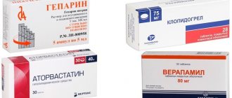
Antiplatelet agents
All patients with angina pectoris are prescribed acetylsalicylic acid for life at a dose of 75-150 mg/day in the absence of contraindications. The dose should be minimally effective, since with increasing dose the risk of developing gastrointestinal side effects (bleeding, ulcerogenicity) increases.
If there are contraindications to acetylsalicylic acid, it is possible to prescribe clopidogrel, which in studies has shown greater effectiveness and is less likely to cause gastrointestinal bleeding. However, the high cost of clopidogrel creates certain difficulties. It has also been shown that the addition of esomeprazole (80 mg/day) to acetylsalicylic acid is better than switching to clopidogrel for the prevention of recurrent ulcer bleeding in patients with peptic ulcer disease and vascular disease.
Symptomatic therapy
β-blockers
β-blockers are effective in relieving angina attacks and are recommended as first-line drugs to relieve anginal episodes. Their antianginal effect is due to a decrease in myocardial oxygen demand due to a decrease in heart rate (HR) and blood pressure. Diastole also lengthens and thereby increases the time of blood supply to ischemic areas of the myocardium. The most preferred are cardioselective β-blockers (they are less likely to cause side effects than non-selective ones), among which the most widely used are metoprolol, bisoprolol and atenolol. The effectiveness of taking a β-blocker is judged by the following clinical parameters: heart rate at rest <60/min, and at maximum physical activity <110/min. β-blockers for coronary heart disease, in addition to their symptomatic effects, have a significant impact on the patient’s further prognosis: their use reduces the risk of developing ventricular fibrillation (the main cause of sudden coronary death) and myocardial infarction (including recurrent).
Side effects
:
- cold extremities;
- symptomatic bradycardia;
- exacerbation of symptoms of bronchial asthma and COPD;
- sexual dysfunction;
- nightmares;
- general weakness.
Calcium channel blockers
There are 2 subgroups of calcium channel blockers: non-dihydropyridine derivatives (for example, verapamil and diltiazem) and dihydropyridine derivatives (for example, nifedipine and amlodipine). The mechanism of action of these subgroups differs, but they all have antianginal effects and are effective in the treatment of angina pectoris. All calcium channel blockers are prescribed in long-acting forms, which are taken once a day. Dihydropyridine derivatives can be added to β-blockers in patients who cannot achieve the desired effect. The combination of non-dihydropyridine calcium channel blockers and beta-blockers is not recommended as excessive bradycardia may occur. Non-dihydropyridine calcium channel blockers can replace beta-blockers if there are contraindications to the latter (for example, bronchial asthma, COPD, severe atherosclerosis of the lower extremities).
Among the side effects, the most common is peripheral edema of the legs (especially when taking dihydropyridine derivatives). Diltiazem can lead to symptomatic bradycardia, verapamil - to constipation and hyperemia, which in turn can lead to a deterioration in myocardial contractility, which should be taken into account when combined with beta-blockers.
Nitrates
Currently, 3 drugs in this group are used: nitroglycerin, isosorbide dinitrate and isosorbide mononitrate. When prescribing these drugs, you need to know that nitrates are classified into dosage forms of short action (<1 hour), moderate long action (<6 hours) and significant long action (6-24 hours).
For exertional angina of functional class I, short-acting nitrates (tablets, capsules, aerosols of nitroglycerin or isosorbide dinitrate) are prescribed, which are taken 5-10 minutes before the expected physical activity to prevent the development of an attack of angina. If an attack of angina is not controlled by taking short-acting nitrates, it is necessary to suspect myocardial infarction or non-cardiac pain.
For exertional angina of functional class II, in addition to short-acting nitrates, moderate long-acting forms can be used.
For exertional angina of functional class III, isosorbide mononitrate (significantly prolonged action) is prescribed. It is taken continuously throughout the day with a nitrate-free period of 5-6 hours (usually at night) to avoid nitrate tolerance.
With angina pectoris of functional class IV, angina attacks can also occur at night. At the same time, extended forms of nitrates are prescribed to ensure their round-the-clock effect and, more often, in combination with other antianginal drugs (for example, β-blockers).
Side effects
(due to vasodilation):
- headache;
- facial hyperemia.
An overdose of drugs can lead to orthostatic hypotension and reflex activation of the sympathetic nervous system, which leads to tachycardia, leading to an attack of angina.
Tolerance to nitrates develops when taking extended forms. There is a drug called molsidomine, the mechanism of action of which is similar to organic nitrates, but tolerance does not develop to it.
Other antianginal drugs
They are additionally used for tolerance to other traditionally prescribed drugs (slow calcium channel blockers, β-blockers, long-acting nitrates).
Nikorandil
Nicorandil is a hybrid compound containing an activator of ATP-dependent potassium channels and nitrate fragments. The drug dilates both stenotic and non-stenotic coronary vessels. Its effectiveness as an additional drug has been proven. Prescribed at a dose of 20 mg twice a day. Side effect: headache.
Ivabradin
Ivabradine is the first inhibitor of If channels with selective and specific action, a pulse-lowering drug. Unlike other drugs that reduce heart rate, ivabradine preserves myocardial contractility and diastolic function, without affecting the electrophysiological parameters of the heart, peripheral vascular resistance, metabolism of carbohydrates and fats and without reducing blood pressure. Study results showed a significant reduction in cardiovascular mortality and frequency of hospitalizations due to worsening CHF, additional to what has already been achieved with ACE inhibitors, β-blockers and AMKR.
Ivabradine, whether prescribed as monotherapy or combination therapy, has also been shown to be effective against both angina symptoms and exercise test scores. The addition of ivabradine to optimal therapy was associated with a significant reduction in the risk of hospitalization due to myocardial infarction. Side effect: slight change in light perception when taking high doses. The starting dosage is 5 mg 2 times a day for 2 weeks, then 7.5 mg 2 times a day.
Trimetazidine
Trimetazidine is a metabolic drug that maintains energy balance and prevents the development of ionic disorders during ischemia. Trimetazidine also stimulates glucose oxidation and acts as an inhibitor of fatty acid oxidation. The efficiency is extremely low. The mechanism of its action is not fully understood. Side effects: weakness and drowsiness.
Ranolazine
Ranolazine is a selective inhibitor of the late current of sodium ions, slows down the voltage-dependent release of calcium from the cell and reduces the negative effect on cardiomyocytes. Ranolazine at a dosage of 500-1500 mg twice a day or extended-release ranolazine at a dosage of 750-1000 mg twice a day, increasing exercise tolerance and reducing attacks of angina and myocardial ischemia, complements symptomatic treatment. Side effects: constipation, dizziness, nausea and fatigue.
Treatment of stable angina
Treatment of the disease includes general measures, medications and cardiac surgery. General measures boil down to lifestyle modification, correction of elevated blood pressure, and prescription of sedatives of plant origin (valerian, St. John's wort, motherwort). Sometimes these measures in patients with FC I are enough to stop pain attacks for a long time.
Drug treatment of stable angina consists of prescribing the following groups of drugs:
- beta-blockers (atenolol, carvedilol, propranolol, etc.). They are prescribed to reduce heart rate, reduce vascular tone, reduce the load on the heart muscle and reduce its need for oxygen. In the absence of contraindications (bronchial asthma, chronic obstructive pulmonary disease) are taken daily. - nitrates are peripheral vasodilators, dilate the coronary arteries and veins, reducing blood flow to the heart and the load on the heart muscle. Short-acting drugs are taken to relieve pain attacks in the form of tablets (nitroglycerin) and aerosol (nitromint, nitrospray). They begin to act in 1 – 2 minutes, the duration of action is no more than 15 minutes. Long-acting drugs (isosorbide, cardiquet, monocinque) are used to prevent angina attacks in patients with FC III - IV daily or before physical activity. - calcium channel antagonists (amlodipine, verapamil) reduce the load on the heart, reducing vascular tone and blood pressure. If there are contraindications to beta blockers, they can be taken daily. - antiplatelet agents (thrombo Ass, aspicor, aspirin cardio) are prescribed to prevent platelet aggregation and their deposition on atherosclerotic plaques. Covered with an enteric film that protects the stomach wall from the irritating effects of aspirin. Taken daily after meals once a day. - lipid-lowering drugs (lovastatin, atorvastatin, rosuvastatin) reduce cholesterol levels in the blood, preventing further formation of new plaques. Taken once a day at night. - ACE inhibitors (perindopril, quadripril) are used to correct arterial hypertension and to protect blood vessels, kidneys, brain, heart from high blood pressure in the vessels.
Surgical treatment methods include:
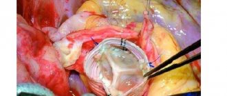
- stenting of the coronary arteries - installation of a metal structure in the artery - a stent that mechanically expands the vessel - balloon angioplasty of the coronary arteries. It is carried out by introducing a catheter with a balloon at the end through the femoral vein, expanding at the site of narrowing of the vessel and “crushing” the plaque, due to which the patency of the artery is restored. After angioplasty, stenting can be performed immediately due to the frequent development of restenoses (repeated narrowings); after it - aorto-coronary bypass surgery - the creation of a shunt (ostium) between the aorta and the affected artery, bypassing the site of narrowing
Indications for operations are the ineffectiveness of drug therapy, the presence of high functional class angina in young people, critical narrowing of the artery lumen (more than 75%), post-infarction angina, and others. Indications and contraindications are determined by the attending physician on an individual basis.
Surgery
Surgical treatment involves performing coronary artery bypass grafting (CABG) or balloon angioplasty (angioplasty) and stenting of the coronary arteries.
When performing CABG, a bypass graft is placed between the aorta and the coronary artery. Autografts (the patient's own veins and arteries) are used as a shunt. The most “reliable” shunt is considered to be a shunt from the internal mammary artery (mammary-coronary bypass).
A less traumatic method of surgical treatment is balloon angioplasty and stenting, the meaning of which is to dilate the affected area of the coronary artery with a special balloon and implantation of a special metal structure - a stent. Due to its low efficiency, balloon vasodilation in its pure form (without subsequent stent implantation) is practically not used today. The implanted stent can be “bare” (bare metal stent), or carry on its surface a special medicinal substance - a cytostatic (drug eluting stent). Indications for a particular method of surgical treatment are determined individually in each specific case after mandatory coronary angiography.
Stem cell treatment
Multipotent stem cell therapy is a promising method for treating many diseases, but is currently in the stage of clinical and pre-clinical trials. The main idea of this therapy is that when stem cells are introduced into the patient’s body, they themselves will go to the site of injury and turn into cells that need to be replaced. However, such a result is not at all guaranteed, and the cell can follow any of the differentiation paths. Specific markers that control the direction of cell differentiation have been poorly studied. All currently existing methods of stem cell therapy do not have evidence of effectiveness performed in accordance with the standards of evidence-based medicine.
Causes of angina pectoris
It has been proven that the most important cause of the occurrence and development of angina pectoris is the rapid narrowing of the coronary vessels of the heart. Their lumen is greatly reduced, which impairs blood supply. In turn, the narrowing of the coronary vessels can be caused by the formation of cholesterol plaques in their thickness.
Risk factors
But there are also risk factors that can affect the development of this disease. There are 2 forms: removable and irremovable. It is worth considering each form in more detail.
Avoidable Risk Factors
Avoidable risk factors include those that directly depend on a person’s activities and lifestyle. The patient can influence them, completely eradicating them or, conversely, developing them.
Avoidable risk factors include:
- Obesity. Excess pounds not only greatly limit a person’s activity, but also lead to the development of many cardiovascular diseases. With obesity, the heart endures a greater load, and blood supply is significantly impaired.
- Smoking tobacco.
- Frequent consumption of alcoholic beverages. They destabilize the myocardium.
- Lack of regular physical activity.
- High or low blood pressure.
Unavoidable risk factors
Fatal risk factors, on the contrary, do not depend on the patient himself. In most cases, they lead to the development of chronic forms of angina pectoris.
Unavoidable risk factors include:
- Bad heredity. Many diseases and illnesses can be acquired from parents. The form of such diseases, as mentioned above, is usually chronic. With each generation they appear and become less acute.
- Floor. Oddly enough, gender also affects the development of angina pectoris. Mostly men suffer from this disease. This is due to the presence in the female body of a special hormone that is responsible for the elimination of low-density lipoproteins and other residual elements.
- Age. It has already been mentioned above that the older a person is, the higher the risk of developing heart and myocardial diseases.
After the age of 70, both men and women have the same risk of developing coronary diseases. This is due to the cessation of estrogen production in women after menopause.