Fascioliasis is a parasitic disease that mainly affects ruminants (cows, sheep), but it can also infect humans. The causative agent of the disease is the giant trematode (Fasciola gigantica) and the liver fluke (F. Hepatica). The causative agents of fascioliasis affect internal organs - the bile ducts and liver tissue. Water contaminated in reservoirs and plants growing near the water are carriers of the causative agent of the disease fascioliasis, which, in turn, is eaten by animals and even people.
The disease is common in South America, Central Asia, and Transcaucasia. The disease belongs to an extremely dangerous category all over the world, all facts of invasion are clearly recorded, and if the growth of the disease increases, appropriate preventive measures are immediately taken. In rare cases, quarantine is declared when the causative agent of fascioliasis is detected in people.
Course of the disease
A person can suffer from the disease, both acute and chronic. The initial and widespread signs are considered to be a severe allergic reaction that occurs in the patient’s body as a result of the release of toxic waste products by worms. Adult parasites cause enormous harm to the body, forming a chronic form of fascioliasis, parasitizing in the liver tissues and in the walls of the bile ducts, disrupting their work with their suction cups and spines, causing serious mechanical damage.
There is a malfunction of the bile ducts and a violation of the outflow of bile is observed, as a result of this process bacterial invasion develops. An undiagnosed disease leads to serious consequences, or rather to the death of vital liver cells.
Fascioliasis in the acute stage of the disease can be successfully treated with prescribed antiparasitic drugs. The advanced chronic stage of the disease is very difficult to cure, almost impossible
Routes of infection
Fascioliasis is a typical anthropozoonosis. The mechanism of infection is predominantly oral and nutritional. This means that a person can become ill by eating contaminated meat, watercress or drinking contaminated water. After this, the helminth larvae enter the intestinal cavity, where they begin their development in the patient’s body.
The prevalence of fascioliasis is greatest in Asia, Africa and Latin America.
The main source of infection for humans are sheep and cattle, which under natural conditions are the final hosts for the parasite in fascioliasis. However, the entire development cycle of the helminth is much more complex. It begins from the moment eggs enter the water with feces. There, larvae are released, which penetrate the body of the intermediate host - a mollusk, where they transform into cercariae. After emerging, they attach themselves to the leaves and stems of underwater plants. They are the ones that are eaten by cattle or humans. Additionally, some of the helminths remain on the surface of the water.
Transmission routes
Infection of a person with fascioleosis depends on many factors:
- living in a region where intermediate hosts (snails) live,
- presence of domestic herbivores,
- climatic conditions,
- nutrition.
Pathogens primarily prefer animals (goats, sheep, cattle) for reproduction; they are natural reservoirs for parasites. Animals of other species, being infected, cannot infect humans with fascioliasis. Although there is information that in Bolivia, donkeys and pigs have the ability to spread the infection. The transmission of fascioliasis disease by wild gray rats (Rattus Rattus) has been established; they are carriers of the parasite in Corsica (region of France). The natural reservoir for the liver fluke in France is considered to be nutria (Myocastor coypus).
Basically, people become infected not from an infected animal, but from eating plants containing invasive cercariae (free-swimming larvae). A number of aquatic plants are susceptible to infection and are sources of infection for humans. In European countries, such sources are considered to be: common watercress (watercress), forest watercress, amphibian watercress (wild cress), dandelion, field lettuce, spearmint.
Liver fluke cercariae are found in an encapsulated state on the surface of natural bodies of water, so a person can easily become infected by drinking such water. During the study and experiments to study the disease, it turned out that a person can easily become infected by eating raw or poorly cooked liver of sick animals, by eating immature liver parasites.
Etiology
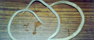
The causative agent of human fascioliasis can be two microorganisms - the liver fluke and the giant fluke. They belong to the same subspecies, as they have a large number of common morphological characteristics. In addition, they can mate with each other. Their main difference is their volumes - the liver fluke is up to three centimeters long and up to 1.3 cm wide, and the giant fluke reaches eight centimeters in length and twelve millimeters in width.
Agricultural livestock act as carriers, but only of the herbivorous group. Fasciola parasitizes the hepatobiliary system, in which helminths lay eggs, and they, in turn, enter the environment along with the animal’s feces, and further develop in fresh water bodies.
Developmental biology of fasciolae
The ways of infection with fascioliasis are as follows:
- consumption of contaminated wild plants - this includes sorrel and watercress, garden greens and wild onions;
- drinking raw water from questionable reservoirs;
- poor sanitary treatment of garden herbs and vegetables;
- eating fascioliasis liver of an animal - in this case, transient eggs enter the human intestine, which do not provoke the development of the disease, but are detected during laboratory examination of feces;
- accidental entry of contaminated water into the oral cavity while bathing.
In all cases, except the last one, the pathogen larvae are released from their membranes in the gastrointestinal tract, after which they penetrate the peritoneum through the intestinal wall. Then fasciola penetrates the liver parenchyma and penetrates the bile ducts. Another route of migration of such a microorganism cannot be ruled out – hematogenous, i.e. with the blood flow.
About four months after entering the human body, fasciolas reach sexual maturity and can lay eggs. The period of parasitism can vary from five to ten or more years.
Symptoms in humans
Symptoms from the moment the pathogen enters may appear on the eighth day, but this is not a fact; perhaps the disease will manifest itself in a few months. Signs of fascioleosis are in many ways similar to allergic reactions of the body, these are:
- a sharp increase in body temperature (more than 40C);
- rashes on the skin;
- swelling and change in skin color (urticaria);
- endless itching of skin rashes;
- Jaundice often appears.
Fascioliosis in humans is accompanied by symptoms from the nervous system and gastrointestinal tract, such as:
- headache,
- weakness,
- general malaise,
- incomprehensible general abdominal pain,
- chills,
- feeling of nausea,
- prolonged vomiting,
- flatulence,
- belching,
- presence of hotness in the mouth,
- diarrhea.
During a medical examination by a specialist of an infected patient, a significant enlargement of the liver is detected; the person feels discomfort and pain when pressing on this organ. But even with other diseases, these sensations are not excluded.
Depending on the patient’s body and his immunity, additional possible clinical symptoms of myocarditis can be added to the above symptoms, which are accompanied by high blood pressure, severe chest pain, as well as tachycardia.
In the chronic stage of the disease, the symptoms are slightly weaker than in the acute stage, but the patient also suffers from heaviness and aching pain in the right hypochondrium. Stages of fascioliasis in humans
There are 4 main phases of fascioliasis in humans:
- The incubation phase is from the pathogen entering the body to the first symptoms. This phase depends on the host’s immune system and on the number of metacercariae that have entered the body (the developmental stage of a mature larva of the class of trematodes), development lasts from several days to three months.
- Infectious or acute phase - at this stage, parasites make their way to the bile ducts. Helminths infect liver tissue and the abdominal cavity and release toxic mechanical substances. The affected body suffers from severe allergic reactions and gastrointestinal disorders.
- Latent (hidden) phase - lasts secretly and asymptomatically for months and even several years; the disease can be discovered completely by accident: during a medical examination or any medical research.
- Chronic phase - develops and progresses over several years. During this time, helminths become adult flukes, parasitizing the bile ducts, causing inflammation and proliferation (hyperplasia) of the epithelium. As a result, cholangitis or cholecystitis develops; due to the large number of parasites and their rather large size, the bile ducts become clogged. At this stage, the patient has an enlargement of not only the liver, but also the gall bladder, ascites (fluid accumulation in the abdominal cavity) is possible, swelling appears, the inflammatory process of the gall bladder forms adhesions of the outer walls, which press on neighboring organs. In parallel with inflammation, small and numerous stones form in the gall bladder, as well as in the bile ducts.
Timely diagnosis and competent therapy play an important role in the disease. Late treatment threatens the patient with irreversible complications such as:
- purulent cholangitis,
- jaundice,
- liver abscess.
Fascioliasis affects not only the liver and bile organ; cases of patients with these parasites in the brain, mammary glands, lungs and eyes have been recorded
Treatment methods
It is prohibited to use albendazole during pregnancy and lactation, as it can be toxic to the child’s body.
Also, the drug should not be prescribed if the patient has hypersensitivity to it.
Albendazole should be prescribed with caution in cases of liver dysfunction, since the drug has a moderate hepatotoxic effect. Therefore, if it is necessary to use a medication, it is necessary to control the concentration of AST, ALT, bilirubin and its fractions.
Tablets are taken orally after meals, 400 mg once a day. The therapeutic concentration of the drug is maintained for 20 hours. The drug should be taken with a sufficient amount of water.
The duration of treatment with specific drugs is from one to three days, depending on the severity of the parasitic infestation.
Traditional medicine plays a supporting role in fascioliasis. Choleretic herbs and herbs are of greatest importance, which improves the effectiveness of antiparasitic drugs, reduces the incidence of complications and speeds up the patient’s recovery.
Nutrition is of great importance during therapy. The patient should avoid foods high in fat (especially animal origin), as well as canned food, processed foods and alcoholic beverages.
Diagnostics
It is quite difficult to identify the disease in the early stages. Even diagnostics with microscopic equipment cannot detect parasite eggs, since the causative agents of fascioleosis are not able to lay eggs for a certain time and this is their characteristic feature. After three months from the moment the pathogen enters the host’s body, it is possible to detect the presence of helminth eggs in a laboratory way.
Experts recommend that if symptoms of the disease appear, do not delay visiting a doctor and be sure to undergo an ultrasound diagnosis of the liver and bile ducts. This is the only way to detect the presence of leaf-shaped pathogens in the patient’s body.
Treatment of fascioliasis
When the diagnosis of fascioliasis is confirmed, the attending physician selects the necessary medications and does not resort to surgical intervention. Laboratory diagnosis of fascioliasis is carried out by conducting an ultrasound examination of the liver and gallbladder, as well as using a CT scan of the lungs. Usually, for fascioliasis, the drugs Biltricid and Chloxil are prescribed. Before starting treatment with anthelmintic drugs, it is customary to use symptomatic therapy, the purpose of which is to relieve pain and other acute forms of manifestation of this disease. Symptoms of the disease are treated with:
- Antipyretics;
- Antihistamines;
- Proton pump blockers;
- Sorbents;
- Antispasmodics;
- Enzyme medications;
- Antibiotics;
- Choleretic and hepatoprotective drugs.
There are cases where fascioliasis became the cause of hepatitis. In this situation, glucocorticosteroids are prescribed for treatment.
At the end of treatment, the patient must undergo general blood and stool tests. The diagnosis is repeated after 3-4 months.
Treatment
Various methods are used in the treatment of fascioleosis in humans; therapy is selected individually and depends on the stage of the disease, as well as on the pathological process in the patient’s body. In the acute stage, a gentle diet is prescribed in addition to the main treatment to reduce the load on the liver; it is recommended to completely eliminate fatty, fried, sweet, and spicy foods. If a patient has myocardial symptoms or hepatitis, additional glucocorticosteroids are prescribed. A full range of antiparasitic drugs are prescribed to remove the organism from the acute stage. Choleretic medications will help cleanse and expel helminths from the bile ducts.
Antiparasitic drugs are effective for humans and pets. The main anthelmintic drug is triclabendazole. Adults and children over 6 years of age are prescribed 10 mg per 1 kg of body weight, a single dose, in case of severe disease - twice with a break of 12 or 24 hours.
Albendazole is considered an alternative drug, mainly in veterinary medicine.
Therapy with the drug praziquantel is ineffective. In medical practice, cases of treatment of fascioleosis in humans with nitazoxanide in Mexico are described; it is considered an expensive remedy and in modern times is not recommended by specialists.
At the beginning of the 21st century, the Egyptian drug Mirazid, made from a special tree resin (myrrh), was developed; this antiparasitic drug has proven itself to be highly effective. Further research into it for the treatment of fascioliasis did not result in much progress.
In case of purulent complications of the disease, specialists prescribe antibacterial agents, the dosage is calculated individually for each patient. If a liver abscess develops, surgical intervention is possible.
After undergoing therapy, the patient is under medical supervision for six months, laboratory tests are carried out on stool for the presence of parasites, and material from the bile ducts is also examined.
Symptoms and causes of fascioliasis
The helminths that cause fascioliasis can parasitize, in addition to humans, many wild and domestic animals, but the hosts always remain definitive. Typically, the life span of the parasite lasts about 5 years, during which time the worm lays approximately 2 million eggs. Eggs caught in unfavorable soil quickly die. Eggs develop normally only in water, where the embryo goes through several stages of development and finds its intermediate host - a small pond snail, a type of mollusk. In it, the parasite completes its development and returns back to the water, where it attaches to plants growing on the shore and becomes covered with a shell. With water or food, the parasite enters the human or animal body, where after 3 months it becomes a sexually mature individual.
Fascioliasis manifests itself quite acutely in humans: body temperature rises, muscles and joints ache, itchy skin and a rash appear all over the body. The temperature rise can reach up to 40C°, while the fever can be either constant or intermittent. Then the stomach begins to hurt, the size of the liver increases, yellowing of the skin develops, and diarrhea occurs.
After a few weeks, these symptoms subside and the disease enters the subacute phase. From 3 months to six months after infection, fascioliasis enters the chronic phase. The penetration of worms into various organs of the body is accompanied by the presence of various symptoms, including:
- With helminths in the brain, headaches, dizziness, epileptic seizures, unsteadiness when gait are observed;
- When the worms reach the lungs, coughing, chest pain, shortness of breath, and hemoptysis are typical;
- When parasites get into the eyes, visual acuity usually decreases.
Prevention
Avoiding infection is simple - you just need to follow the rules of personal hygiene and food hygiene.
- It is forbidden to use water from natural reservoirs, much less consume it as food; the water must be boiled properly;
- wash fruits, vegetables, herbs;
- buy proven meat that has passed veterinary control;
- it is important to monitor and conduct a veterinary examination of cattle to avoid the spread of this disease;
- carry out sanitary education work among residents of dangerous regions.
Prevention of this pathology
To avoid infection with liver fluke, a number of preventive measures are taken:
- The possibility of untreated water from reservoirs entering the body is excluded; simple boiling is used for this, and in cases where it is not possible to limit the risk of infection in this way, it is necessary to filter the water through a cloth.
- Eat only clean vegetables and herbs that have been thoroughly washed.
- Carrying out veterinary preventive measures aimed at reducing the incidence of disease in livestock, and which involve the use of fresh hay, the fight against various mollusks in reservoirs near grazing animals.
- Ensuring detection and timely treatment of disease in animals and humans.
In most cases, with timely treatment, fascioliasis can be completely cured.
Symptoms in animals
The number of metacercariae entering the body affects the clinical symptoms of fascioleosis. The most common definitive hosts are sheep, and their clinical symptoms are divided into 4 types:
- Acute type 1: an invasive dangerous dose is more than 5000 metacercariae eaten. Clinical symptoms do not have time to manifest themselves in full force, as the sheep die. In rare cases, they experience ascites, abdominal bleeding, jaundice, weakness, and pale skin.
- Acute type II: invasive and dangerous dose of 1000 to 5000 ingested metacercariae. In the second acute type, sheep die; symptoms manifest themselves in the form of pallor, loss of consciousness and ascites.
- Subacute type: invasive dose of 800 to 1000 metacercariae eaten. With such a dose of parasites, sheep become lethargic, anemia progresses, and death may occur. Basically the weight of the animal is lost.
- Chronic fascioliasis: invasive dose of 200 to 800 ingested metacercariae. Initially, the disease is asymptomatic, and then the body becomes exhausted, loses weight, and develops swelling under the lower jaw and ascites.
A complete blood test determines anemia, hypoalbuminemia (decreased blood albumin levels), and increased eosinophils (eosinophilia). With this disease, liver enzymes in the blood increase significantly - glatamate dehydronenesis (GLDH), gamma-glutamyl transferase (GGT), lactate dehydrogenase (LDH), they can be detected 12-15 weeks after metacercariae enter the body, or in acute and chronic types of fascioliasis.
The disease causes great economic loss for sheep breeders, since most animals quickly lose weight and die.
Almost the same clinical signs occur in cattle. Adult representatives are more resistant to liver fluke infection.
The consequences of fascioliasis in livestock include economic losses caused by liver disposal after slaughter and weight loss.
Diagnosis of fascioliasis
Diagnosing diseases in the early stages (in the acute phase) is quite problematic. The presence of fascioliasis is assumed by careful study of data from epidemiological, anamnestic and clinical studies.
The possibility of mass invasion of certain groups of people (geologists, tourists, etc.) is allowed.
At the same time, the presence or absence of cases of the disease in a given region is determined.
At the moment, diagnosis of the disease involves the use of immunological methods, such as serological test systems, RSK, RIF, REMA. Confirmation of the diagnosis is possible only by studying stool and duodenal contents for the presence of parasite eggs in them.
This examination is carried out no earlier than 3 months after possible infection.
The diagnosis is made on the basis of: 1) Epidemiological data (the fact of bathing or drinking water from stagnant bodies of water, washing salad herbs with this water, eating unwashed salad herbs).
2) Clinical data (symptoms of the acute phase of the disease or the chronic phase of parasitosis). 3) Laboratory diagnostics are carried out depending on the stage of the disease.
In the early phase, coproovoscopy is difficult, since eggs are produced after 3-3.5 months, so serological diagnostic methods, that is, blood testing for the presence of antibodies - RNGA reactions, ELISA, become of primary importance.
In the chronic phase - copro-ovoscopy or duodeno-ovoscopy (detection of fasciolae eggs in stool and duodenal contents). Stool examination (coproovoscopy) is carried out twice at an interval of 7-10 days to exclude the detection of not true eggs, but transient ones (when fascioliasis liver is consumed in canned food, pates).
Until re-examination, calas by-products are excluded from the diet. Research is carried out using the enrichment method (due to the small number of eggs).
Differential diagnosis is carried out with the following diseases: allergic conditions and reactions, gastroduodenitis, hepatitis, cholecystitis, cholangitis, helminthiases of other etiologies (opisthorchiasis, clonorchiasis, trichinosis), liver cirrhosis and others.
Identifying the disease at an early stage is quite a difficult task; for this, the patient needs to consult an infectious disease specialist. Diagnosis is based on various data.
Epidemiology
In many regions, cases of human and animal infection with liver fluke have been reported. Countries that raise cattle, sheep, and goats are especially affected by the disease. In Western Europe, cases of human infection are excluded, but in developing countries many people become infected with fascioliasis.
In recent years, based on the results of the study, experts have drawn the following conclusion: human fascioliasis is a significant problem for global health. The incidence is increasing, statistics show that 51 countries on five continents have reported cases of infection. These are the countries of Europe, America, Africa and Oceania. The epidemic spread of a disease in humans in a certain area has virtually no effect on diseases in animals. In South America, in the regions of Bolivia and Peru, pathogens are often found in humans, and no outbreaks of the disease have been recorded in veterinary medicine. Or in countries such as Uruguay, Argentina, Chile, which are leaders in cattle breeding, outbreaks of fascioleosis in humans are extremely rare.
Europe
In European countries, fascileosis in humans occurs in France, Spain, Portugal, as well as in post-Soviet countries. France is considered the endemic region. From 1970 to 1982, nine French clinics recorded 5,863 infected people.
In the countries of the former USSR, the main source of infection was Tajikistan. According to the latest indicators, Turkey was added to this list, having conducted medical serological studies of fascioleosis in people, in some areas of this country, antibodies were detected in blood tests of patients in 3.01% in Antalya, from 0.9 to 6.1% in the Mediterranean region of Turkey (Isparta). In many European countries, the disease fascioleosis in humans is random, and the disease develops upon arrival from countries with an endemic threshold where helminth pathogens are common.
North and South America
This disease is rare in North America. In regions of Mexico, 53 cases were recorded. Central America regularly records cases of fascioliasis in humans, especially in the Caribbean islands, in the Puerto Rico area and in Cuba. The endemic foci are the provinces of Pinar del Rio and Villa Clara. The most famous hyperendemic areas include the high plains (plateaus) of the Altiplato. In a medical examination of residents of the northern Bolivian Altiplano, infection rates ranged from 72% to 90%.
In Peru, the liver fluke is distributed throughout the country in humans. The countries of Uruguay, Brazil, Venezuela, and Colombia have high rates of infection in animals; in humans, infection is a rare case.
Africa
In Africa, cases of infection occur in the northern regions, in the Nile Delta, which flows in Egypt.
Asia
Among Asian countries, it is Iran that suffers from fascioliasis infection; more than 10 thousand people are registered there. The pathogen can rarely be found in East Asia. Several cases of the disease have been reported in Japan, Vietnam, and Thailand.