About anatomy and topography
The aorta is the main trunk of arteries in the systemic circulation. It originates in the cavity of the left ventricle of the heart. Consists of 3 parts:
- ascending;
- average;
- descending.
The aortic arch is the middle part. It is a derivative of the 4th left arterial arch. Topographically located between the manubrium of the sternum and the IV thoracic vertebra. The arc moves backward and to the left. Then it spreads over the apex of the left bronchus, where the descending part of the aorta begins.
Conventionally, the structure consists of 2 parts:
- concave;
- convex.
From the concave side of the aortic arch, blood vessels depart from the bronchi and the thymus gland. 3 trunks originate from the convex part, located from right to left:
- Brachiocephalic (brachiocephalic).
- General carotid (carotid) left.
- Left subclavian.
The branches of the aortic arch extend upward from its middle part. All of these arteries supply the upper half of the body, including the brain.
Types of cardiosclerosis
There are several types of cardiosclerosis. Classification is carried out according to affected areas.
- The diffuse form is characterized by uniform damage to the entire heart muscle.
- Cicatricial cardiosclerosis forms scar connective tissue in focal areas of the heart muscle.
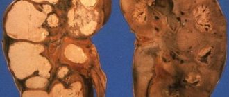
Atherosclerotic cardiosclerosis is a consequence of ischemic disease. Slow replacement of connective tissue occurs due to impaired blood supply to the myocardium and oxygen starvation of the heart muscle.
With this disease, contractility decreases. The heart increases in size, and left ventricular hypertrophy occurs. An X-ray of the heart determines the condition of the scar tissue and the degree of its growth.
Anomalies, defects and diseases
Blood vessel pathology can be divided into 2 large groups:
- Congenital.
- Acquired.
In the first case, disturbances occur at the stage of embryogenesis. This depends on hereditary predisposition and the action of aggressive factors in the early stages of pregnancy. Changes may be found in other parts of the aorta. If such a situation arises, then they talk about combined and combined defects.
With acquired pathology, the aortic arch initially does not have anatomical defects or abnormalities. The lesion is a consequence of the underlying disease.
Congenital defects and anomalies include:
- Hypoplasia.
- Atresia.
- Pathological tortuosity (Kinking syndrome).
- Coarctation.
- Defects of the middle section system, among which are:
- complete double aortic arch;
- malformations of the right and left arches;
- anomalies in length, size, continuity;
- anomalies of the pulmonary trunk and arteries.
Of the acquired diseases, the middle part is affected by:
- atherosclerosis;
- gunshot and knife wounds;
- Takayasu aortoarteritis;
- aneurysms.
Such a variety of possible lesions of this part of the circulatory system ensures that doctors are interested in early diagnosis and timely treatment.
Signs and symptoms
Today, thanks to the availability of modern equipment, it is possible to diagnose atherosclerosis of the abdominal aorta in the early stages. This disease is asymptomatic and can only be detected through random examinations. Computed tomography shows changes occurring at the very beginning of the disease. But externally recognizing atherosclerosis of the abdominal region is very difficult; a person can live with this pathology for years and not even suspect it. But as it progresses, typical signs of atherosclerosis of the abdominal aorta will begin to appear:
- Discomfortable sensations in the abdominal area.
- Frequent pain inside the abdominal cavity, which becomes worse after eating.
- Digestive system disorders – constipation or diarrhea.
- Pulsating contractions from the left side of the abdomen and around the navel.
- Belching, heartburn, nausea after eating.
- Weight loss.
Brief characteristics of individual species
Hypoplasia is a uniform tubular narrowing. This limitation in the diameter of the blood vessel prevents the full outflow of blood from the left ventricle. In this case, not only the arch itself, but also the descending aorta and the ascending section can be involved in the pathological process.
In most cases it is combined with other defects. Most of these patients die at an early age. Treatment is only surgical.
Atresia or break is called Steidel's anomaly. In this case, one of the segments of the vessel is completely missing. The consequence of this is that the descending aorta does not communicate with the ascending aorta.
They are isolated from each other. Blood supply is provided by the open ductus arteriosus. Children with this defect die in the first month of life without surgical intervention.
Pathological tortuosity is called Kinking syndrome. Its essence is that the aortic arch at its distal end has an abnormal length and curvature. Patients with this defect do not make any complaints.
When Kinking syndrome is detected in children, doctors choose a wait-and-see approach. As the child grows, the defect may go away on its own.
This anomaly is more often diagnosed in women. It is a narrowing of any part of a blood vessel. When the branches of the aortic arch are affected, there are several options:
- Stenosis or atresia of the left subclavian artery.
- Stenosis of the right subclavian artery.
- Anomalous origin of the right subclavian artery:
- distal;
- proximal.
The narrowing can be localized, but usually consists of a pathological process spread over several cm. Often combined with other congenital anomalies. Included in tetralogy of Fallot, Turner syndrome. The defect is detected from birth.
With adequate drug support and a small degree of severity of the anomaly, patients have a favorable prognosis. Early surgical correction can significantly increase life expectancy (up to 35–40 years) and its quality.
Manifestations of atherosclerosis
Quite often, atherosclerosis occurs without obvious symptoms, which is associated with the large size of the aorta (as well as sections and branches of the aorta), developed muscle and elastic layers. The growth of plaques leads to overload of the heart, which is manifested by pressure surges, fatigue, and increased heartbeat.
As the pathology progresses, the process spreads to the branches of the aortic arch of the descending and ascending sections, including the arteries that feed the heart. In this case, the following symptoms occur: angina pectoris (retrosternal pain that radiates to the shoulder blade or arm, shortness of breath), impaired digestion and kidney function, surges in blood pressure, cold extremities, dizziness, headaches, frequent fainting, weakness in the arms.
Defects of the aortic arch system
This group includes anomalies of the position, size, shape, course, relationship and continuity of arterial vessels. Such defects are most often asymptomatic.
Complaints appear when there are pronounced changes and the anomaly spreads to the proximal part of the descending section. Dysphagia or respiratory phenomena may occur due to close pathological contact of the aortic arch and its branches with the trachea and esophagus.
In this case, surgical intervention is necessary to prevent the development of severe complications.
The most common type is a complete double aortic arch. A distinctive feature of this defect is the presence of both arches (right and left), from which branches also extend. They all then merge with the descending artery behind the esophagus.
The prognosis for life in such patients is extremely favorable. In most cases, they do not require medication support.
Urgent Care
Emergency care provided by a general team and a special team is symptomatic, depending on the presence and nature of the leading symptoms. Anesthesia - morphine 0.5 IV and 0.5 IM 1% solution, for collaptoid reactions - dopamine (400 mg in 400 ml 0.9% saline under blood pressure control) or mezaton (1 ml 1% solution -ra) s.c. or i.v. In the conditions of a special team, in the presence of increased A/D, antihypertensive drugs can be administered under close monitoring to prevent further dissection of the aneurysm - sodium nitroprusside - 1.0-1.5 mcg/kg/min IV; magnesium sulfate (10 ml of 25% solution) i.v.
Acquired vices
The most important secondary lesions of the blood vessel are:
- atherosclerosis;
- aneurysm.
In the first case, the lumen narrows due to the forming “fatty” plaques. It is easily diagnosed due to the compacted, emphasized contour of the vessel during an ultrasound of the heart or an x-ray of the lungs.
Compliance with the principles of proper nutrition and rational pharmacotherapy will help slow down the process and prevent complications.
An aneurysm is an area of dilation of a blood vessel. The consequence of this is a narrowing of the lumen of its branches at the place of their origin. The cause of this situation is most often injury or atherosclerotic changes.
For a long time, the pathology may not reveal itself. When the ascending or descending aorta is involved in the process, the aneurysm is large in size and the first symptoms appear.
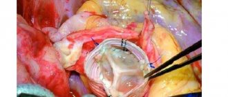
The main method of treatment is surgical. The treatment regimen before surgery must include medications that lower blood pressure to prevent dissection or rupture of the aneurysm.
Top main symptoms
Despite the variety of pathology options in the aortic arch system and its branches, most patients note the following complaints:
- shortness of breath;
- cough;
- hoarseness of voice;
- swallowing disorders;
- headache;
- dizziness;
- temporary paralysis of the limbs;
- swelling of the face.
The listed complaints are due to the involvement of the main branches of the middle part of the aorta in the pathological process. Only a doctor can determine what kind of disease or defect occurs.
To do this, a whole range of various instrumental examinations is performed. Treatment regimens are selected individually, taking into account the type of pathology.
The aortic arch occupies an important place in the process of blood supply to organs and systems. The presence of a defect or disease in its system can lead to serious consequences, death.
Therefore, it is important to undergo medical examination, contact a specialist in a timely manner, and follow all his recommendations.
Aorta, its parts
The aorta is the largest arterial vessel in the human body. All the arteries that form the systemic circulation depart from the aorta. The aorta is divided into the ascending aorta, aortic arch and descending aorta.
I.
Ascending aorta
- emerges from the left ventricle of the heart and in its initial section has an extension - the aortic bulb. The length of the ascending aorta is about 6 cm, all of it is located within the pericardium and goes in the ascending direction. From the ascending aorta depart the right and left coronary (coronary) arteries, which supply blood to the heart.
II.
The aortic arch
is convex upward and goes from front to back from right to left, passing into the descending part. Three large vessels depart from the aortic arch: the brachiocephalic trunk (innominate artery), the left common carotid and left subclavian arteries. The brachiocephalic trunk is a large vessel up to 40 cm long, which goes up and to the right and at the level of the sternoclavicular joint is divided into 2 branches: the right common carotid and right subclavian arteries.
III.
The descending aorta
is a direct continuation of the arch. This is the longest part of the aorta, running from the level of the 3rd – 4th thoracic to the 4th lumbar vertebrae, where it divides into the right and left common iliac arteries, and this place is called aortic bifurcation. The thin median sacral artery departs from the bifurcation into the pelvic cavity. The descending aorta has two parts: up to the diaphragm, the descending aorta is called the thoracic aorta, and below the diaphragm is called the abdominal aorta.
Arteries of the head and neck
Common carotid artery
The left common carotid artery arises from the aortic arch, the right - from the brachiocephalic trunk. Coming out through the upper aperture of the chest, the right and left common carotid arteries are located on the lateral surfaces of the neck and have no branches along their entire length. At the level of the upper edge of the thyroid cartilage of the larynx, each of them is divided into the external and internal carotid arteries.
I.
The external carotid artery
branches many times.
a) The anterior group of branches is formed by:
– superior thyroid artery – supplies blood to the thyroid gland, larynx and neck muscles lying below the hyoid bone, the sternocleidomastoid muscle.
– lingual artery – supplies blood to the tongue, parotid and sublingual salivary glands, neck muscles lying above the hyoid bone.
– facial artery – goes from the angle of the lower jaw to the medial edge of the eye. Supplies blood to facial muscles, pharynx, tonsils, submandibular salivary glands.
b) The back group consists of:
– occipital artery – supplies blood to the skin and muscles of the back of the head, the dura mater.
– Posterior auricular artery – supplies blood to the auricle and mastoid process.
c) The medial group includes the ascending pharyngeal artery, which gives branches to the wall of the pharynx, deep muscles of the neck, dura mater, middle ear, tonsils, soft palate, and auditory tube.
d) Group of terminal branches:
– superficial temporal artery – passes in front of the auricle to the temporal region, giving branches to the parotid salivary gland, auricle, temporal muscle and then divides into frontal and parietal branches that supply the skin and muscles of the calvarium.
– maxillary artery – reaches the infratemporal and pterygoid fossa, supplying blood to the deep areas of the face and head: the cavity of the middle ear, teeth, mucous membrane of the oral cavity, nose and its paranasal sinuses and the soft palate, the dura mater of the brain.
II.
The internal carotid artery
does not give branches in the neck. It rises up and through the carotid canal of the pyramid of the temporal bone passes into the cranial cavity, where it branches into branches:
1) Ophthalmic artery - goes through the optic canal into the orbit and supplies blood to the eyeball and its muscles, eyelids, and lacrimal gland.
2) Arteries of the brain: the anterior cerebral artery, the middle cerebral artery and the posterior communicating artery - supply blood to the brain.
Arteries of the trunk and upper limbs
I.
The subclavian artery
is a paired vessel. The right subclavian artery arises from the brachiocephalic trunk, the left - from the aortic arch. First, the subclavian artery goes under the collarbone, then passes into the gap between the anterior and middle scalene muscles, goes around the 1st rib and passes into the axillary cavity, where it continues under the name of the axillary artery. On its way to the armpit, the subclavian artery gives off a number of large branches:
1. Vertebral artery - rises up through the openings in the transverse processes of the cervical vertebrae, giving branches along the way to the spinal cord and deep muscles of the neck, passes through the foramen magnum into the cranial cavity, merges with the vertebral artery of the opposite side into one basilar artery, from which it branches branches supplying the brain.
2. Internal mammary artery - descends from the subclavian artery into the chest, reaching the diaphragm. Its branches supply blood to the thymus, trachea, bronchi, pericardial sac, diaphragm, chest muscles, and mammary gland.
3. Thyrocervical trunk - goes upward, giving branches to the thyroid gland, muscles of the neck and back.
4. Costocervical trunk - departs from the subclavian artery in the interstitial space, where it immediately divides into branches that supply blood to the muscles of 1-2 intercostal spaces, deep muscles of the head and neck, as well as the spinal cord.
5. Transverse artery of the neck - stretches in the transverse direction from the subclavian artery to the upper part of the trapezius muscle. Nourishes the muscles of the neck and upper back.
II.
The axillary (axillary) artery
is a continuation of the subclavian and lies deep in the axillary fossa. Its main branches:
1. Superior thoracic artery - supplies blood to the mammary gland, intercostal muscles, pectoralis major and minor muscles.
2. Thoracromial artery – clavicle, shoulder and acromioclavicular joints, skin and muscles of the shoulder.
3. Lateral thoracic artery – axillary lymph nodes, chest muscles, mammary gland.
4) Subscapular artery – skin and muscles of the shoulder, shoulder girdle, back.
5) The anterior circumflex artery of the humerus and the posterior circumflex artery of the humerus - supply blood to the muscles of the shoulder, shoulder girdle and shoulder joint.
III.
The brachial artery
is a continuation of the axillary artery and gives off branches to the skin and all muscles of the shoulder, as well as to the elbow joint. In the cubital fossa, the brachial artery is divided into the ulnar and radial, with the ulnar artery lying on the medial side of the forearm, and the radial artery on the lateral side. They supply blood to the skin and muscles of the forearm and elbow joint, and then descend to the hand and form the superficial and deep palmar arches, from which branches extend to the muscles of the palm and fingers.
Arteries of the thoracic and abdominal cavities
Descending aorta and its branches
I.
The thoracic aorta
is adjacent to the vertebral column and has visceral and parietal branches. Visceral branches include:
1. Bronchial - supply blood to the trachea, bronchi, and lungs.
2. Esophageal - to the wall of the esophagus.
3. Mediastinal – to the mediastinal organs.
4. Pericardial - to the pericardial sac.
The parietal branches of the thoracic aorta include:
1. Superior diaphragmatic - supply blood to the upper surface of the diaphragm.
2. Posterior intercostals - 10 pairs pass through 3-12 intercostal spaces - supply blood to the intercostal muscles, mammary glands, skin of the chest and back, and partly the spinal cord.
II.
The abdominal aorta
is a continuation of the thoracic aorta and lies on the anterior surface of the lumbar vertebrae to the left of the midline. At the level of 4–5 lumbar vertebrae, the abdominal aorta is divided into the right and left common iliac arteries (aortic bifurcation). The unpaired median sacral artery departs from the bifurcation site. Along the course of the abdominal aorta, parietal and visceral branches depart from it.
The parietal branches include:
1. Lower diaphragmatic – nourish the lower surface of the diaphragm, partially the adrenal glands.
2. Lumbar arteries - 4 pairs supply blood to the skin and muscles of the abdomen and back, and partially to the spinal cord.
The visceral branches include paired and unpaired. Paired branches supply blood to the paired abdominal organs. These include: renal, adrenal, ovarian (in women) and testicular (in men) arteries. The unpaired visceral branches of the abdominal aorta include: the celiac trunk, as well as the superior and inferior mesenteric arteries.
I.
The celiac trunk
departs from the aorta at the level of the 12th thoracic vertebra and immediately divides into 3 branches:
1. Left gastric artery - goes to the left of the celiac trunk along the lesser curvature of the stomach, feeding its walls, and connects with the right gastric artery.
2. Common hepatic artery - goes to the right from the celiac trunk and is divided into:
a) The proper hepatic artery - nourishes the liver, the gallbladder and gives off a branch to the stomach - the right gastric artery.
b) Gastroduodenal - to the pancreas and duodenum.
c) Right gastroepiploic - runs along the greater curvature of the stomach, supplying blood to its walls and the greater omentum. Connects with the left gastroepiploic artery.
3. Splenic artery - goes to the left of the celiac trunk and supplies the spleen, and also gives branches to the stomach - the gastric arteries and to the greater omentum - the left gastroepiploic artery.
II.
The superior mesenteric artery
- departs from the abdominal aorta at the level of the 12th thoracic - 1st lumbar vertebrae and gives branches to the pancreas, all parts of the small intestine (duodenum, jejunum and ileum), as well as the upper parts of the large intestine.
III.
Inferior mesenteric artery
- departs from the aorta at the level of the 3rd lumbar vertebra and gives branches to all parts of the large intestine (cecum, colon, rectum).
Arteries of the pelvis and lower extremities
Common iliac artery and its branches
The common iliac arteries (right and left) are formed by dividing the abdominal aorta (at the level of the 4th lumbar vertebra). Each of the common iliac arteries at the level of the sacroiliac joint then divides into the external and internal iliac arteries.
I. Internal iliac artery
- descends into the pelvis, supplying blood to its walls and organs. Its parietal branches include:
1. Iliopsoas artery – supplies the muscles of the lumbar region of the back and abdomen.
2. Lateral sacral arteries - go to the bones, skin and muscles of the sacral region.
3. Superior and inferior gluteal arteries - go to the skin and muscles of the gluteal region, pelvis and thigh.
4. Obturator artery - through the obturator foramen it enters the thigh, supplies blood to the muscles of the pelvis, thigh, hip joint, ischium, skin of the perineum and external genitalia.
The visceral branches of the internal iliac artery include:
1. Umbilical artery - to the ureter, bladder, seminal vesicles, vas deferens, epididymis.
2. Uterine artery (in women) – to the walls of the uterus, vagina, fallopian tube and ovary.
3. Middle rectal - to the rectum, seminal vesicles, prostate gland.
4. Internal genital artery to the penis (in men), clitoris (in women), urethra, perineal muscles.
II. External iliac artery
- carries blood to the entire lower limb. In the pelvic area, branches extend from it to the muscles of the pelvis, abdomen, testicular membranes and labia majora. Then the external iliac artery passes under the inguinal ligament and is located on the thigh called the femoral artery, which gives branches to the skin and muscles of the thigh, groin area, and knee joint. A continuation of the femoral artery is the popliteal artery, which lies in the popliteal fossa and weaves its branches around the knee joint. It then divides into the anterior and posterior tibial arteries. The anterior tibial artery gives off branches to the skin and muscles of the anterior surface of the leg, and then continues into the dorsal artery of the foot, going to the muscles of the metatarsus and toes.
The posterior tibial artery - supplies blood with its branches to the posterior surface of the leg and on the foot continues into the medial and lateral plantar arteries - to the skin and muscles of the plantar surface of the foot.
An important role in the functioning of the body is played by the circulatory system, consisting of the heart - a kind of muscle pump that uninterruptedly pumps blood, and vessels, which, according to their structure and functions, are divided into arterial, venous and capillary.
The main trunk, which gives rise to all arterial vessels, is the aorta. And which vessels depart from the aortic arch: we’ll look at it in our review and video in this article.
Branches of the descending department
The descending aorta, in turn, is divided into sections:
- Thoracic, located above the diaphragm;
- Abdominal, located below the diaphragm.
Thoracic section:
- Parietal arterial vessels supply blood to the chest walls: the superior phrenic arteries, the branching surfaces of the diaphragm from the chest cavity, and the posterior intercostal arterial vessels, which supply blood to the intercostal and rectus abdominal muscles, mammary gland, spinal cord, and soft tissues of the back.
- Visceral vessels extending from the thoracic region branch in the organs of the posterior mediastinum.
Abdominal:
- Parietal branches branching in the walls of the abdominal cavity (four pairs of lumbar arteries supplying blood to the muscles and skin of the lumbar region, abdominal walls, lumbar spine and spinal cord) and the lower surface of the diaphragm.
- Visceral arterial branches going to the abdominal organs are paired (to the adrenal glands, kidneys, ovaries and testicles; the names of the arteries correspond to the names of the organs they supply with blood) and unpaired. The names of the visceral arteries correspond to the names of the organs they supply.
Anatomy and topography of the aorta
The aorta is the largest arterial trunk of the systemic (main) circulation. It starts in the left ventricle of the heart and continues to the level of the body of the fourth lumbar vertebra.
The structure of the aortic wall is divided into:
- endothelium - the inner lining that ensures selective permeability of substances from the blood into the vascular wall;
- muscle layer consisting of smooth muscle cells;
- the outer layer, which contains large quantities of elastic fibers, nerve endings and feeding vessels.
Thanks to control by the central nervous system, regular contraction and relaxation of smooth muscle fibers occurs. This promotes the physiological flow of blood and its active distribution from the center to the peripheral tissues. You can find out in more detail which vessels depart from the aortic arch only by reading this article in full.
This is interesting. The speed of blood movement through the aorta is on average 0.5-1.3 m/s.
In anatomy and clinical medicine, it is customary to distinguish the following parts in the structure of the aorta:
- ascending part about 6 cm long;
- arc;
- descending part:
- thoracic region, which lasts 16-17 cm;
- abdominal section
The ascending aorta emerges from the left ventricle of the heart, continuing its conus arteriosus. At its very beginning, the vessel forms an extension - a bulb (bulbus) with a diameter of 25-30 mm. Behind the sternum it goes up and gradually turns into an arch.
The aortic arch is characterized by deviation to the left and posteriorly. At the level of the fourth thoracic vertebra, it narrows slightly, forming an isthmus, and passes into the thoracic region.
The descending aorta is the longest section of the arterial vessel. It continues from the thoracic (IV vertebra) to the lumbar (IV vertebra) spine and, when passing the diaphragm, is divided into two parts - the thoracic and abdominal.
All vessels leaving the aorta are presented in the table below.
| Part of the aorta (see photo) | Outgoing vessels | ||
| Coronary arteries (right, left) | |||
| Brachiocephalic trunk | |||
| General a. carotis (left) | |||
| Subclavian a. (left) | |||
| Descending | Intercostal branches | ||
| Esophageal branches | |||
| Tracheal branches | |||
| Pleural branches | |||
| Pericardial branches, etc. | |||
| Celiac trunk | |||
| Mesenteric a. | |||
| Renal a. and etc. | |||
Note! The vessels of the ascending aorta nourish the heart muscle itself, ensuring its stable functioning. Narrowing of the coronary arteries leads to such serious pathologies as coronary artery disease, angina pectoris, acute coronary syndrome (heart attack).
Causes of the disease
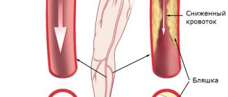
The main cause of the development of atherosclerosis is a disorder of lipid metabolism. It is cholesterol, or rather low-density cholesterol, that is deposited on the vascular wall and gradually forms a plaque. Its formation is facilitated by a violation of the integrity of the artery walls.
Damage to blood vessels is provoked by the following factors:
- high blood pressure – leads to thinning of the arterial wall and loss of elasticity;
- age – over the years, the walls of blood vessels become brittle;
- increased concentration of cholesterol and low-density lipoproteins in the blood;
- smoking - chemical compounds from tobacco smoke damage the walls;
- excess weight;
- excess fatty foods in the diet;
- genetic predisposition – the presence of atherosclerosis in close relatives;
- sedentary lifestyle;
- alcohol abuse;
- prolonged stress;
- chronic inflammatory diseases;
- taking certain medications.
Hormonal disorders and metabolic pathologies also contribute to the accumulation of dangerous cholesterol in the body. High blood pressure favors thrombus formation, which is a condition predisposing to atherosclerosis.
Outgoing arteries
According to the anatomical structure of the human body, three large arteries depart from the arch (see).
Brachiocephalic trunk
Truncus (tr.) brachiocephalicus, or brachiocephalic trunk, is the first and largest artery arising from the aortic arch. This is a short and thick vessel: its length is on average 3-5 cm, and its diameter is 7 mm.
The vessel is located approximately along the midline of the body and goes upward; at the level of the sternoclavicular joint it is divided into the right arteries - brachiocephalic, subclavian.
Left common carotid artery
Arteria carotis communis sinistra, or left carotid artery (common) is an artery, unlike its right pair, that arises directly from the arch. It is responsible for sufficient blood supply to the brain, eyeball, tissue of the neck and head.
A. carotis communis goes strictly upward, without giving branches along its entire length, and at the level of the upper border of the thyroid cartilage it is divided into the following arteries:
- external carotid;
- internal sleepy.
Note! The indicator of normal blood flow in the vessels of the brain is 55 ml/100 g. The main reason for its decrease (in more than 90% of cases) is considered to be atherosclerotic damage to the arteries.
A. subclavia sin.
Arteria subclavia sinistra, left subclavian artery, is the main vessel supplying blood to the upper shoulder girdle, as well as the organs of the neck and head.
Has three departments:
- the first, starting from the aortic arch, bending around the dome of the pleura, extending to the neck and ending in the interscalene space;
- the second, passing along the interscalene space and the groove of the same name on the right rib;
- the third, starting at the exit from the interscalene space and at the level of the edge of the first rib continuing into the axillary artery.
From the first section a.subclavia departs:
- vertebral artery, which supplies the spine and spinal cord;
- internal mammary artery, supplying blood to the thyroid gland, mediastinal organs, large bronchi, pericardium, diaphragm, sternum and other organs of the thoracic and upper abdominal cavity;
- the thyroid trunk, which ensures an uninterrupted supply of oxygen and nutrients to the thyroid gland, tissues of the neck and partly the back.
In approximately 30% of all people, the thyroid artery (inferior) also branches off from the arch, supplying the accessory pyramidal part of the thyroid gland. If it is necessary to perform a conicotomy or tracheotomy, there is a high probability of damage, so it is important that these manipulations are performed by an experienced doctor.
The vessels arising from the aortic arch play a key role in the blood supply to the organs of the head and neck, including the brain. Any changes in their work lead to symptoms of hypoxia and functional disorders of the central nervous system.
That is why medical instructions recommend that patients with headaches, attacks of dizziness, memory impairment and other cognitive disorders undergo examination as early as possible and begin treatment for vascular problems, because the deterioration of the condition - the price of delay - progresses every day.
Aorta
The aorta is the largest arterial vessel in the human body. It leaves the left ventricle; its beginning is the opening of the aorta, ostium aortae. All the arteries that form the systemic circulation depart from the aorta.
The aorta is divided into the ascending part of the aorta (ascending aorta), pars ascendens aortae (aorta ascendens), the aortic arch, arcus aortae, and the descending part of the aorta (descending aorta), pars descendens aortae (aorta descendens). The latter, in turn, is divided into the thoracic part of the aorta (thoracic aorta), pars thoracica aortae (aorta thoracica), and the abdominal part of the aorta (abdominal aorta), pars abdominalis aortae (aorta abdominalis).
Ascending aorta
, pars ascendens aortae, originates in the left ventricle from the opening of the aorta. Behind the left half of the sternum, at the level of the third intercostal space, it goes up, slightly to the right and forward and reaches the level of the cartilage of the second rib on the right, where it continues into the aortic arch.
The beginning of the ascending aorta is expanded and is called the aortic bulb, bulbus aortae. The wall of the bulb forms three protrusions - the aortic sinuses, sinus aortae, corresponding to the position of the three semilunar valves of the aorta.
Just like valves, these sines represent: right, left and rear.
A originates from the right sinus. coronaria dextra, and from the left - a. coronaria sinistra.
Aortic arch
, arcus aortae, convex upward and directed from front to back, passing into the descending part of the aorta. At the junction there is a noticeable slight narrowing - the isthmus of the aorta, isthmus aortae. The aortic arch has a direction from the cartilage of the second rib on the right to the left surface of the bodies of the III-IV thoracic vertebrae.
Three large vessels depart from the aortic arch: the brachiocephalic trunk, truncus brachiocephalicus, the left common carotid artery, a. carotis communis sinistra, and left subclavian artery, a. subclavia sinistra.
The brachiocephalic trunk, truncus brachiocephalicus, arises from the initial part of the aortic arch. It is a large vessel up to 4 cm long, which goes up and to the right and at the level of the right sternoclavicular joint is divided into two branches: the right common carotid artery, a. carotis communis dextra, and the right subclavian artery, a. subclavia dextra. Sometimes the inferior thyroid artery departs from the brachiocephalic trunk, a. thyroidea ima.
Development options are rare: 1) the brachiocephalic trunk is absent, the right common carotid and right subclavian arteries arise in this case directly from the aortic arch; 2) the brachiocephalic trunk extends not from the right, but from the left; 3) there are two brachiocephalic trunks, right and left.
Descending aorta
, pars descendens aortae, is a continuation of the aortic arch and lies along the length from the body of the III - IV thoracic vertebra to the level of the IV lumbar vertebra, where it gives off the right and left common iliac arteries, aa. iliacae communes dextra et sinistra, and itself continues into the pelvic cavity in the form of a thin stem - the median sacral artery, a. sacralis mediana, which runs along the anterior surface of the sacrum.
At the level of the XII thoracic vertebra, the descending aorta passes through the aortic opening of the diaphragm and descends into the abdominal cavity. Before the diaphragm, the descending part of the aorta is called the thoracic part of the aorta, pars thoracica aortae, and below the diaphragm - the abdominal part of the aorta, pars abdominalis aortae.
The aorta,
aorta
, is divided into three sections: the ascending aorta, the aortic arch and the descending aorta, which in turn is divided into the thoracic and abdominal parts.
Why is this variant of atherosclerosis dangerous?
Any illness does not bring anything good to a person. However, atherosclerosis of the coronary arteries and aorta is the worst version of this disease.
The branches extending from the aorta ensure adequate blood flow to the brain and, accordingly, the full performance of all its functions. Atherosclerosis of the thoracic aorta leads to a significant and progressive deterioration of blood flow throughout the brain with a gradual decrease in all its functional capabilities.
The coronary, or coronary, arteries are responsible for the blood supply to the heart. The worse the blood flow to the heart muscle, the worse the blood supply to all other organs and tissues of the human body. Combination with other pathologies, as well as coagulation disorders, several times increases the likelihood of developing a heart attack and stroke in people with atherosclerosis of the aorta and coronary arteries.
Ascending aorta
pars
ascendens aortae ,
exits the left ventricle behind the left edge of the sternum at the level of the third intercostal space;
in the initial section it has an extension - the aortic bulb, bulbus aortae .
At the location of the aortic valve, there are three sinuses on the inner side of the aorta,
sinus aortae .
From the beginning of the ascending aorta, the right and left coronary arteries depart.
Aortic arch
arcus
aortae ,
turns left and back from the posterior surface of the second costal cartilage to the left side of the body of the fourth thoracic vertebra, where it passes into the descending part of the aorta.
In this place there is a slight narrowing - the isthmus of the aorta, isthmus aortae .
The edges of the corresponding pleural sacs approach the anterior semicircle of the aorta on its right and left sides. The left brachiocephalic vein is adjacent to the convex side of the aortic arch, and the right pulmonary artery begins under the aortic arch, below and slightly to the left is the bifurcation of the pulmonary trunk. Behind the aortic arch is the bifurcation of the trachea. Three large arteries begin from the convex semicircle of the aortic arch: the brachiocephalic trunk, the left common carotid and the left subclavian arteries.
Descending aorta
pars
descendens aortae ,
divided into right and left common iliac arteries;
this place is called the bifurcation of the aorta, bifurcatio aortae .
The descending aorta is in turn divided into thoracic and abdominal parts.
The thoracic part of the aorta, pars
thoracica aortae ,
is located in the chest cavity in the posterior mediastinum. In the chest cavity, the thoracic part of the aorta gives off paired parietal branches; posterior intercostal arteries, as well as visceral branches to the organs of the posterior mediastinum.
The abdominal part of the aorta, pars
abdomindlis aortae ,
is located on the anterior surface of the lumbar vertebral bodies. The abdominal part of the aorta gives off paired parietal branches to the diaphragm and to the walls of the abdominal cavity. The visceral branches of the abdominal aorta are the celiac trunk, the superior and inferior mesenteric arteries (unpaired branches) and the paired renal, middle adrenal and testicular (ovarian) arteries.
Diagnostics
Atherosclerotic cardiosclerosis can be diagnosed at the initial stage of development. To make a diagnosis, a number of studies are prescribed. The sooner the patient sees a doctor, the more effective the treatment will be.
- A biochemical blood test determines changes in cholesterol, LDL, HDL, and triglycerides.
- Electrocardiography allows you to determine ischemia of individual areas of the heart muscle and the formation of scar connective tissue of the myocardium.
- Echocardiography shows the pathological state of the myocardium and detects disturbances in its contractions.
- Angiography of the coronary arteries is performed in a hospital setting.
- In some cases, x-rays of the chest and abdominal area are prescribed.
- X-rays are taken in several projections to assess the condition of the connective tissue and the size of the heart.
- Bicycle ergometry and step test are prescribed to determine the latent form of coronary insufficiency.
After a complete examination, treatment is prescribed.
The goal of the treatment procedures is to improve the patency of the coronary vessels, reduce the level of cholesterol, LDL and triglycerides, and increase HDL. With proper treatment, the risk of repeated attacks of myocardial infarction is reduced. In particularly difficult cases, surgery is performed.