Blood vessels of the human body (diagram)
Blood vessels
- elastic tubular formations in the body of animals and humans, through which the force of a rhythmically contracting heart or a pulsating vessel carries out the movement of blood throughout the body: to organs and tissues through arteries, arterioles, capillaries, and from them to the heart - through venules and veins.
System structure
There are different types of blood vessels in the body. Each of them has its own purpose. Thus, the system includes arteries, veins and lymphatic vessels. The first of them are designed to ensure that blood enriched with nutrients flows to tissues and organs. It is saturated with carbon dioxide and various products released during the life of cells, and returns through the veins back to the heart. But before entering this muscular organ, the blood is filtered in the lymphatic vessels.
The total length of the system, consisting of blood and lymphatic vessels, in the body of an adult is about 100 thousand km. And the heart is responsible for its normal functioning. It is this that pumps about 9.5 thousand liters of blood every day.
What are the pulmonary and systemic circulations?
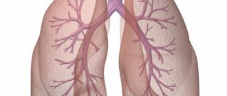
The pulmonary circulation is, in fact, the circulatory system of the lung. The small circle begins with the largest vessel - the pulmonary trunk. Through this vessel, blood flows from the right ventricle into the circulatory system of the lung tissue. Next, the vessels branch, first into the right and left pulmonary arteries, and then into smaller ones. The arterial vascular system ends with alveolar capillaries, which, like a mesh, envelop the air-filled alveoli of the lung. It is at the level of these capillaries that carbon dioxide is removed from the blood and oxygen is added to the hemoglobin molecule (hemoglobin is located inside red blood cells). After enrichment with oxygen and removal of carbon dioxide, the blood returns through the pulmonary veins to the heart - to the left atrium.
The systemic circulation is the entire set of blood vessels that are not part of the circulatory system of the lung. Through these vessels, blood moves from the heart to peripheral tissues and organs, as well as reverse blood flow to the right side of the heart. The systemic circulation begins from the aorta, then the blood moves through the vessels of the next order. The branches of the main vessels direct blood to the internal organs, the brain, and limbs.
It makes no sense to list the names of these vessels, but it is important to regulate the distribution of blood flow pumped by the heart to all tissues and organs of the body. Upon reaching the organ supplied with blood, strong branching of vessels occurs and the formation of a circulatory network of tiny vessels - the microvasculature. At the level of capillaries, metabolic processes occur and the blood, which has lost oxygen and part of the organic substances necessary for the functioning of organs, is enriched with substances formed as a result of the work of organ cells and carbon dioxide. As a result of such continuous work of the heart, pulmonary and systemic circulation, continuous metabolic processes occur throughout the body - the integration of all organs and systems into a single organism takes place.
Thanks to the circulatory system, it is possible to supply organs distant from the lung with oxygen, remove and neutralize (by the liver, kidneys) decay products and carbon dioxide. The circulatory system allows hormones to be distributed throughout the body in the shortest possible time and immune cells to reach any organ and tissue. In medicine, the circulatory system is used as the main element distributing drugs.
Principle of operation
The circulatory system is designed to provide life support to the entire body. If there are no problems, then it functions as follows. Oxygenated blood exits the left side of the heart through the largest arteries. It spreads throughout the body to all cells through wide vessels and tiny capillaries, which can only be seen under a microscope. It is the blood that enters the tissues and organs.
The place where the arterial and venous systems connect is called the “capillary bed.” The walls of the blood vessels in it are thin, and they themselves are very small. This allows oxygen and various nutrients to be fully released through them. The waste blood enters the veins and returns through them to the right side of the heart. From there it enters the lungs, where it is again enriched with oxygen. Passing through the lymphatic system, the blood is cleansed.
Veins are divided into superficial and deep. The first ones are close to the surface of the skin. They carry blood into the deep veins, which return it to the heart.
Regulation of blood vessels, heart function and general blood flow is carried out by the central nervous system and local chemicals released in the tissues. This helps control the flow of blood through arteries and veins, increasing or decreasing its intensity depending on the processes taking place in the body. For example, it increases with physical activity and decreases with injury.
The main groups of blood vessels and their properties in the body
The structure of blood vessels in the circulatory system can be divided into three main components:
- Capillaries are continuations of blood vessels and supply tissues and all elements of the body with oxygen. Everyone is familiar with the phrase “a capillary in the eye has burst,” and it really won’t be that difficult to injure it. Due to the fact that the capillary shell is especially thin, nutrients can easily reach all parts of the body. The number of capillaries in the human body varies, for example, in the heart muscle there are many more of them than in others. Capillaries also do not all function at once; while some of them are working, some rest while in sleep mode, and come into operation in the event of sudden strain on the body, or against a background of stress. Capillaries are an integral system that can be divided into 5 units: arterioles, precapillaries (play a connecting role in the system), true capillaries, postcapillaries, and venules (the place where the capillary passes into the vein). Each link is a special mechanism in the blood vascular system and plays an important role in proper functioning.
- Veins are the main element of blood circulation in the body. It is through them that blood flows from tissues and organs to the heart, closing the shape of a circle. The lining of veins is thin, the elasticity of such a lining is much thinner than that of arteries; veins are usually accompanied by several arteries, and those not accompanied are often involved in the connecting process under the skin.
- Arteries are a membrane that is more durable and elastic than veins and capillaries. Their main function is to carry oxygenated blood from the heart to the organs and tissues. There are two groups of arteries: elastic type and muscular artery. The first group is located closer to the heart muscle (for example, the aorta); the membrane of this group is the strongest to allow uninterrupted ejection of blood (heartbeat). The second category, when the smooth muscles of the inner layer of the arteries contract, contract, and when they relax, they expand, this ensures constant blood circulation and continuity of its movement through the vessels.
These three main components of blood vessels, which represent a blood highway that supplies the entire body with blood and useful elements. Therefore, it is very important to monitor the condition of your vessels, maintain them in proper form, so that there are no malfunctions.
In addition to supplying oxygen to internal organs, blood vessels enrich and nourish all layers of the skin. Blood vessels not only enrich skin cells, they participate in the process of skin regeneration in case of damage, and skin cells also become more elastic.
There are two types of cutaneous vessels, superficial and deep. The scheme of how deep vessels work looks like this: blood from the arteries flows through channels to the hair follicles and sweat glands, enriching the surface layer of the skin. The operating pattern of the superficial vessels ensures that blood flows to the skin in a direction perpendicular to the skin. There are also two types of lymphatic vessels located in the skin. You should be wary if you notice a slight gap on the skin of your face; this is a place of thinning.
A whole section of anatomy is devoted to the structure, functions of the vascular system and blood circulation. This system is based on a complex complex of veins, vessels, capillaries, arteries and aortas. The properties and functions of each element cannot be replaced by any other analogue. The main function in the circulatory system is the uninterrupted transportation of blood through all elements of the system to certain organs. Some elements in the system are in reserve, which allows, in the event of an emergency failure, to continue supplying blood to internal organs and skin cells.
How does blood flow
The spent “depleted” blood enters the right atrium through the veins, from where it flows into the right ventricle of the heart. With powerful movements, this muscle pushes the incoming fluid into the pulmonary trunk. It is divided into two parts. The blood vessels of the lungs are designed to enrich the blood with oxygen and return it to the left ventricle of the heart. In every person this part of him is more developed. After all, it is the left ventricle that is responsible for how the entire body will be supplied with blood. It is estimated that the load that falls on it is 6 times greater than that to which the right ventricle is exposed.
The circulatory system includes two circles: small and large. The first of them is designed to saturate the blood with oxygen, and the second is to transport it throughout the orgasm, delivering it to every cell.
Requirements for the circulatory system
In order for the human body to function normally, a number of conditions must be met. First of all, attention is paid to the condition of the heart muscle. After all, it is the pump that drives the necessary biological fluid through the arteries. If the functioning of the heart and blood vessels is impaired, the muscle is weakened, this can cause peripheral edema.
It is important that the difference between low and high pressure areas be maintained. This is necessary for normal blood flow. For example, in the area of the heart the pressure is lower than at the level of the capillary bed. This allows you to comply with the laws of physics. Blood moves from an area of higher pressure to an area where it is lower. If a number of diseases arise due to which the established balance is disturbed, then this is fraught with stagnation in the veins and swelling.
The release of blood from the lower extremities is carried out thanks to the so-called muscular-venous pumps. This is the name of the calf muscles. With each step, they contract and push blood against the natural force of gravity towards the right atrium. If this functioning is disrupted, for example, as a result of injury and temporary immobilization of the legs, then edema occurs due to a decrease in venous return.
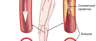
Another important link responsible for ensuring that human blood vessels function normally are the venous valves. They are designed to support fluid flowing through them until it enters the right atrium. If this mechanism is disrupted, perhaps as a result of injury or due to wear and tear of the valves, abnormal blood collection will occur. As a result, this leads to an increase in pressure in the veins and squeezing out the liquid part of the blood into the surrounding tissues. A striking example of a violation of this function is varicose veins in the legs.
Classification of vessels
To understand how the circulatory system works, you need to understand how each of its components functions. Thus, the pulmonary and vena cava, pulmonary trunk and aorta are the main routes for the movement of the necessary biological fluid. And everyone else is able to regulate the intensity of blood inflow and outflow to tissues due to the ability to change their lumen.
All vessels in the body are divided into arteries, arterioles, capillaries, venules, and veins. They all form a closed connecting system and serve a single purpose. Moreover, each blood vessel has its own purpose.
Arteries
The areas through which blood moves are divided depending on the direction in which it moves in them. So, all arteries are designed to transport blood from the heart throughout the body. They come in elastic, muscle and muscle-elastic types.
The first type includes those vessels that are directly connected to the heart and emerge from its ventricles. These are the pulmonary trunk, pulmonary and carotid arteries, and aorta.
All of these vessels of the circulatory system consist of elastic fibers that stretch. This happens with every heartbeat. As soon as the contraction of the ventricle has passed, the walls return to their original form. Due to this, normal pressure is maintained for a period until the heart fills with blood again.
Blood enters all tissues of the body through arteries that arise from the aorta and pulmonary trunk. At the same time, different organs need different amounts of blood. This means that the arteries must be able to narrow or expand their lumen so that fluid passes through them only in the required doses. This is achieved due to the fact that smooth muscle cells work in them. Such human blood vessels are called distributive. Their lumen is regulated by the sympathetic nervous system. Muscular arteries include the cerebral artery, radial, brachial, popliteal, vertebral and others.
Other types of blood vessels are also distinguished. These include muscular-elastic or mixed arteries. They can contract very well, but are also highly elastic. This type includes the subclavian, femoral, iliac, mesenteric arteries, and celiac trunk. They contain both elastic fibers and muscle cells.
Excerpt characterizing Blood Vessels
“Oh, my God, Count, there are moments when I would marry anyone,” Princess Marya suddenly said to herself, with tears in her voice. “Oh, how hard it can be to love a loved one and feel that... nothing (she continued in a trembling voice) you can’t do for him except grief, when you know that you can’t change it.” Then one thing is to leave, but where should I go?... - What are you, what’s wrong with you, princess? But the princess, without finishing, began to cry. – I don’t know what’s wrong with me today. Don't listen to me, forget what I told you. All Pierre's gaiety disappeared. He anxiously questioned the princess, asked her to express everything, to confide in him her grief; but she only repeated that she asked him to forget what she said, that she did not remember what she said, and that she had no grief other than the one he knew - the grief that Prince Andrei’s marriage threatens to quarrel with his father son. – Have you heard about the Rostovs? – she asked to change the conversation. - I was told that they would be here soon. I also wait for Andre every day. I would like them to see each other here. – How does he look at this matter now? - Pierre asked, by which he meant the old prince. Princess Marya shook her head. - But what to do? There are only a few months left until the year ends. And this cannot be. I would only like to spare my brother the first minutes. I wish they would come sooner. I hope to get along with her. “You have known them for a long time,” said Princess Marya, “tell me, hand on heart, the whole true truth, what kind of girl is this and how do you find her?” But the whole truth; because, you understand, Andrei risks so much, doing this against the will of his father, that I would like to know... A vague instinct told Pierre that in these reservations and repeated requests to tell the whole truth, Princess Marya’s ill will towards her future daughter-in-law was expressed, that she I wanted Pierre not to approve of Prince Andrei’s choice; but Pierre said what he felt rather than thought. “I don’t know how to answer your question,” he said, blushing, without knowing why. “I absolutely don’t know what kind of girl this is; I can't analyze it at all. She's charming. Why, I don’t know: that’s all that can be said about her. “Princess Marya sighed and the expression on her face said: “Yes, I expected and was afraid of this.” – Is she smart? - asked Princess Marya. Pierre thought about it. “I think not,” he said, “but yes.” She doesn't deserve to be smart... No, she's charming, and nothing more. – Princess Marya again shook her head disapprovingly. - Oh, I so want to love her! You will tell her this if you see her before me. “I heard that they will be there one of these days,” said Pierre. Princess Marya told Pierre her plan about how, as soon as the Rostovs arrived, she would become close to her future daughter-in-law and try to accustom the old prince to her. Boris did not succeed in marrying a rich bride in St. Petersburg and he came to Moscow for the same purpose. In Moscow, Boris was indecisive between the two richest brides - Julie and Princess Marya. Although Princess Marya, despite her ugliness, seemed more attractive to him than Julie, for some reason he felt awkward courting Bolkonskaya. On her last meeting with her, on the old prince’s name day, to all his attempts to talk to her about feelings, she answered him inappropriately and obviously did not listen to him. Julie, on the contrary, although in a special way, unique to her, willingly accepted his courtship. Julie was 27 years old. After the death of her brothers, she became very rich. She was now completely ugly; but I thought that she was not only just as good, but even much more attractive than she was before. She was supported in this delusion by the fact that, firstly, she became a very rich bride, and secondly, that the older she became, the safer she was for men, the freer it was for men to treat her and, without taking on any obligations, take advantage of her dinners, evenings and the lively company that gathered at her place. A man who ten years ago would have been afraid to go every day to the house where a 17-year-old young lady was, so as not to compromise her and tie himself down, now went to her boldly every day and treated her not as a young lady’s bride, but as a acquaintance who has no gender. The Karagins' house was the most pleasant and hospitable house in Moscow that winter. In addition to parties and dinners, every day a large company gathered at the Karagins, especially men, who dined at 12 o'clock in the morning and stayed until 3 o'clock. There was no ball, party, or theater that Julie missed. Her toilets were always the most fashionable. But, despite this, Julie seemed disappointed in everything, told everyone that she did not believe in friendship, nor in love, nor in any joys of life, and expected peace only there. She adopted the tone of a girl who had suffered great disappointment, a girl as if she had lost a loved one or had been cruelly deceived by him. Although nothing of the sort happened to her, they looked at her as if she were one, and she herself even believed that she had suffered a lot in life. This melancholy, which did not prevent her from having fun, did not prevent the young people who visited her from having a pleasant time. Each guest, coming to them, paid his debt to the melancholy mood of the hostess and then engaged in small talk, dancing, mental games, and Burime tournaments, which were in fashion with the Karagins. Only some young people, including Boris, delved deeper into Julie’s melancholic mood, and with these young people she had longer and more private conversations about the vanity of everything worldly, and to them she opened her albums covered with sad images, sayings and poems.
Arterioles and capillaries
As blood moves along the arteries, their lumen decreases and the walls become thinner. Gradually they turn into the smallest capillaries. The area where the arteries end is called arterioles. Their walls consist of three layers, but they are poorly defined.
The thinnest vessels are capillaries. Together they represent the longest part of the entire circulatory system. They are the ones that connect the venous and arterial beds.
A true capillary is a blood vessel that is formed as a result of the branching of arterioles. They can form loops, networks that are located in the skin or synovial bursae, or vascular glomeruli located in the kidneys. The size of their lumen, the speed of blood flow in them and the shape of the networks formed depend on the tissues and organs in which they are located. For example, the thinnest vessels are located in skeletal muscles, lungs and nerve sheaths - their thickness does not exceed 6 microns. They form only flat networks. In mucous membranes and skin they can reach 11 microns. In them, the vessels form a three-dimensional network. The widest capillaries are located in the hematopoietic organs and endocrine glands. Their diameter reaches 30 microns.
The density of their placement is also uneven. The highest concentration of capillaries is observed in the myocardium and brain, for every 1 mm3 there are up to 3,000 of them. At the same time, in skeletal muscle there are only up to 1,000 of them, and in bone tissue even less. It is also important to know that in an active state, under normal conditions, blood does not circulate through all capillaries. About 50% of them are in an inactive state, their lumen is compressed to a minimum, only plasma passes through them.
Human arterial system: structural features and main functions
The aorta of the cardiac circulation begins with an extension - the aortic bulb, from which the right and left coronary arteries depart. The bulb passes into the ascending aorta. Curving to the left, the aortic arch passes into the descending aorta. From the concave side of the aortic arch branches extend to the trachea, bronchi and thymus; Three large vessels depart from the convex side of the arch: on the right - the brachiocephalic trunk, on the left - the left common carotid and left subclavian arteries. The brachiocephalic trunk is divided into the right common carotid and subclavian arteries.
The common carotid artery (right and left) runs up next to the trachea and esophagus, dividing into the external carotid artery, which branches outside the cranial cavity, and the internal carotid artery, which passes inside the skull and goes to the brain. The external carotid artery supplies blood to the external parts and organs of the head and neck. The internal carotid artery enters the cranial cavity, where it divides into a number of branches that supply blood to the brain and organ of vision. The human arterial system also includes the subclavian artery and its branches, which supply blood to the cervical spinal cord with its membranes and the brain, part of the muscles of the back of the head, back and scapula, diaphragm, mammary gland, larynx, trachea, esophagus, thyroid gland and thymus. The subclavian artery in the axillary region becomes the axillary artery, which supplies blood to the upper limb.
The descending aorta is divided into thoracic and abdominal. The thoracic part of the aorta is located asymmetrically on the spine, to the left of the midline, and supplies blood to the internal organs located in the chest cavity and its walls. From the thoracic cavity, the aorta passes into the abdominal cavity through the aortic opening of the diaphragm. At the level of the IV lumbar vertebra, the aorta divides into two common iliac arteries. The main function performed by the arteries of the abdominal aorta is the blood supply to the abdominal viscera and abdominal wall.
The structure and main functions of veins in the circulatory system
The veins of the systemic circulation are divided into three systems: the superior vena cava system; the inferior vena cava system, including the hepatic portal vein system; system of cardiac veins that form the coronary sinus of the heart. The main trunk of each of these veins opens with an independent opening into the cavity of the right atrium. The veins of the superior and inferior vena cava systems are connected to each other. The main functions of the veins are to collect blood: the superior vena cava collects blood from the upper half of the body, head, neck, upper limb and chest cavity; The inferior vena cava collects blood from the lower extremities, walls and viscera of the pelvis and abdomen.
The main function of the portal vein in the blood supply is to collect blood from the unpaired organs of the abdominal cavity: the spleen, pancreas, greater omentum, gall bladder and other organs of the digestive tract. Unlike all other veins, the portal vein, having entered the portal of the liver, again breaks up into smaller and smaller branches, up to the sinusoidal capillaries of the liver, which flow into the central vein in the lobule. From the central hepatic veins flow into the inferior vena cava.
Venules and veins
Capillaries, into which blood flows from arterioles, unite and form larger vessels. They are called postcapillary venules. The diameter of each such vessel does not exceed 30 microns. At the transition points, folds are formed that perform the same functions as valves in the veins. Blood elements and plasma can pass through their walls. Postcapillary venules unite and flow into collecting venules. Their thickness is up to 50 microns. Smooth muscle cells begin to appear in their walls, but often they do not even surround the lumen of the vessel, but their outer membrane is already clearly defined. The collecting venules become muscular. The diameter of the latter often reaches 100 microns. They already have up to 2 layers of muscle cells.
The circulatory system is designed in such a way that the number of vessels draining blood is usually twice as large as the number of those through which it enters the capillary bed. In this case, the liquid is distributed like this. The arteries contain up to 15% of the total amount of blood in the body, the capillaries contain up to 12%, and the venous system contains 70-80%.
By the way, fluid can flow from arterioles to venules without entering the capillary bed through special anastomoses, the walls of which include muscle cells. They are found in almost all organs and are designed to allow blood to be discharged into the venous bed. With their help, pressure is controlled, the transition of tissue fluid and blood flow through the organ are regulated.
Veins are formed after the fusion of venules. Their structure directly depends on location and diameter. The number of muscle cells is influenced by their location and the factors under which fluid moves into them. Veins are divided into muscular and fibrous. The latter include the vessels of the retina, spleen, bones, placenta, soft and hard membranes of the brain. The blood circulating in the upper part of the body moves mainly under the force of gravity, as well as under the influence of the suction action during inhalation of the chest cavity.
The veins of the lower extremities are different. Each blood vessel in the legs must withstand the pressure created by the column of fluid. And if the deep veins are able to maintain their structure due to the pressure of the surrounding muscles, then the superficial ones have a more difficult time. They have a well-developed muscle layer, and their walls are much thicker.
Another characteristic feature of veins is the presence of valves that prevent the reverse flow of blood under the influence of gravity. True, they are not in those vessels that are located in the head, brain, neck and internal organs. They are also absent in the hollow and small veins.
The functions of blood vessels vary depending on their purpose. So, veins, for example, serve not only to move fluid to the heart area. They are also designed to reserve it in separate areas. Veins are used when the body works hard and needs to increase the volume of circulating blood.
Classification of blood vessels[ | ]
Among the vessels of the circulatory system, arteries
,
veins
and vessels of the
microcirculatory
;
the latter carry out the relationship between arteries and veins and include, in turn, arterioles
,
capillaries
,
venules
and
arteriole-venular anastomoses
[1]. Vessels of different types differ not only in their diameter, but also in tissue composition and functional features [2].
- Arteries are vessels through which blood moves away from the heart. Arteries have thick walls that contain muscle fibers as well as collagen and elastic fibers. They are very elastic and can contract or expand, depending on the amount of blood pumped by the heart. The blood flowing through the arteries is saturated with oxygen (the exception is the pulmonary artery, through which venous blood flows)[3][4].
- Arterioles are small arteries (less than 300 microns in diameter), immediately preceding capillaries in the blood flow. Smooth muscle fibers predominate in their vascular wall, thanks to which arterioles can change the size of their lumen and, thus, resistance. The smallest arterioles - precapillary arterioles
, or
precapillaries
- retain only single smooth muscle cells in their walls [5] [6]. - Capillaries are tiny blood vessels that are so thin that substances can easily pass through their walls. The diameter of their lumen ranges from 3 to 11 microns, and the total number in the human body is about 40 billion. Through the wall of the capillaries (no longer containing smooth muscle cells), nutrients and oxygen are released from the blood into the cells and carbon dioxide and other waste products are transferred from cells into the blood[7][8].
- Venules are small blood vessels that provide in a large circle the outflow of oxygen-depleted blood saturated with waste products from the capillaries into the veins. postcapillary venules
adjacent to the capillaries (
postcapillaries
) with a diameter of 8 to 30 microns and collecting venules with a diameter of 30-50 microns, flowing into the veins [9]. - Veins are vessels that carry blood to the heart. As the veins enlarge, their number becomes less and less, and in the end only two remain - the superior and inferior vena cava, which flow into the right atrium. The walls of veins are thinner than the walls of arteries and contain correspondingly fewer muscle fibers and elastic elements[10][11].
- Arteriolo-venular anastomoses
are vessels that provide direct blood flow from the arteriole to the venule, bypassing the capillary bed. They contain in their walls a well-defined layer of smooth muscle cells that regulate such flow [12] [13].
Structure of arterial walls
Each blood vessel consists of several layers. Their thickness and density depend solely on what type of veins or arteries they belong to. This also affects their composition.
For example, elastic arteries contain a large number of fibers that provide stretching and elasticity of the walls. The inner lining of each such blood vessel, which is called the intima, makes up about 20% of the total thickness. It is lined with endothelium, and underneath there is loose connective tissue, intercellular substance, macrophages, and muscle cells. The outer layer of the intima is limited by an internal elastic membrane.
The middle layer of such arteries consists of elastic membranes; with age they thicken and their number increases. Between them are smooth muscle cells that produce intercellular substance, collagen, and elastin.
The outer shell of the elastic arteries is formed by fibrous and loose connective tissue; elastic and collagen fibers are located longitudinally in it. It also contains small vessels and nerve trunks. They are responsible for feeding the outer and middle shells. It is the outer part that protects the arteries from ruptures and overextensions.
The structure of the blood vessels, which are called muscle arteries, is not much different. They also consist of three layers. The inner shell is lined with endothelium, it contains an internal membrane and loose connective tissue. In small arteries this layer is poorly developed. Connective tissue contains elastic and collagen fibers, they are located longitudinally in it.
The middle layer is formed by smooth muscle cells. They are responsible for contracting the entire vessel and pushing blood into the capillaries. Smooth muscle cells connect with the intercellular substance and elastic fibers. The layer is surrounded by a kind of elastic membrane. The fibers located in the muscle layer are connected to the outer and inner membranes of the layer. They seem to form an elastic frame that prevents the artery from sticking together. And muscle cells are responsible for regulating the thickness of the lumen of the vessel.
The outer layer consists of loose connective tissue, which contains collagen and elastic fibers; they are located obliquely and longitudinally in it. It also contains nerves, lymphatic and blood vessels.
The structure of mixed type blood vessels is an intermediate link between muscular and elastic arteries.
Arterioles also consist of three layers. But they are expressed rather weakly. The inner lining is the endothelium, a layer of connective tissue and elastic membrane. The middle layer consists of 1 or 2 layers of muscle cells that are arranged in a spiral.
Vein structure
In order for the heart and blood vessels called arteries to function, it is necessary that the blood can flow back up, bypassing the force of gravity. Venules and veins, which have a special structure, are intended for these purposes. These vessels consist of three layers, just like arteries, although they are much thinner.
The inner lining of the veins contains endothelium, it also has a poorly developed elastic membrane and connective tissue. The middle layer is muscular, it is poorly developed, and there are practically no elastic fibers in it. By the way, it is precisely because of this that the cut vein always collapses. The outer shell is the thickest. It consists of connective tissue and contains a large number of collagen cells. It also contains smooth muscle cells in some veins. They help push blood towards the heart and prevent it from flowing back. The outer layer also contains lymphatic capillaries.