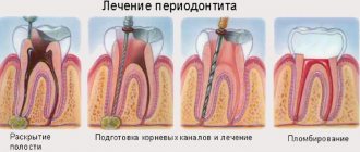
Acute odontogenic osteomyelitis of the jaw
Acute odontogenic osteomyelitis of the jaw is a serious disease with major disturbances in both local and general status of the patient.
This is a purulent-necrotic process of the jaw bone tissue, the cause of which is acute or exacerbation of chronic periodontitis. The spread of the inflammatory process into the bone marrow spaces adjacent to the periapical lesion and into the thickness of the bone leads to necrosis of the latter. The development and course of the osteomyelitic process depend on the state of the body's reactivity; reduced immunity predetermines a hypoergic reaction. In the clinical course, odontogenic osteomyelitis has three stages : acute, subacute and chronic; may be a limited and diffuse process.
Pathogenesis of dental inflammation
Infection occurs through a damaged tooth or micro-wounds in the gums and palate.
Odontogenic osteomyelitis of the jaw is provoked by the following pathogenic microorganisms:
- staphylococcus;
- streptococcus;
- Escherichia coli or Pseudomonas aeruginosa;
- Klebsiella;
- fusobacteria;
- proteas.
The pathogenesis of odontogenic osteomyelitis has the following features. Invading the tissue, bacteria provoke purulent inflammation of the bones, periosteum and bone marrow of the jaw, which leads to necrosis of all jaw structures. In children, the disease develops as a complication after scarlet fever, diphtheria or improper teething. The main causes of dental inflammation are as follows:
The disease may be a consequence of previous otitis media.
- any diseases of the tooth and its tissues (pulpitis, caries, alveolitis);
- acute and chronic inflammation of the ENT organs (otitis media, sinusitis, tonsillitis);
- jaw injuries and gunshot wounds;
- foci of infection that spreads throughout the body through the bloodstream;
- drug use.
Symptoms
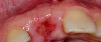
Acute osteomyelitis is locally detected in the form of pronounced swelling due to swelling of soft tissues in the affected area and periostitis from the vestibular surface along the transitional fold and from the oral side of the jaw. The causative tooth and its neighbors become mobile, percussion is painful. Vincent's symptom may appear, characterizing the involvement of the neurovascular bundle in the mandibular canal. Enlarged, painful regional lymph nodes are identified. Characteristic signs of acute osteomyelitis are chills and a sharp increase in body temperature (up to 38-40 ° C), in contrast to periostitis, in addition, the patient notes general weakness, sleep disturbance, decreased appetite, and loss of ability to work. These phenomena are accompanied by deviations from normal values in the blood and urine (leukocytosis, neutrophil shift, increase in ESR to 40-50 mm/h, protein in the urine, etc.). On an x-ray in the acute period, as a rule, no clear changes in bone tissue are detected.
In the subacute period, which occurs after 10-12 days, general manifestations of inflammation begin to subside, body temperature drops to low-grade fever, sleep and appetite gradually improve. Locally, subperiosteal ulcers are opened with the formation of fistulas, from which pus is released. There is a tendency for adjacent teeth to strengthen, and the teeth involved in the process become more mobile. During this period, radiographs can reveal foci of bone tissue destruction in the form of resorption without clear boundaries.
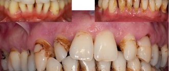
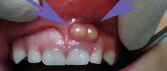
Gradually, after about a month, the process enters the chronic stage. From the formed fistulas, pus is released and pathological granulations bulge. The mobility of teeth in the area of inflammation increases. Not only new subperiosteal abscesses can form, but also abscesses and phlegmons of the soft tissues of the maxillofacial area. The general condition of the patient during the chronic stage of osteomyelitis improves, however, in case of exacerbation of the process, the above symptoms reappear (chills, high body temperature, weakness, etc.). The course of chronic osteomyelitis is long, the process ends after sequestration of dead bone areas. Depending on the prevalence of inflammation, sequestration occurs within 1-3 months. and more. Gradually, laboratory parameters return to normal.
The focal form of osteomyelitis is relatively easy, in contrast to the diffuse process. The focal form also includes osteomyelitis of the socket - a complication of alveolitis after tooth extraction, accompanied by the disintegration of a blood clot. With the wrong tactics of the doctor, who repeatedly within 2-3 weeks. carries out curettage of the socket to “form” a blood clot in it, the demarcation shaft is disrupted, the cortical plate is destroyed, the process spreads to the bone walls of the socket and purulent-necrotic inflammation occurs - osteomyelitis, leading to sequestration. In this case, pathological granulations form in the socket, thick pus is released, and upon probing in the depths of the socket, a mobile small sequestrum or small bone chips in the form of sand can be detected.
Limited osteomyelitis is often observed as a complication of a jaw fracture. The course of traumatic osteomyelitis differs from odontogenic osteomyelitis, and this is due to the fact that the inflammatory process does not develop in a closed focus, but there is communication with the external environment in the area of damage, and the outflow of exudate occurs freely from the first days of the disease. Sequestration occurs somewhat faster. If the outflow of exudate is disrupted, the process is accompanied by the formation of abscesses, followed by the formation of fistulas with purulent discharge.
Diffuse odontogenic osteomyelitis affects a large area of the jaw, often part of the alveolar process with teeth. Gradually, the process is limited, a sequestral cavity is formed, in which necrotic areas of the bone are located. When probing through the fistulous tract, the mobility of the separated sequestrum can be determined. The radiograph reveals sequestration in the form of a dense shadow of irregular shape, foci of bone tissue resorption and thickening of the jaw due to the hysterosteal reaction (Fig. 2).
In the clinic of surgical dentistry, the so-called primary chronic odontogenic osteomyelitis is encountered, in which the patient cannot indicate the acute initial stage of the disease. Children and adolescents are most often affected, but adults are no exception. It is characteristic that with this inflammation the proliferative process prevails over the destructive one, thickening of the jaw occurs, and an odontogenic focus in the form of chronic periodontitis is detected. Fistulas are usually absent. The radiograph reveals small foci of destruction against the background of normal bone with a pronounced periosteal reaction. This form of osteomyelitis must be differentiated from cancer and specific inflammatory processes (actinomycosis, etc.).
Forms of the disease
Depending on the intensity of inflammation and clinical indicators, the following forms of the disease are distinguished:
- Spicy. The body reacts sharply to the infection. Weakness, insomnia, headaches, and elevated body temperature are observed. The pain first appears in the area of the infected tooth. With mechanical impact on the tooth, the pain increases and a swaying sensation occurs. Swelling of the gums next to the tooth is pronounced, and an abscess may develop. The pain spreads to neighboring teeth and can radiate to the eye socket, ear, temple. As a result of osteomyelitis of the lower jaw, the sensitivity of the mucous membrane, skin and part of the lower lip is impaired.
- Subacute. Short-term positive dynamics are possible due to the breakthrough of pus from the bone tissue. At the same time, inflammation and bone destruction do not stop.
- Chronic. Although external health may improve, in reality there is an exacerbation of osteomyelitis, accompanied by the appearance of a new fistula. The mobility of the teeth becomes more noticeable, and the jaw in the area of the disease becomes flat. Osteomyelitis involves a long-term course of the disease. Self-treatment of osteomyelitis of the jaw in this case is impossible.
With purulent inflammation, the pain extends beyond the jaw. The lymph nodes in the neck are easily palpable and painful. The appearance of the body changes greatly when the body is intoxicated: the face becomes pointed, the skin turns grey, the eye sclera turns yellow (if the liver and spleen are affected). Pressure surges are typical. The difficulty of diagnosing osteomyelitis of the jaw is due to the fact that general symptoms that occur in other diseases also dominate over the characteristic local signs.
Chronic odontogenic osteomyelitis of the jaws. Principles of diagnosis and treatment.
⇐ PreviousPage 18 of 21Next ⇒Answer: Chronic osteomyelitis is a purulent or proliferative inflammation of bone tissue, characterized by the formation of sequestration, characterized by an improvement in the general condition of the body, a decrease in pain and swelling of soft tissues. The clinical picture is determined by the localization, nature and extent of the process. Most often, a diffuse nature of the process is observed, with its spread to several parts of the jaws, the death of significant areas of bone and the rudiments of permanent teeth. This course of the process is more typical for children aged 3-7 years. In some cases, a more favorable course of the disease is noted, when the process does not tend to spread and is localized within one or two sections of the jaw, often in the area of the body and chin. This course of chronic odontogenic osteomyelitis is also typical for the localization of the process in the upper jaw.
I feel satisfactory. Signs of chronic intoxication are revealed: pale skin, decreased appetite, sleep disturbance, lethargy and apathy.
The chronic destructive process is characterized by the presence of fistulas (external and intraoral), often single, although often multiple. The discharge from the fistula is purulent. Juicy granulations bulge from the mouth of the fistula. Sometimes there are no fistulas. In chronic osteomyelitis of the upper jaw, fistulas are noted only in the oral cavity, which are localized in the area of the alveolar process and along the transitional fold.
Mobility of intact teeth located at the site of inflammation is often observed. Due to inflammatory infiltration of the soft tissues surrounding the jaw, opening the mouth may be difficult.
The process is prone to exacerbations, in which patients complain of pain in the jaw area and the appearance of swelling. Fistulas and trismus occur. Sometimes abscesses and phlegmons form in the surrounding tissues. Regional lymph nodes are moderately enlarged in the remission stage, but during exacerbation of the process they are sharply enlarged, painful on palpation, and less mobile.
Diagnosis of Chronic osteomyelitis: The first radiological signs of changes in the intraosseous structure appear on the 7-10th day from the onset of the disease. Thinning and vagueness of the cortical layer along the lower edge of the body, angle and posterior surface of the branch are noted. Fine focal destruction is characteristic. During the 3-4th week, the destructive process increases in intensity and extent, becoming diffuse in nature. Initial signs of necrotization of bone tissue are radiologically determined at 3-4 weeks. By the 4-6th week, sequestra are formed, most often in the area of the cortical layer of the lower edge of the body, the angle and the posteroexternal surface of the branch. The destruction, which increases over time, involves the cortical plates of several tooth primordia. The formation of a total sequestrum (condylar process with part of the branch) and a pathological bone fracture are detected already at the beginning of the 2nd month.
X-ray diagnosis of sequestration is based on identifying an area of intense bone compaction, surrounded on all sides by a rarefaction zone. The width of this zone is 4-5 mm or more. At the same level, massive periosteal layers are revealed on the surface of the bone, covering the dead area of the bone from all sides and creating a sequestration box. The severity and density of periosteal layers depend on the duration of the process.
With a pathological fracture of the condylar process, the fracture line passes at the base of the condylar process, much less often - at the level of the neck of the jaw. Radiologically, a pathological fracture is characterized by the presence of a rarefaction band, which is differentiated along its entire length by a typical displacement of the process downward, anteriorly and inwardly, with the formation of angular deformation along the posterior edge of the branch. In the phase of developed consolidation, moderate periosteal layers are detected. The cortical layer in the area of destruction is partially destroyed, fibered, and at this level periosteal layers appear, merging with the bone in a short time.
Treatment of Chronic osteomyelitis: Treatment is complex. Both conservative and surgical methods are used.
Conservative treatment methods are effective when the disease is 1-1.5 months old. Success is facilitated by the inclusion in the complex of measures of the use of antibiotics of bone-tropic action, immunotherapy agents (staphylococcal toxoid, autovaccine), stimulants of the reticuloendothelial system (pentoxyl, methyluracil). Conservative methods are effective in the treatment of patients with limited, non-spreading, chronic osteomyelitis with a short period of limitation.
Indications for surgical intervention are the presence of sequesters that do not tend to resolve, long-standing fistulas and dead buds, as well as impaired renal function. Considering the timing of sequestration formation in children (4-6 weeks from the onset of the process), sequestrectomy should be prescribed no earlier than 1.5-2 months from the onset of the disease. The size of sequesters and the tendency for their resorption are also important when determining treatment tactics.
When a total sequester forms, there are absolute indications for its removal. In the preoperative period, given the low rates of nonspecific protective factors and immunological reactivity, drugs are prescribed that stimulate specific and nonspecific resistance of the body, which promotes rapid wound healing and a reduction in postoperative suppurative complications. Antibiotics are prescribed in the postoperative period or 2-3 days before surgery. They carry out desensitizing, vitamin and physical therapy.
94. Surgical methods of complex treatment of periodontal diseases.
Surgical treatment is of great importance in the complex therapy of periodontitis. It is carried out after conservative therapy and is aimed at eliminating local causes that support inflammation: removal of dental plaque, including subgingival plaque, curettage of granulations and de-epithelialization of the gingival pocket. Surgical methods include curettage and vacuum curettage of periodontal pockets (Junger-Sachs-Znamensky method), gingivotomy, gingivectomy, Widmann-Neumann flap surgery, frenulotomy, frenulectomy and microosteoplasty. For a localized form of periodontitis in the central group of the lower teeth, operations are used - vestibuloplasty, gum surgery. They are aimed at eliminating the tension of the highly attached tendons of the muscles of the lower lip and cords from the mucous membrane to the alveolar process, which lead to detachment of the gums from the neck of the tooth, the development and aggravation of periodontal lesions. Traditional surgical methods are complemented by diathermocoagulation and cryodestruction.
Curettage is one of the main types of treatment. There are simple and subgingival curettage. It is carried out at a pocket depth of 3-4 mm. For curettage, a special set of instruments is used, which includes excavators of various sizes, hooks for removing stones, bone spoons, etc. Curettage is carried out under local anesthesia, for example, under intraligamentary anesthesia. They begin by removing visible supragingival deposits, then gradually move to the neck and under the gum, plunging into the periodontal pocket. Granulations and deep-growing epithelium are scraped out of the pathological pocket. The resulting blood clot is a reliable biological protection against infection and promotes successful tissue scarring. The pocket can also be filled with emulsions, pastes with biologically active drugs and bioceramic substances. For pockets larger than 4 mm, vacuum curettage is performed using a special vacuum apparatus, and diathermocoagulation is also used, which allows blood to clot and tissue to curdle. The hemostatic effect is provided by cryodestruction with liquid nitrogen, freon, carbon dioxide, and oxygen.
Gingivotomy - dissection of the periodontal pocket and its curettage - is more often used in the development of periodontal abscess. After the operation, a pathological pocket remains, which is covered with a therapeutic bandage, introducing biologically active substances and biomaterials. Plastic surgery and pocket closure of the exposed neck and tooth root are possible. Gingivectomy consists of excision of the wall of deep periodontal pockets to the level of their bottom and performing curettage. With a simple gingivectomy, a triangular excision of tissue is performed in the area of one tooth; with a radical one, the gingival margin is excised to the bottom of pathological pockets within a group or all teeth, and the alveolar edge is leveled to convert vertical bone resorption into horizontal. The resulting wound is closed with an iodoform swab. At the same time, the possibility of filling the wound with biomaterials and bioceramics has a beneficial effect on the outcome of the operation.
The Widmann-Neumann operation (flap operation) is indicated for degree II tooth mobility and the presence of periodontal pockets not exceeding ½ of the root length (moderate and severe damage). Two vertical incisions are made to the bone from the edge of the interdental papilla to the transitional fold on the vestibular side within 6 teeth. Both cuts are connected by cutting the tops of the papillae between the teeth. The mucoperiosteal flap is folded back along with the gingival margin from the vestibular side and 0.5 cm from the intraoral side. Curettage of pathological pockets is performed, the gingival margin is excised to the level of their bottom. The flap is mobilized and sutured with knotted sutures through the interdental spaces to the edge of the wound from the oral cavity. Various therapeutic dressings made from dentin and zinc oxide, containing aspirin, corticosteroids, heparin, heparoid, etc., can be applied to the postoperative area.
Microosteoplasty is used to stimulate osteogenesis and consists of filling one or several deep bone pockets with crushed bone (autograft and allograft), which helps reduce their size. In contrast to the Widmann-Neumann operation, during bone grafting, the mucoperiosteal flap is separated only to the level of the bottom of the pathological pockets and the gingival margin is not excised. After careful curettage, removal of granulations and removal of the epithelial cover of the pocket wall, bone is introduced into the pocket in the form of lyophilized bone meal, crushed stone, shavings, formalized bone, cartilage, brefobone, collagen osteoplate, bioceramics, etc. The operation is completed by applying sutures in each interdental space .
Frenulotomy is performed for a shortened frenulum of the tongue by cutting it. It is advisable to perform this operation as early as possible - in infancy and childhood. frenulectomy is performed when there is a short frenulum of the tongue or lip, as a result of which a diastema subsequently develops. Two semi-oval vertical incisions are made, excising the frenulum along with the compactosteotomy. Having mobilized the mucous membrane along the edges of the wound, the latter is sutured tightly.
Vestibuloplasty for a shallow vault of the oral vestibule consists of deepening it and closing the wound surface by borrowing a flap from the lower lip. On the mucous membrane of the lower lip, a rectangular flap is cut out with the base facing the gingival edge, not reaching it 0.5-0.7 cm, so as not to disrupt the nutrition of the flap. The width of the flap corresponds to the size of the shallow vault, its length is up to 1 cm. The muco-submucosal flap is dissected from the underlying tissues. Through the resulting wound, the place of high attachment of muscle tendons (m. mentalis, i.e. incisivus labii inferioris) is separated from the periosteum to the level of the bottom of the vestibule. The flap is shifted towards the alveolar process, lining the created depression. A mucosal defect appears on the lip in the form of a narrow strip, which is sutured by mobilizing the surrounding tissue. If there are cords in the vestibule of the oral cavity, caused by an anomaly of muscle attachment, the mucous membrane is dissected to the crest of the cord up to 1 cm long, the place of attachment of the tendons is separated from the bone and the tissue is displaced to the bottom of the arch of the vestibule. The wound is sutured. A similar operation is performed in the area of each cord. In vestibuloplasty, a thin, split-thickness skin flap and other free tissue grafting techniques can be used to deepen the oral vestibule. After vestibuloplasty, the formation of an in-depth vestibule of the oral cavity with orthopedic or orthodontic devices is required for a period of up to 10-14 days.
There are many types of vestibuloplasty - according to Edlan-Meyhar, Glickman, Kruchinsky and Artyushevich and their modifications, as well as in combination with patch surgery. The choice of each of them depends on the severity of the lesion, the degree and type of atrophy of the alveolar process, etc.
The success of surgical treatment of periodontitis depends on a set of measures, including conservative therapy and rational dental prosthetics.
95 Dislocations and fractures of teeth. Fractures of the alveolar process. Principles of diagnosis and treatment.
Tooth luxation is a change in the spatial relationship of a tooth with its alveolus. Incomplete tooth luxation is a change in the position of the tooth crown in the dentition and displacement of the tooth root in relation to the walls of the alveolus. Impacted tooth dislocation is the penetration of the tooth root into the thickness of the bone tissue of the alveolar process. Complete tooth luxation is the complete loss of a tooth from the alveolus. Etiology: mechanical impact (impact, fall, etc.), careless use of elevators for tooth extraction, increased load on the tooth during biting or chewing food. Clinical picture : Incomplete tooth dislocation: pain in the tooth, which intensifies when you touch it, inability to bite and chew food, incorrect position of the tooth, mobility. Swelling, abrasions, hemorrhages, wounds of the lips or cheeks are detected. The mouth is sometimes half open. X-ray: narrowing or complete absence of the periodontal fissure on the side of the inclination of the tooth, and its expansion on the opposite side. Impacted tooth dislocation : pain, “shortening” of the tooth crown, bleeding from the gums, no tooth mobility; Only part of the tooth crown is located above the gum; the tooth root can be located in soft tissues or in the thickness of the bone. X-ray: the height of the crown is less than the adjacent teeth, a fracture of the bone substance of the socket, the root of the tooth is in the bone. In primary occlusion, injury to the permanent tooth germ may occur. Complete dislocation of a tooth: the absence of a tooth is objectively observed, the socket is empty, bleeding or filled with a blood clot. X-ray: empty alveolus of the tooth, the internal cortical plate is not damaged. Treatment of incomplete luxation is aimed at preserving the tooth. 1. Simultaneous tooth reposition after anesthesia, followed by immobilization using a splint. It is carried out against the background of anti-inflammatory, desensitizing and antibiotic therapy. 2. Long-term reposition with orthodontic devices when the patient approaches when the tooth has already been strengthened in the wrong position. Impacted tooth dislocation. 1. Waiting tactics (the tooth may move to its original position). 2. Simultaneous reposition with tooth immobilization. 3. Long-term reposition with orthodontic devices. 4. Tooth extraction followed by replantation - returning the tooth to its socket. 5. Tooth extraction followed by prosthetics. Tooth fractures Tooth fractures are damage to a tooth that disrupts the integrity of its crown or root part. Dental trauma may be accompanied by destruction of the tooth socket, fractures of the alveolar process or jaws. There are: 1) incomplete fractures (without opening the pulp): a) cracks in enamel and dentin; b) marginal fracture of the crown in the enamel zone; c) marginal fracture of the crown in the area of enamel and dentin; 2) complete fractures (with exposure of the pulp) - open and closed: a) neck of the tooth; b) root; c) root apex. Clinical picture: pain in the tooth during injury, increasing with load, pink coloration of the crown, tooth mobility, crown defects. X-ray: the presence of a band of clearing (fracture line), sometimes displacement of fragments. Treatment : the amount of assistance is determined by the level and nature of the fracture. A. If the enamel and dentin are damaged without opening the tooth pulp, the sharp edges of the crown are ground off. B. In case of a fracture of the crown with exposure of the pulp, conservative treatment is carried out (if the patient applied within a period not exceeding 12 hours), or the coronal part of the pulp is removed and the root canal of the tooth is filled (if applied at a later date), followed by restoration of the anatomical shape of the tooth with filling material , crown, pin tooth. B. If there is significant damage, the teeth are removed.
Diagnostics:
Tooth dislocations
Diagnosis of tooth dislocation is carried out on the basis of examination, tooth displacement, palpation and x-ray examination.
Dental fractures
Clinical diagnosis includes: anamnesis, examination of the soft tissues of the lips and cheeks, teeth, manual examination of the teeth, alveolar processes. To clarify the diagnosis and draw up a treatment plan, it is necessary to conduct x-ray studies of the alveolar process and electroodontic diagnostics.
⇐ Previous18Next ⇒