Most abdominal organs are supplied with blood from the celiac trunk, the superior and inferior mesenteric arteries and their branches. This ensures the flow of nutrients and oxygen to the tissues.
The danger of the disease lies in the fact that its symptoms are significantly similar to the clinical picture of many diseases of the digestive tract, which is what makes diagnosis difficult. Exacerbation of the disease can have tragic consequences, so timely diagnosis and treatment are extremely important.
The mechanism of development of the disease is long-term disruption of blood flow and ischemic processes. To varying degrees, this disease occurs in more than half of elderly patients. The acute stage of the disease is intestinal infarction or acute occlusion of abdominal vessels. The pathology is also known as abdominal toad or tonsillitis.
The disease is divided into celiac, superior and inferior mesenteric forms. It depends on the vessel that is involved in the process. Each form has its own characteristic symptoms, which arise depending on the location of the outbreak. Also, abdominal ischemia occurs in several stages. The first stage is compensation, when the disease manifests itself slightly, and the vessels continue to provide blood flow. The subcompensated stage is accompanied by a deterioration in the function of the abdominal vessels, which is manifested by a pronounced clinical picture. Decompensation occurs when the damage to the vessel is so great that it completely disrupts the blood supply to the tissue.
Etiology
Among the causes of ischemia:
- disturbance of central hemodynamics (changes in blood pressure, heart rate)
- blood loss
- local artery spasm
- atherosclerosis
- thrombosis and embolism
- compression of the artery from the outside, for example, by a tumor
- blood diseases
- organ frostbite
ISCHEMIA -i, g. (specialist.). Local bleeding of tissue as a result of narrowing of the lumen of the artery feeding it. Ischemia
(from the Greek ischo - I delay, stop and háima - blood), local anemia, insufficient blood content in an organ or tissue caused by a narrowing or complete closure of the lumen of the adductor artery. Transient I. (like hyperemia) can arise as a result of physiological regulation of blood supply, for example, during a reflex spasm of an artery caused by mental influence (fear), the influence of pain, cold, chemicals (adrenaline, ergotine, etc.).
), biological irritants (bacteria, toxins), a consequence of blockage of an artery by a thrombus or embolus (see Thrombosis, Embolism), narrowing of the lumen of a vessel due to atherosclerotic or inflammatory processes in its wall, compression of the artery by a tumor, scar, foreign body, etc. The consequences of I. depend on the degree of disruption of blood flow, the speed of development and duration of I.
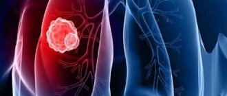
Necrosis (from the Greek Νεκρός - dead) is a pathological process expressed in the local death of tissue in a living organism as a result of any exo- or endogenous damage. Necrosis manifests itself in swelling, denaturation and coagulation of cytoplasmic proteins, destruction of cellular organelles and, finally, the entire cell.
Causes and risk factors
Unfortunately, the exact causes have not yet been studied . It is assumed that most often the disease is caused by the high sensitivity of the cells of the coronary vessels to a variety of active substances.
Vasospastic angina is also caused
by dysfunction of the internal walls of blood vessels and arteries .
The inner walls of the heart vessels or endothelium, when damaged, increase the production of substances in the body that contribute to vascular spasms and reduce the production of others that lead to vasodilation.
Another reason is spasm of the coronary arteries. In this case, the arteries are blocked, but their structure does not change. Risk factors in this case may be:
- hypothermia;
- smoking;
- autoimmune diseases;
- violation of the composition of electrolytes.
The disease can also occur when the blood supply to the heart muscle decreases . The number of heartbeats decreases, the myocardium becomes electrically unstable and conduction and rhythm disturbances occur.
Also, the cause of this disease is the appearance of stenosing coronal atherosclerosis . It occurs when there are problems with the arteries that supply the heart muscles - their lumen narrows, and the blood supply to the heart decreases.
Ventricular extrasystole: treatment and main symptoms. Find out more about what it is and how it is treated.
Is moderate sinus tachycardia dangerous for a child’s health? You asked - we answer!
Find out what sinus arrhythmia means on an ECG and what treatment your doctor will prescribe. All the details are here.
Microcirculation during ischemia
A significant increase in resistance in the adductor arteries causes a decrease in intravascular pressure in the microvessels of the organ and creates conditions for their narrowing. The pressure drops primarily in small arteries and arterioles to the periphery from the site of narrowing or blockage, and therefore the arteriovenous pressure difference throughout the microvasculature decreases, causing a slowdown in the linear and volumetric velocities of blood flow in the capillaries.
As a result of narrowing of the arteries in the area of ischemia, such a redistribution of red blood cells occurs in the branching vessels that blood that is poor in formed elements (low hematocrit) enters the capillaries. This causes the transformation of a large number of functioning capillaries into plasma capillaries, and a decrease in intracapillary pressure contributes to their subsequent closure. As a result, the number of functioning capillaries in the ischemic tissue area decreases.
The resulting weakening of microcirculation during ischemia causes a disruption in tissue nutrition: the delivery of oxygen decreases (circulatory hypoxia occurs) and energy materials. At the same time, metabolic products accumulate in the tissues.
Due to a decrease in pressure inside the capillaries, the intensity of fluid filtration from the vessels into the tissue decreases, creating conditions for increased resorption of fluid from the tissue into the capillaries. Therefore, the amount of tissue fluid in the intercellular spaces is significantly reduced and lymph flow from the ischemic area is weakened until it stops completely.
Cardiac ischemia
Coronary heart disease is by far the most common disease, which tends to affect an increasing number of people of different ages, but mostly older people, often leading to disability and often resulting in the death of the patient. Cardiac ischemia is caused by insufficient blood supply to the heart muscle.
Signs of cardiac ischemia manifest themselves long before an exacerbation. A person experiences a feeling of heaviness in the heart area, followed by paroxysmal pain. The pain radiates to the arms, chest, and lower jaw. The patient experiences shortness of breath, irregular heartbeat, and often nausea and vomiting. The presence of angina pectoris is the first sign of the development of cardiac ischemia.
With angina pectoris, pain is localized in the retrosternal region. There are nervous breakdowns, attacks of unreasonable fear, increased sweating and weakness. A characteristic sign of cardiac ischemia is the development of myocardial infarction, the symptoms of which are somewhat different from the manifestations of angina pectoris.
Cardiac ischemia often leads to death, so if you experience any pain in the heart, you must consult a specialist and undergo a full examination.
Classifications of necrosis
By etiology
Toxigenic
Trophoneurotic
Ischemic
Allergic
In case of necrosis, it is necessary to prevent the development of wet gangrene. For this purpose, the area of necrosis is treated with an open method designed to dry the tissue.
With wet gangrene, you must try to convert it into dry gangrene. To do this, alcohol dressings are applied, and dead tissue is lubricated with an iodine solution. When a demarcation line appears, a necrectomy is performed (removal of the necrosis zone).
Symptoms and signs
The disease is characterized by the following symptoms:
- burning or pressing pain in the area behind the sternum, lasting no more than 5 minutes, occurring mainly in the morning and night hours;
- Irregular heart function and attacks of rapid heartbeat;
- lack of connection between the occurrence of pain and physical or emotional stress;
- rapid disappearance of pain after taking nitroglycerin tablets;
- cyclicity of attacks.
Additional symptoms of vasospastic angina:
- arrhythmia;
- increased blood pressure;
- fainting;
- pale skin;
- cold sweat;
- in rare cases, nausea.
If signs of vasospastic angina are observed, you should urgently consult a cardiologist. The sooner he clarifies the diagnosis, the sooner treatment will be prescribed and the more favorable the prognosis. Complications are also less likely to occur.
Why is a stroke of the cerebellum dangerous, the consequences of the disease, differences from other forms of stroke - find out all the details on our website.
Why can a single atrial extrasystole occur and will it turn into a persistent form? We will tell you all the details.
What is supraventricular extrasystole and why is it dangerous? How to treat this condition? All the details are in this review.
Compensation for impaired blood flow during ischemia
With ischemia, complete or partial restoration of blood supply to the affected tissue often occurs (even if the obstruction in the arterial bed remains). This depends on collateral blood flow, which can begin immediately after ischemia occurs. The degree of such compensation depends on the anatomical and physiological factors of the blood supply to the corresponding organ.
Anatomical factors include features of arterial branching and anastomoses. There are:
- Organs and tissues with well-developed arterial anastomoses (when the sum of their lumens is close in size to that of the blocked artery) are the skin and mesentery. In these cases, blockage of the arteries is not accompanied by any disturbance of blood circulation in the periphery, since the amount of blood flowing through the collateral vessels is sufficient from the very beginning to maintain normal blood supply to the tissue.
- Organs and tissues whose arteries have few (or no) anastomoses, and therefore collateral blood flow into them is possible only through a continuous capillary network. Such organs and tissues include the kidneys, heart, spleen, and brain tissue. If an obstruction occurs in the arteries of these organs, severe ischemia occurs in them, and as a result, a heart attack.
- Organs and tissues with insufficient collaterals. They are very numerous - these are the lungs, liver, and intestinal wall. The lumen of the collateral arteries in them is usually more or less insufficient to ensure collateral blood flow.
A physiological factor promoting collateral blood flow is active dilatation of the arteries of the organ. As soon as a deficiency of blood supply occurs in the tissue due to blockage or narrowing of the lumen of the afferent arterial trunk, a physiological regulatory mechanism begins to operate, causing an increase in blood flow through the preserved arterial pathways.
https://www.youtube.com/watch?v=ytdevru
This mechanism causes vasodilation, since products of impaired metabolism accumulate in the tissue, which have a direct effect on the walls of the arteries, and also excite sensitive nerve endings, resulting in a reflex dilatation of the arteries. At the same time, all collateral pathways of blood flow into the area with circulatory deficiency expand, and the speed of blood flow in them increases, facilitating blood supply to the tissue experiencing ischemia.
If the blood flow in the collateral arterial pathways supplying blood to the ischemic area remains increased for a relatively long time, then the walls of these vessels are gradually rebuilt in such a way that they turn into arteries of a larger caliber. Such arteries can completely replace a previously blocked arterial trunk, normalizing blood supply to tissues.
Diagnostics
The following techniques help identify circulatory disorders:
- clinical and biochemical blood tests that detect inflammation, cholesterol, lipids, hemoglobin;
- magnetic resonance angiography;
- coronary angiography of vessels - radiographic examination with a contrast agent;
- Doppler ultrasound;
- magnetic resonance, computed tomography of organs.
Additional methods for diagnosing ischemia depend on the affected area:
- brain - radiography of the spine, ophthalmoscopy - examination of the fundus;
- intestines – sigmoidoscopy, irrigoscopy, colonoscopy;
- myocardium – ultrasound of the heart, electrocardiography at rest and during physical activity;
- limbs - Doppler measurement of peripheral pressure, ankle-brachial index.
Article on the topic: Focusin - instructions for use, dosage, indications, dosage regimen, composition, release form and price
Forecast
Vasospastic angina is a rare form of the disease. It is diagnosed in only 1.3-2.2% of situations.
The outcome depends on the severity of the pathological process. In general, the case fatality rate is 25%. In terms of what time it is not known: it could be a day, a week, a year or more. Therefore, delaying treatment is not recommended.
Death from heart attack, stroke, or cardiac arrest is likely. At a minimum, a dangerous arrhythmia develops. With comprehensive treatment, it is possible to bring the disease under control.
Treatment tactics
Treatment involves both non-surgical and surgical treatment. , all factors that can provoke the disease are first :
Nitroglycerin is used as medicine . It can be used in any form, both tablets and spray.
To prevent attacks, you can use nitroglycerin in the form of a patch, as well as prolonged nitrates (cardiquet, nitrosorbide). If the vascular endothelium is damaged as a result of spasms, the likelihood of thrombosis increases. In this case, antiplatelet agents , such as aspirin, are added to the drugs already in use.
For permanent treatment,
calcium antagonists .
The mechanism of action of these drugs is associated with a decrease in calcium content in smooth muscle cells. Thus, spasm of the coronary vessels is eliminated. Such drugs are used daily. In rare cases, alpha-blockers are used to treat variant angina . They reduce the effect of the nervous system on the muscular layer of the arteries.
Surgery is only allowed in certain cases . Angioplasty with stenting is performed. This procedure allows you to open clogged and narrowed blood vessels using a thin metal tube.
Coronary artery bypass grafting is also used to treat the disease. In this case, the patient’s vessel is sutured to the coronary artery slightly above and below its narrowing.
In cases where the disease is arrhythmic, the patient is fitted with a cardioverter-defibrillator , which, if necessary, restarts the heart using an electrical discharge.
Learn more about the disease from the video:
source
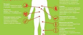
Symptoms
Manifestations are generally standard for coronary insufficiency. However, the vasospastic form is determined by a more severe course, and its duration is also longer. If a standard episode lasts from 5 to 30 minutes, even an hour is not enough.
- Severe pain behind the sternum. Burning, pressing. It radiates to the arm, forearm, neck, shoulder blade. The intensity does not change when standing up or breathing. At the same time, it is impossible to distinguish the condition from a heart attack. The course of the attack is atypical. It is recommended to call an ambulance for hospitalization and urgent diagnosis.
- Dyspnea. In a state of complete rest. The episode develops spontaneously, usually at night or early in the morning while the patient is still sleeping.
- Sweating. Hyperhidrosis. The patient, relatively speaking, can be squeezed out.
- Darkening in the eyes, dizziness, vertigo. Signs of malnutrition of cerebral structures. They pose a great danger and can cause a stroke.
- Fainting state. It happens more than once, during the same attack. This is a poor prognostic sign and worsens overall outcome.
- Tachycardia. Increased heart rate. Dangerous forms of arrhythmias, such as fibrillation or extrasystoles, are not presented unless there are concomitant cardiac problems.
- Paleness of the skin.
- Cyanosis or blue discoloration of the nasolabial triangle.
- Panic attack. The patient experiences severe fear for no apparent reason. Possible sudden psychomotor agitation. A person at such a moment is inadequate and can pose a danger to himself.
- Nausea, extremely rarely - vomiting.
- Instability of blood pressure: sometimes high, sometimes low and vice versa.
How dangerous is the condition?
The main threat from vasospastic angina is sudden death from one or another consequence. Among the possible complications of the pathological process:
- Heart attack. Acute death of cardiac structures. The myocardium dies rapidly, the cells destructure within a matter of hours, sometimes even faster. Necrosis is extensive, which leads to the rapid death of a person. An attack of Prinzmetal's angina ends with a similar outcome in 25% of cases. Recovery requires urgent hospitalization, assistance under the supervision of a group of specialists.
- Heart failure. Occurs in another 25-30% of cases. It is preceded by severe symptoms of an emergency condition. Emergency measures and resuscitation are a chance to save a person’s life. It requires special knowledge that almost no one has.
- Ischemic stroke. In the long term. The result of insufficient cerebral circulation. The essence is the death of neurons, without the possibility of their restoration. The process ends with a neurological focal deficit. Speech, vision, touch, muscle activity and other functions suffer.
Dangerous complications can be prevented through timely treatment and following the doctor’s recommendations. But even this is not a guarantee.
Causes of vasospastic angina, symptoms, treatment and life prognosis
Azospastic angina is a special case of coronary insufficiency and represents a sharp, critical narrowing of the corresponding arteries by more than 70%. Occurs in a state of complete rest, usually at night.
Pathology does not develop on its own, but against the background of third-party phenomena, which ones need to be looked at. This could be hypertension, long-term smoking, endocrine abnormalities, all of these are high-risk factors that should be corrected urgently.
It is impossible to cure vasospastic angina completely. But there are chances to take control of the process and improve the patient’s quality and life expectancy. For this purpose, both surgical and medicinal techniques are used.
Prognosis depends on the duration of the pathology and the frequency of relapses.
Causes
Development factors are not always cardiac. Some can be corrected by the patient himself, while others are objective.
Despite the rich empirical material, doctors still cannot say exactly why such a dangerous form of angina occurs.
The only thing that can be pointed out with one hundred percent certainty is that the main reason is the narrowing of the coronary arteries.
In 90% of cases, an attack of vasospastic angina occurs against the background of complete rest. Moreover, at night, when all processes are slowed down, blood pressure is lowered, as is the heart rate.
What else can trigger an episode of violation:
- Intense physical activity inadequate to the level of training. Running, climbing stairs, carrying heavy objects, especially heavy ones. This is a relatively rare factor. This is how exertional angina develops, but not the Prinzmetal form. Although a similar scenario is possible. At the very least, it increases the likelihood of another night attack.
Treatment
In a hospital setting at an early, acute stage. Then outpatient with regularly scheduled hospitalizations. The most commonly used medicinal tactics are:
- Nitroglycerin for urgent relief of pain. These are the only approved drugs for self-help at home (1 tablet under the tongue, if there is no effect after 20 minutes, then another one). Against the background of a severe episode, the drug helps partially. No complete correction is observed.
- Cardio- and cytoprotectors to improve metabolism, protect against oxidation, and negative external influences. Mildronate, Trimetazidine in combination.
- Beta blockers. Metoprolol. It affects both blood pressure and heart rate.
- Calcium antagonists. Verapamil or Diltiazem.
- Antiplatelet agents to improve the rheological properties of blood and increase its fluidity. Aspirin-Cardio, Heparin and others. In strictly controlled dosages.
- Anti-arrhythmia medications are prescribed as needed. Amiodarone as the main one.
Attention:
The regimen should be determined only by a doctor. Many drug combinations lead to rapid development of heart or kidney failure. It is not recommended to take risks.
If organic defects are present, surgical treatment is performed. Stenting, ballooning of the coronary artery (mechanical expansion of the lumen), prosthetics of the affected area of the vessel. Bypass surgery, the creation of an artificial source of blood supply bypassing the natural one, is also possible.
Treatment of vasospastic angina has three objectives: eliminating the root cause, relieving symptoms, and preventing attacks (relapses).
As part of additional feasible measures, mandatory cessation of smoking, alcohol (alcohol), and fatty foods is indicated. It is recommended to adjust the diet in accordance with table No. 10; if possible, consultation with a nutritionist is indicated.
Prevention
The following activities are shown:
- Quit smoking, alcohol, drugs, and self-administration of any medications, including non-steroidal anti-inflammatory drugs.
- Diet correction.
- Moderate physical activity to eliminate physical inactivity.
- Avoid stress whenever possible.
- Timely treatment of diseases that can provoke a dangerous form of coronary insufficiency.
This is not a guarantee, but the risks are significantly lower. Once a condition is detected, recommendations remain relevant.