Gestosis during pregnancy is a complication in which the functioning of physiological systems and organs is disrupted, and the pathological condition disappears after delivery. According to studies, it is formed in the third trimester of pregnancy, but can occur earlier, from 4 to 20 weeks.
- Reasons for the formation of gestosis
- The first signs of gestosis
- Symptoms of gestosis in pregnant women
- Diagnostics
- Treatment of gestosis
Consequences of gestosis
Preeclampsia, what is it and how does it manifest?
Preeclampsia has three characteristic symptoms, which are not difficult to make a primary diagnosis:
- Swelling. Hidden and obvious, noticeable on the limbs and face.
- Proteinuria. In urine analysis, the protein content is increased.
- Hypertension. Increased blood pressure.
The pathology negatively affects the functioning of the cardiovascular, nervous and endocrine systems, and changes in blood composition occur. Preeclampsia develops after the 18th week and manifests itself by the end of the 27th week.
About a third of pregnant women experience manifestations of this complication. In some cases, it causes the death of the mother or baby.
Clinical manifestations are often consistent, so early gestosis occurs in the first half of pregnancy. The patient notes constant nausea, vomiting, and increased salivation (not to be confused with “normal” toxicosis during pregnancy).
At a later stage, nephropathy, hydrops of pregnancy, preeclampsia and eclampsia appear. In the presence of a diseased liver, skin and nervous diseases, rare forms of pathology are formed.
Preeclampsia of the combined form appears with hypertension, endocrinopathy, biliary tract and kidney disease, and impaired lipid metabolism in the body.
Treatment
Treatment of mild gestosis is carried out on an outpatient basis, at home. For more severe forms, treatment is carried out in a hospital. First of all, non-drug treatment methods are used - physiotherapy, acupuncture, psychotherapy, electrosleep, hypnosis. Drug treatment of gestosis should be comprehensive. Drugs that normalize the function of the central nervous system and suppress the gag reflex are used. Intravenous infusions of saline solutions, glucose, and vitamins are performed. Usually up to 2-2.5 liters of solutions are administered per day. In some cases, medications are prescribed to replace regular meals. When treating pregnant women suffering from early gestosis, it is necessary to monitor biochemical blood parameters and urine tests. In rare cases, with excessive (so-called indomitable) vomiting, which is accompanied by general exhaustion, it is necessary to raise the question of terminating the pregnancy.
Recommendations from experts
Some advice for women who have symptoms of early gestosis:
- Listen to your taste desires, eat only what you want. Food should be easily digestible and contain enough vitamins;
- Eat small meals every 3-4 hours. For nausea, relief comes from chewing (dried fruits, nuts, crackers, salty crackers, lemon, etc.);
- If nausea begins in the morning, after getting up, then you can organize breakfast in bed. In any case, it is recommended to eat something - a piece of white bread or a roll, a cracker, sweet tea. Don’t rush to brush your teeth first thing after getting up!;
- It is recommended to eat dry: food separately, drinks by themselves. In some cases, mixtures and purees for baby food, protein mixtures for pregnant women (femilak, berlamin-modular, enipit) help. Be sure to include alkaline mineral water in your diet. The total amount of liquid drunk can be increased to 2-2.5 liters.
- If you are drooling, it is good to rinse your mouth with an infusion of mint, sage, and chamomile.
Reasons for the formation of gestosis
There is not yet a unified theory about what contributes to gestosis during pregnancy, but some theories of the etiology of the disease have been proposed.
The cortico-visceral theory suggests the formation of neurosis in pregnant women, when there is a failure in the interaction between the cortex and the subcortical structure of the brain, leading to reflex changes. In practice, this theory is often confirmed; gestosis occurs as a consequence of nervous tension.
According to the immunological theory, with gestosis there is improper hormonal control of body functions. The immunological conflict between the fetus and mother is considered as the main cause. At the same time, vasospasm increases blood pressure and reduces the amount of blood necessary for the nutrition and functioning of cells.
Genetic theory suggests that women whose mothers suffered from preeclampsia are susceptible to complications. A lack of B vitamins and folic acid increases the content of homocysteine, an amino acid that increases blood clot formation. From its influence, microholes are formed in the vessels, through which plasma protein and liquid fall into the tissue.
Preeclampsia during pregnancy leads to swelling, the signs of which are not visible at the onset of the disease, but weight increases significantly. Plasma penetrates and stagnates in the tissues, resulting in swelling, increased blood pressure and decreased frequency of urination.
Similar microholes appear in the vascular walls of the kidneys, through which protein penetrates into the urine. A pregnant woman is regularly prescribed a general urine test. This helps in accurate diagnosis and allows treatment to begin immediately after pathology is detected.
When the inner lining of the vessel, the endothelium, is damaged, its permeability increases, which promotes the effusion of fluid into the body tissue. This changes the density of the blood, its thickness and coagulability. The risk of blood clots increases.
Preeclampsia is dangerous due to disorders in the brain. Blood clots and minor hemorrhages form, intracranial pressure increases and nervous tissue degeneration occurs.
For the baby to be born healthy...
What makes gestosis during pregnancy so insidious is that the expectant mother may feel quite normal. It is important to pay attention to sudden changes in weight in time - this is one of the first signs of gestosis and evidence that the kidneys are no longer able to cope with the increasing load. This is when hidden swelling begins to appear.
Outpatient treatment of gestosis during pregnancy is possible only at the first stage - hydrops of pregnancy. If the expectant mother has edema and is diagnosed with mild or moderate gestosis, she will be hospitalized. If gestosis is severe, there are signs of preeclampsia, or the patient has suffered an eclamptic attack, then she is hospitalized immediately in the intensive care unit. In any case, the patient is carefully examined (blood and urine tests, fetal blood circulation studies and ultrasound). Consultations with an ophthalmologist, neurologist and therapist are also necessary.
The first signs of gestosis
The appearance of gestosis in the early stages is difficult to notice; the condition is easily confused and mistaken for a mild ailment in pregnant women. Blood pressure increases slightly, causing headache, weakness, nausea and fatigue.
- Increased protein is found in the urine; the higher its content, the worse the manifestation of the pathology.
- Blood pressure exceeds 140/90 mm Hg. Art.
- Edema - when protein is detected in the urine, they indicate gestosis.
Classification
Assessment of the severity of gestosis in points (Goeeke, modification by G. M. Savelyeva). Up to 7 points - mild; 8-11 points - average degree; 12 points or more - severe.
ICD-10 classification
According to the International Classification of Diseases, 10th revision (ICD-10, 1995), gestosis is classified as follows:
- Pre-existing hypertension complicating pregnancy, childbirth and the postpartum period.
- Pre-existing hypertension with associated proteinuria.
- Pregnancy-induced edema and proteinuria without hypertension.
- Pregnancy-induced hypertension without significant proteinuria.
- Pregnancy-induced hypertension with significant proteinuria is moderate preeclampsia.
- Eclampsia during pregnancy, childbirth, and the postpartum period.
Russian classification
In Russia, the classification of gestosis includes 4 forms, which can transform into each other under the influence of various reasons. These four forms can be considered as stages of a single pathological process:
- Dropsy.
- Nephropathy (mild, moderate and severe).
- Preeclampsia.
- Eclampsia.
There are also pure gestosis (developed in full health) and combined (against the background of various diseases).
Classification of the American Society of Obstetricians and Gynecologists (ACOG)
I. Hypertension due to pregnancy.
1. Preeclampsia.
A. Mild degree. Mild preeclampsia is diagnosed if there are no signs of severe preeclampsia. B. Severe degree. The diagnosis of severe preeclampsia is made when one or more of the following criteria are present:
- increase in systolic blood pressure more than 160 mm Hg. or diastolic blood pressure more than 110 mm Hg, recorded twice with an interval of more than 6 hours,
- loss of protein in urine more than 5 g/day;
- oliguria (urine amount less than 400 ml per day);
- neurological and/or visual disorders (headache, disturbances of consciousness, blurred vision, etc.);
- signs of pulmonary edema and cyanosis.
2. Eclampsia.
II. Chronic hypertension of any etiology, not related to pregnancy.
III. Preeclampsia or eclampsia combined with chronic hypertension.
IV. Transient hypertension.
V. Unclassified hypertensive disorders.
NHBPEP classification
The classification was developed by the working group of the National High Blood Pressure Education Program in 2000.
1. Gestational hypertension (previously referred to as pregnancy-induced hypertension and included transient hypertension).
2. Preeclampsia.
Minimum criteria: blood pressure >140/90 mm Hg. Art. after 20 weeks of pregnancy. Proteinuria >300 mg/24 hours. Increased likelihood of eclampsia. Blood pressure >160/110 mm Hg. Art. Proteinuria 2.0 g/24 hours. Serum creatinine >1.2 mg/dl. Platelets <100,000/mm3. Microangiopathic hemolysis (increased LDH). Increased ALT or AST. Persistent headache or other brain or visual disturbances. Persistent pain in the epigastric region.
3. Eclampsia.
4. Preeclampsia due to chronic hypertension.
5. Chronic hypertension.
Symptoms of gestosis in pregnant women
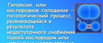
The disease affects most organs: kidneys and liver, heart, placenta and nervous system. The formation of constant hypoxia is possible, which leads to intrauterine growth retardation.
In the initial stage of gestosis (toxicosis), women vomit up to several times a day. There is constant nausea, loss of appetite, desire to eat spicy and salty foods. Bouts of vomiting do not affect weight. Temperature is within normal limits. These symptoms appear in the first months of pregnancy and then disappear on their own.
Rarely, vomiting may stop, and then becomes uncontrollable (more than 20 times per day). The patient is weakened, has an aversion to food, the pulse is thready, and the blood pressure drops. Acetone and protein are found in the urine. In serious cases, the temperature rises sharply and arrhythmia is possible.
At the end of pregnancy, gestosis develops gradually. Initially, dropsy forms, over time nephropathy develops, then severe forms: preeclampsia, eclampsia. In cases of dropsy, women experience swelling due to fluid retention. At this time, hidden and visible swelling occurs. Given the slowdown in diuresis, body weight increases very quickly.
The tumor is noticeable in the ankle joint, then spreads higher. Swelling of the face is noticeable. By evening, the limbs and lower abdomen swell.
Three symptoms of gestosis with nephropathy:
- edema;
- protein in urine;
- hypertension.
A woman may have a combination of any symptoms. Nephropathy occurs simultaneously with dropsy. Increased diastolic pressure is dangerous because it reduces placental blood flow. The fetus does not receive enough oxygen. Later, nephropathy can develop into a serious complication - eclampsia.
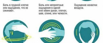
With preeclampsia, the central nervous system is affected. In addition to the three main symptoms of gestosis, heaviness in the back of the head, pain in the abdomen, head, nausea, and vomiting occur. A pregnant woman experiences visual impairment (flickering of spots), memory, and insomnia.
Symptoms of preeclampsia:
- pressure is more than 160/110 mm Hg. Art.;
- decreased urine output (<500 ml), worse blood clotting due to decreased platelets, noticeable impairment of liver function.
Eclampsia is the worst phase of gestosis. The woman may experience attacks of painful cramps. Light and any sharp sounds are irritating, this lasts for several minutes, after which loss of consciousness is possible. There is a danger of developing a deep coma - this threatens placental abruption, bleeding, fetal hypoxia and premature birth. The life of the fetus is in danger.
It should be noted that with gestosis, the pregnant woman feels well for some time, complaining only of minor swelling and weight gain. However, edema does not only form in the extremities. The placenta suffers from it - this impairs the supply of oxygen to the fetus.
Preeclampsia during pregnancy is a serious condition; the first symptoms are the reason for an immediate visit to the doctor.
Preeclampsia during pregnancy is a serious disease; it is in order to recognize it in the early stages that doctors weigh expectant mothers at every examination.
1. What is it 2. Symptoms 3. Causes 4. Signs 5. Classification 6. Treatment 7. Second half 8. Late 9. After childbirth 10. Prevention 11. Consequences
What is this
Statistics vary widely from source to source. This is mainly due to three reasons:
- Incomplete coverage of all pregnant women with dispensary observation.
- Late referral of pregnant women to antenatal clinics, when mild forms of gestosis become more severe (preeclampsia and eclampsia).
- Change in the general name and classification of pathological disorders.
In connection with the last point, some clarity should be provided. This will avoid substitution of concepts and confusion in ideas.
Previously, all pathological symptoms throughout pregnancy and deviations from the norm in the results of laboratory and instrumental studies were called toxicosis of pregnant women. Depending on the duration and form, it was divided into toxicosis of the first and second half of pregnancy.
In accordance with this classification, the term “toxicosis” is replaced by the term “preeclampsia”. Some experts believe that it can be early (in the first trimester of pregnancy) and late (in the second half). Others recognize only gestosis in the second half of pregnancy, and those disorders that appear in the first half are considered either physiological or not related to the etiology (cause) and pathogenesis (mechanism of development) of gestosis. The first option seems more convenient, allowing one to get an idea of any pathological deviations from the normal course of pregnancy.
They are associated with pregnancy and disappear after delivery. This is the basis for the assumption by all scientists that the cause of gestosis lies in:
- the negative role of the fertilized egg and fetus;
- a disorder of a woman’s adaptive mechanisms designed to provide the fetus with the opportunity for normal development.
There are many factors contributing to the occurrence of gestosis during pregnancy. However, among them, the greatest attention is paid to concomitant overt or hidden endocrine diseases, impaired renal and liver function, high blood pressure, multiple pregnancies, hydatidiform moles and some others.
There are many versions and theories about the initial and subsequent mechanisms of the development of pathology, but all of them, except the autoimmune one, raise great doubts. Rather, they reflect individual links in the cascade development of a single mechanism.
How is gardnerellosis treated in women?
Causes of vulvitis in girls here
Causes of vaginitis during pregnancy /bolezni/vgn/vaginit-pri-beremennosti.html
Symptoms
Preeclampsia during pregnancy usually appears in the later stages, the occurrence of which is indicated by the following symptoms:
- Increased blood pressure above 140/90 mmHg. You may not be aware of this, but accompanying nausea, headache and blurred vision indicate changes for the worse.
- The appearance of protein in the urine, which is detected by doctors when taking tests before each scheduled examination. This phenomenon indicates a violation of the kidneys, without which gestosis does not appear.
- Convulsive seizures, which can occur in severe cases of gestosis.
- Placental abruption.
- Developmental delay and fetal death.
In 90 percent of cases, the disease begins after 34 weeks of pregnancy and is more often observed in primiparous women. Also, the risk of gestosis increases with multiple pregnancies and when carrying a child under the age of twenty or over thirty-five years. Sometimes there may be an early stage of this disease, when it appears at twenty weeks. In this case, gestosis is more severe, and the first signs of the disease are clearly expressed.
Causes
Despite numerous studies, the causes and mechanism of development of gestosis have not yet been fully established.
Several theories are currently considered relevant:
- The cortical-visceral theory considers gestosis as a kind of neurosis of pregnant women with a disruption of the physiological relationships between the cortex and subcortical formations of the brain, leading to changes in the functioning of the vascular system and circulatory disorders.
- According to supporters of the endocrine theory, dysregulation of the cardiovascular system, blood supply to internal organs, metabolism in organs and tissues occurs due to changes in the function of endocrine organs that produce biologically active substances that regulate vascular tone, fluidity and blood clotting.
- Proponents of the immunological theory believe that changes in blood vessels, organs and tissues, characteristic of gestosis, arise due to an inadequate reaction of the mother’s immune system to certain antigens of fetal tissue, which is not observed during a normal pregnancy.
- The advancement of the genetic theory is based on a statistically confirmed increase in the frequency of gestosis in those pregnant women whose mothers suffered from gestosis during pregnancy. The existence of “preeclampsia genes” is also not rejected.
- The provisions of the placental theory are based on the fact that during gestosis there are no changes in the vessels of the uterus that feed the placenta, characteristic of a physiological pregnancy. As a result, active substances are released, leading to disruption of the normal functioning of the entire vascular system of the mother’s body.
- Currently, most researchers have come to the conclusion that there is no single mechanism for the development of gestosis, but a combined effect of various factors is observed, ultimately leading to dysfunction of internal organs.
Signs
There are three main signs of gestosis during pregnancy, which usually appear together or in pairs: swelling, protein in the urine and increased blood pressure.
The appearance of protein in the urine (proteinuria). The first and main criterion that indicates kidney damage. Preeclampsia almost never occurs without proteinuria, and the stronger it is, the worse it is. Although the identification of this sign alone does not indicate gestosis.
Normally, there should be no protein in the urine. Small amounts, around 0.033 g/l, in combination with leukocytes may be a sign of kidney inflammation (pyelonephritis). 0.8 g/l or more is more likely to indicate gestosis. Proteinuria in combination with an increase in blood pressure over 140/90 always indicates gestosis.
A urine test must be taken before each visit to the doctor at the antenatal clinic. If you feel that your urine has become cloudy, dark in color, or covered with foam, get tested without waiting until the appointed day.
Increased blood pressure more than 140/90 mmHg. Art. This is the second main sign of gestosis, which may go unnoticed, or may manifest itself as headache, nausea, flashing spots before the eyes, and dizziness.
The combination of high blood pressure with protein in the urine is called preeclampsia, and indicates the initial stage of brain damage to the expectant mother. This is why your blood pressure needs to be measured at every doctor's visit.
In severe cases, untreated high blood pressure can cause serious damage to the nervous system, including loss of consciousness, seizures (eclampsia), and bleeding in the brain (stroke). This danger occurs when the upper figures of blood pressure exceed 160 and the lower 110 millimeters of mercury.
Edema. They often occur during normal pregnancy, and in themselves are not a sign of gestosis, but only in combination with proteinuria or high blood pressure. Moreover, gestosis without edema (“dry”) is more severe.
Whether you have swelling can be easily determined by performing a simple test. With your thumb, press on the inner surface of the shin in the area of the bone and hold for several seconds. If a hole remains at the point of pressure, then there is swelling. This test can also be performed on any other part of the body.
Another sure sign of edema is that slippers or shoes have become too small, and the wedding ring cannot be removed from the finger. In some cases, there is hidden swelling. They can be identified by too much weight gain compared to the norm.
Classification
One of the classifications of pathology is divided into types:
- Early gestosis;
- Late gestosis.
The disease becomes more severe at the end of pregnancy.
Depending on the signs and form, the disease can be divided into the following degrees of severity:
1st degree Preeclampsia of the 1st degree includes dropsy of pregnancy. This stage is characterized only by edema of varying severity. Usually they are less pronounced in the morning, and in the evening the condition worsens.
2nd degree With 2nd degree gestosis, all three symptoms of OPG are noted. In diagnosing hypertension, the most important indicators are diastolic pressure. The fact is that it is directly related to placental blood flow: the higher the diastolic pressure, the less oxygen the child receives. It is noteworthy that it is not so much the increase in pressure that is dangerous as its abrupt changes. This stage is especially difficult for pregnant women with concomitant diseases.
Complications develop:
- Placental abruption;
- Fetal hypoxia;
- Bleeding;
- Premature birth.
The main danger is that with complicated gestosis, the fetus is at risk of death.
Nephropathy is diagnosed simply by urine analysis. If things go wrong, it is important to monitor the condition of the fundus. Changes may indicate a violation of cerebral circulation.
Stage 3, preeclampsia
As the condition worsens, stage 3 of gestosis develops. Pain and heaviness in the head indicate the onset of preeclampsia. Possible blurred vision, vomiting, and pain in the liver area. Memory deterioration, apathy, insomnia, irritability and other signs of changes in blood circulation in the brain are possible. Edema has a damaging effect on the liver, as evidenced by pain on the right side. There are even hemorrhages in this organ. “Floaters” and “veils” before the eyes may indicate problems with the retina.
Main signs of preeclampsia:
- The amount of urine decreases to 0.4 liters or less;
- Blood pressure – 160/110 or more;
- Protein in urine;
- Blood clotting disorder;
- Decreased platelet count;
- Changes in liver function;
- Nausea, vomiting;
- Symptoms of brain and visual disorders.
Eclampsia An even more severe degree of gestosis is eclampsia. In addition to all of the above symptoms, convulsions are present. Typically, attacks are caused by external stimuli: loud sound, bright light, stress, pain. The attack does not last long - about 2 minutes. The danger of this condition is cerebral hemorrhage, cerebral edema, and death. Despite the similarities between gestational seizures and epileptic seizures, they have a number of differences. In epilepsy, urine tests are normal, there is no hypertension, and a characteristic epileptic aura is noted before a seizure.
HELLP syndrome One of the dangerous forms is called HELLP syndrome. Its signs include bloody vomiting, jaundice, severe coma, and liver failure. Usually observed in women who have given birth frequently. It can appear even after childbirth (unlike other forms of gestosis). About 80% of women and the same number of unborn children die from this type of pathology.
The most rare forms of gestosis include:
- Eczema;
- Dermatoses;
- Bronchial asthma;
- Pregnancy itch.
Some researchers suggest that all these forms are exacerbations of pre-existing diseases in women.
With different frequencies, pregnant women may suffer from other types of gestosis:
- Osteomalacia. Otherwise - softening of the bones. A pronounced form is rare. More often it manifests itself in tooth decay, bone pain, changes in gait, and neuralgia. The reason for this condition lies in the lack of microelements - especially calcium - and vitamins.
- Ptyalism (salivation). It is often accompanied by vomiting. With excessive saliva production, the body becomes dehydrated, speech is impaired, and the skin and mucous membranes are irritated.
- Hepatosis. Accompanied by jaundice. It is necessary to differentiate with hepatitis. Therefore, a thorough diagnosis is carried out, and the woman is temporarily isolated from others.
- Liver atrophy. If such a complication occurs during early gestosis and cannot be treated, then it is recommended to terminate the pregnancy.
- HELLP syndrome is considered a truly rare form. Still, for most women, pregnancy ends happily - with the birth of a healthy baby.
What you need to know about the symptoms of colpitis
Treatment of ovarian dysfunction with folk remedies here
How to treat uterine bleeding /bolezni/metrorragiya/matoch-krovotecheniya.html
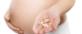
Treatment
The essence of treatment for gestosis is to restore the normal state of women's health. In the second half of pregnancy, in case of development of gestosis, the main rule of behavior is timely contact with a specialist without attempts at self-treatment. Correct treatment of gestosis can only be prescribed by a doctor, taking into account the fact that some medications, as a result of their use, can further aggravate the condition of the pregnant woman and the fetus she is carrying. An example is a situation where a pregnant woman, wanting to get rid of edema, on the advice of relatives, begins to take diuretic pills. However, she will not be able to achieve a positive result, since the cause of edema here is pathological vascular permeability. This incorrect approach to treatment worsens the condition even more.
All doctor’s prescriptions can be easily followed at home if gestosis is mild. However, severe forms require observation by hospital specialists, where they will promptly provide any medical assistance. Timely delivery is also an important stage in the treatment of gestosis. If the pregnant woman’s condition does not improve, fetal hypoxia is detected, and there is no effect of therapy, childbirth becomes the only way out in this situation. In mild forms of gestosis, the birth of a child naturally is quite possible, but there is a risk of deterioration of the woman’s condition during pushing as the load on the mother’s body increases. In most cases, a caesarean section is performed, especially in cases of renal or liver failure, stroke, eclampsia and retinal detachment.
Second half
Gestosis in the second half of pregnancy is also called late gestosis (toxicosis). They pose a great danger because... can lead to serious complications. They most often develop from the 28th week of pregnancy, but can appear at the end of the first and beginning of the second half of pregnancy. In modern medicine, late gestosis is sometimes called OPG-gestosis: O - edema, P - proteinuria (protein in the urine), G - hypertension (increased blood pressure).
The characteristic triad of symptoms (swelling, protein in the urine, increased blood pressure) may not occur in all women. One of them may indicate the development of gestosis. The only visible manifestation of gestosis for a woman is swelling. And increased blood pressure and protein in the urine can only be detected by a doctor. Therefore, it is so important for a pregnant woman to register for pregnancy in a timely manner and regularly attend doctor’s appointments.
The combination of symptoms for gestosis can be different. Currently, all 3 signs of late gestosis are observed only in 15% of cases, edema with increased pressure - in 32% of cases, protein in the urine and increased pressure - in 12% of cases, edema and protein in the urine - in 3% of cases. Moreover, obvious edema is observed in 25%, and hidden edema (indicated by pathological weight gain) - in 13% of cases.
The first stage of late gestosis is edema, or dropsy of the pregnant woman. A woman can notice the appearance of edema by feeling a slight numbness in her fingers. With swelling, it becomes difficult to straighten your fingers and put rings on your fingers.
Swelling does not always mean the development of gestosis. Swelling may be the result of increased production of progesterone (the so-called pregnancy hormone). Edema can also appear as a result of exacerbation of a chronic disease (varicose veins, heart disease, kidney disease). But only a doctor can figure out whether edema is a common manifestation of pregnancy, a symptom of a chronic disease, or a symptom of gestosis.
If there is excessive weight gain in a pregnant woman, but there is no visible edema, then to check, the woman can undergo a Maclure-Aldrich test: a saline solution is injected subcutaneously and the time taken for the “button” to dissolve is observed. If it does not disappear in less than 35 minutes, it means there is hidden swelling.
If swelling becomes visible, it means that 3 liters of excess fluid are retained in the body. First, the feet swell, then the swelling spreads upward, involving the legs, thighs, abdomen, neck and face. Even if a woman does not experience any unpleasant sensations, it is necessary to take urgent measures to prevent gestosis from worsening. It is dangerous to self-medicate and take diuretics, because... this will make the situation even worse. The condition can deteriorate sharply at any time.
The second stage of gestosis – nephropathy – usually develops against the background of dropsy. Its first symptom is an increase in blood pressure. For a pregnant woman, not only an increase in pressure is important, but also sharp fluctuations in it, which can cause placental abruption and fetal death or sudden bleeding.
The third stage of gestosis - preeclampsia - is characterized by the fact that in addition to edema and high blood pressure, protein appears in the urine. At this stage, severe disturbances in the blood supply to the brain may develop, which is manifested by the appearance of severe headaches, a feeling of heaviness in the back of the head, flashing spots before the eyes, nausea and vomiting, visual impairment, memory impairment, and sometimes even mental disorders. Irritability, insomnia, lethargy, pain in the abdomen and right hypochondrium are also noted. Blood pressure is increased - 160/110 mm Hg. Art. and higher.
The fourth, most severe stage of gestosis is eclampsia. Sometimes it, bypassing preeclampsia, develops very quickly after nephropathy. With eclampsia, the function of many organs is impaired, and convulsions may occur. Seizure attacks can be provoked by various factors: a sharp sound, bright light, a stressful situation, pain. The attack of convulsions continues for 1-2 minutes. There may be tonic (“pulling” spasms) and clonic (small muscle twitches). The convulsive attack ends with loss of consciousness. But there is also a non-convulsive form of eclampsia, in which, against the background of high blood pressure, a woman suddenly falls into a coma (loses consciousness).
Eclampsia is fraught with serious complications: placental abruption, premature birth, bleeding, fetal hypoxia and even fetal death. At this stage, it is possible that a heart attack, pulmonary edema, stroke, or renal failure may occur.
Eclampsia most often develops in women with their first pregnancy. When predicting the risk of developing eclampsia, genetic factors should also be taken into account. With hydatidiform mole and multiple pregnancy, the risk of developing eclampsia increases significantly.
In some cases, an asymptomatic or low-symptomatic course of gestosis is possible. But rapid development of this pregnancy complication is also possible. Therefore, at the slightest suspicion that a pregnant woman has gestosis, delay in examination and treatment is dangerous for the life of the mother and child.
Late gestosis can have an unpredictable development. It can progress sharply, and the deterioration of the woman’s condition will rapidly increase with each passing hour. The earlier gestosis develops, the more aggressive its course, and the more severe consequences it will have, especially if treatment is not timely.
Late
There are several theories explaining the reasons for the development of late gestosis in pregnant women:
- Corticesvisceral - the occurrence of gestosis occurs in the body of pregnant women as a neurosis, as a result of which the physiological relationships of the cortex and subcortical elements of the brain are disrupted.
- Endocrine - explains the appearance of gestosis as a result of changes in the functions of endocrine organs.
- Immunological - represents assumptions about changes in blood vessels, organs and tissues due to an inadequate response of the pregnant woman’s immune system to fetal tissue antigens, resulting in signs of late gestosis.
- Genetic - confirmed by statistics on the hereditary appearance of signs of late gestosis.
- Placental - based on the absence of changes necessary to nourish the uterus during pregnancy.
Late gestosis during pregnancy is manifested by the following symptoms:
- protein is detected in the urine, which indicates a violation of the kidneys;
- urination becomes more frequent or scanty;
- blood pressure increases;
- convulsions occur and the kidneys may fail.
Preeclampsia in the later stages can cause preeclampsia, for which characteristic symptoms are swelling of the extremities, the appearance of protein in the urine, hypertension and reflex changes. Also in this case, frequent headaches, nausea and vomiting, and pain in the right hypochondrium may appear.
Also, with gestosis, eclamsia may appear, manifested by individual convulsions, a series of convulsive seizures, as well as a coma of varying duration. Prevention of late gestosis Therefore, if a pregnant woman exhibits late gestosis, then treatment of the disease should be carried out immediately.
After childbirth
After childbirth, some women experience symptoms of gestosis. Such patients are prescribed appropriate treatment, which is continued until their condition stabilizes. The treatment regimen is determined individually.
Prevention
No one is immune from the development of this complication. However, it is possible to prevent the development of a more complex form of the disease.
- The expectant mother's diet should contain foods with a high protein content - cottage cheese, lean meats, eggs, fish. You should limit or completely eliminate the consumption of salty foods and salt. This also applies to confectionery products (ice cream, candies, cakes, pastries, etc.). Sweets can be replaced with fruits. The diet also needs to include fiber, which is found in dried fruits, seaweed, vegetables, and herbs.
- You should not drink a lot of liquid. The maximum amount of liquid you drink per day is no more than 1.5 liters, including first courses.
- Watch your weight. Normal weight gain during pregnancy is 12 kg. If you eat properly and nutritiously, you will not gain excess weight.
- It is recommended that a pregnant woman take a daily walk in the fresh air, which helps improve blood circulation.
- You should not skip visits to the gynecologist and tests, because this will allow you to identify gestosis in pregnant women at an early stage and prevent the development of serious consequences for the mother and child.
Consequences
Preeclampsia causes a very dangerous condition for a pregnant woman, which in medicine is commonly called eclampsia. Eclampsia is characterized by a critical increase in blood pressure and the appearance of a convulsive attack, which begins with twitching of the facial muscles. Then convulsions of the whole body occur, swelling of the brain and lungs occurs, and coma occurs. A woman risks dying from a cerebral hemorrhage, and a child from oxygen deficiency.
When diagnosing eclampsia, doctors urgently send the woman to the hospital and carry out early delivery in order to save both mother and baby.
Tweet
Pin It
Diagnostics
Laboratory tests and the patient’s complaints help determine the condition of the pregnant woman. For diagnosis the following is carried out:
- coagulogram, which determines blood clotting time;
- blood test (biochemistry and general);
- changes in body weight;
- urine analysis (biochemistry and general);
- fundus examination;
- blood pressure dynamics;
- the volume of fluid excreted taking into account its consumption;
To determine the condition of the fetus, ultrasound and Doppler ultrasound are prescribed. To clarify the diagnosis, consultations with a nephrologist, therapist, ophthalmologist and neurologist are carried out.
Etiology and pathogenesis of HELLP syndrome
It is based on an autoimmune mechanism of damage to the vascular endothelium, hypovolemia with blood thickening and the formation of microthrombi, followed by fibrinolysis.
The occurrence of the disease is associated with a decrease in prostacyclin, which causes vasoconstriction in the placenta. This leads to microangiopathic changes in the endothelium, the release of placental thromboplastin and its entry into the maternal bloodstream. The consequence of this is adhesion and aggregation of platelets, thrombocytopenia occurs. When red blood cells pass through altered microvessels, hemolysis occurs. Impaired perfusion in the liver leads to the development of toxic hepatosis with necrosis of the liver parenchyma, in some cases to the formation of subcapsular hematomas. As a result, the level of blood enzymes increases.
MAIN SYMPTOMS OF HELLP SYNDROME
With HELLP syndrome there is a clear clinical picture. Initial manifestations of the disease may be nonspecific and include:
- headache,
- increased fatigue,
- malaise,
- flu-like symptoms
As the disease progresses, pain appears in the epigastric region, a feeling of heaviness and pain in the right hypochondrium, nausea, vomiting as a result of irritation of the peritoneum and irritation of the Glissonian capsule. If intrahepatic pressure increases, this can lead to periportal bleeding, which progresses with the subsequent formation of hematomas under the Glissonian capsule and their fusion. With mechanical damage (increased intra-abdominal pressure during vaginal delivery), the subcapsular hematoma may rupture.
Sometimes HELLP syndrome manifests itself as a clinical picture of PONRP with massive coagulopathic bleeding, jaundice, vomiting mixed with blood, hemorrhage at injection sites, rapid formation of hepatic-renal failure, convulsions, coma.
LABORATORY INDICATORS OF HELLP SYNDROME
- Increased transaminase levels (AST >200 units/l, ALT >70 units/l, LDH >600 units/l;
- Thrombocytopenia less than 100 x 109 thousand;
- Decrease in antithrombin III level below 70%;
- Intravascular hemolysis and increased bilirubin concentration, both due to direct and indirect
- Increased prothrombin time and activated partial thromboplastin time (aPTT)
- Decrease in fibrinogen concentration,
- An increase in the content of nitrogenous waste in the blood,
- Reduced blood glucose levels.
- Morphological examination of the liver indicates that the pathological process is based on intravascular coagulation syndrome and liver necrosis. The most accurate diagnosis is possible using computed tomography of the liver.
Acute fatty hepatosis of pregnant women is an atypical form of gestosis and poses a particular danger to the pregnant woman and the fetus.
Clinic: the disease develops more often in primigravidas in the third trimester, under the age of 25, but can also occur at an older age.
During acute gastrointestinal tract infection, two periods are distinguished: anicteric and icteric.
The first anicteric period can last from several days to 5-6 weeks, is characterized by various complaints, and has few objective symptoms.
The second period is a short period with a stormy clinical course, a rapid progressive course (from several hours to several days) rich in both subjective and objective signs. Within 2-4 days, the jaundice intensifies and becomes more intense. The increase in jaundice is accompanied by the appearance of vomiting, and the vomit acquires an admixture of blood and becomes the color of “coffee grounds.” Belching with rotten eggs appears, hiccups, heartburn, the tongue is coated with a dirty gray coating, bloating, more in the epigastric region, hepatic odor from the mouth, liquid discolored stool. The size of the liver is reduced or unchanged. Signs of heart failure are increasing: tachycardia, arrhythmia, ECG shows diffuse changes in the myocardium, ischemia, respiratory failure, hemorrhagic diathesis, acute renal failure, peripheral edema, fluid accumulation in the serous cavities, antenatal fetal death.
Treatment of gestosis
It is advisable to hospitalize a patient at any stage of pregnancy. This is necessary to preserve the functions of the body systems and successful childbirth.
Outpatient observation is allowed only for stage 1 dropsy. In case of development of nephropathy, preeclampsia and eclampsia, hospitalization is required. Termination of pregnancy is carried out early for health reasons.
Therapy is aimed at preventing the development of complications and the formation of intrauterine disorders in the fetus.
To do this, normalize the work:
- nervous system;
- determine the condition of the vascular wall;
- improve blood circulation;
- normalize water-salt metabolism;
- reduce viscosity and increase blood clotting;
- regularly monitor blood pressure dynamics;
- normalizes metabolic processes in the body.
The duration of treatment directly depends on the severity of gestosis. A mild form will require a two-week hospital stay, while a moderate form will require a long stay. In difficult cases, the pregnant woman will have to remain under daily supervision until delivery.
Premature birth is carried out if:
- Lack of positive dynamics from therapy for persistent nephropathy (moderate severity).
- If the expected effect is not observed during resuscitation in the first 2 hours.
- Disturbances in the development and growth of the fetus (with nephropathy).
- Eclampsia, risk of complications.
Preeclampsia in the second half of pregnancy requires constant medical supervision. Unattended childbirth is allowed only if the woman’s condition is satisfactory, there are no abnormalities in the fetus and if the results of therapy are positive. In all other cases, a caesarean section is prescribed.
Consequences of gestosis
A pregnant woman is at risk of deteriorating kidney and heart function, and pulmonary edema cannot be ruled out. Hemorrhages in internal organs are possible.
Preeclampsia is dangerous due to placental abruption during pregnancy, a lack of oxygen and nutrients to the developing fetus. This threatens developmental delays and dangerous fetal hypoxia.
Diagnosis of gestosis during pregnancy
During pregnancy, especially to detect preeclampsia, the amount of protein in daily urine is determined. The risk of developing preeclampsia appears from the 20th week of pregnancy:
Other laboratory tests:
- Hematocrit number of blood.
- Blood hemoglobin.
- Serum AST.
- The number of platelets in the blood.
- Blood urea nitrogen content.
- The hematocrit number may increase due to blood thickening in preeclampsia.
- An increase in the activity of aminotransferases in the blood serum, a decrease in the number of platelets and an increase in the urea nitrogen content in the blood reflect the development of a pathological process in the body.
Prevention of gestosis
Since the causes of the disease have not been fully determined, the only thing a woman can do to protect herself from this disease is to strictly follow the doctor’s recommendations. If you register in a timely manner and from the first weeks of pregnancy do everything that a specialist says, then the chances of becoming a victim of gestosis will be much less. It seems that the doctor does not advise anything in particular; everyone knows that you should not overwork, lift heavy objects, get hypothermic, or eat junk food. But not everyone wants to follow even these simple recommendations; many believe that they are quite healthy and no disease threatens them. Gynecologists believe that with the first thought of pregnancy, a woman should reconsider her life. Here are the most basic rules that are important for all pregnant women to follow, regardless of their health status:
- Healthy 8-hour sleep;
- No physical stress;
- Rest;
- An attitude of positive emotions, lack of stress;
- Exercise therapy, special gymnastics, breathing exercises;
- Regular walking, swimming, light exercise throughout pregnancy;
- Proper nutrition: eat often, in small portions;
- Drink at least 2 liters of water per day;
- Control your weight;
- Take kidney decoctions to avoid swelling;
- During pregnancy, you can take Canephron, Cyston, Cystenal.
- Magnesium preparations are needed: Magnerot, Magne-B6;
- Curantil, vitamin E, Lipoic acid, Chophytol.
We also recommend reading: Taste in the mouth during pregnancy
It is necessary to think about preventing gestosis at the very beginning of pregnancy. These simple recommendations will allow you to reduce the risk of unpleasant and dangerous symptoms to a minimum, and maintain good health and good mood throughout your pregnancy. And as a result, without any problems, you can become the mother of a beautiful, healthy baby.
Take this seriously, because fetal mortality during preeclampsia is 32%, which is twice the general mortality rate.
Childbirth with gestosis
Even with the possibility of professional treatment, births can become unpredictable for women in labor who suffer from gestosis. This is always a complex and dangerous process that requires the participation of experienced specialists. If the patient’s well-being allows, the pregnancy is maintained until natural delivery or until the fetus becomes completely healthy and viable. Premature birth is accepted in the case of eclampsia or preeclampsia, with moderate gestosis, if the prescribed treatment is ineffective within a week, when the patient’s condition noticeably worsens or placental insufficiency develops.
Natural childbirth with gestosis is possible only if:
- The child is in a cephalic position;
- The mother's pelvis corresponds in size to the fetal head;
- Mother is under 30 years old;
- The cervix is mature.
Specialists should remember that during pregnancy complicated by gestosis, both mother and child react acutely to stress. A woman in labor may suddenly develop high blood pressure. Therefore, often the only correct and safe solution is a caesarean section. The operation is performed for eclampsia and preeclampsia, as well as for complications of gestosis or the presence of cervical pathologies, gestosis that does not respond to treatment within three weeks.
80% of births in women suffering from gestosis are complicated. This requires a team of experienced and qualified specialists. Even those births that began naturally can end in a cesarean section if the development of the situation threatens the death of the mother or child. Parents must understand this need, despite their own beliefs that the child should be born naturally. In such a difficult situation, there is no better way out than to trust specialists, since their experience and knowledge allow you to make the right decision. Educating parents about illnesses that anyone can face is an opportunity to prepare everyone involved for what's to come.
Preeclampsia in pregnancy ranks second after bleeding among the causes of death among mothers.