| Anemia | |
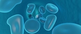
| Histological picture of blood, with iron deficiency anemia | |
| ICD-10 | D50 - D89 |
| ICD-10-CM | D64.9 |
| ICD-9-CM | 285.9[1][2] and 285.8[2] |
| DiseasesDB | 663 |
| MedlinePlus | 000560 |
| eMedicine | med/132 |
| MeSH | D000740 |
| Media files on Wikimedia Commons | |
Anemia
(Greek αναιμία; from the Greek αν - a prefix meaning negation and the Greek αἷμα - blood), synonym -
anemia
, is a condition characterized by a decrease in the number of red blood cells and a decrease in the hemoglobin content per unit volume of blood.
The word “anemia” without specification does not define a specific disease, since anemia is considered one of the symptoms of various pathological conditions of the body. It is necessary to distinguish between hydremia
(pseudoanemia, for example, in pregnant women) and
anemia
- with hydremia, the number of blood cells (erythrocytes, leukocytes, platelets) and hemoglobin remains the same, but the volume of the liquid part of the blood increases [3]. Anemia is the most common blood disorder, affecting about a third of the world's population[4]. Anemia is more common in women than men,[5] during pregnancy, as well as in children and the elderly.[6]
Content
- 1 Features of classification
- 2 Definition of anemia 2.1 By color index 2.1.1 Hypochromic anemia
- 2.1.2 Normochromic anemia
- 2.1.3 Hyperchromic anemia
Classification Features
Anemia is not a disease; it manifests itself as a symptom accompanying a number of diseases and pathologies that can either be associated with a primary lesion of the blood system or not depend on it. To classify anemia, it is customary to use the principle of practical expediency[7]. The current clinical practice corresponds to the classification:
- anemia caused by acute blood loss;
- anemia due to impaired erythrocyte production: aplastic, iron deficiency, megaloblastic, sideroblastic, chronic diseases;
- anemia due to increased destruction of red blood cells: hemolytic[8].
The modern classification of anemia is based on clinical and laboratory signs that allow differential diagnosis of anemia. A special place when conducting a complex of medical studies in the process of identifying the cause of anemic syndrome is given to the indicator “serum ferritin”[9].
Posthemorrhagic anemia
Acute loss of a large amount of blood is accompanied by the development of posthemorrhagic anemia, in which several stages are distinguished. They are aimed at physiological compensation for the restoration of lost blood volume, and are reflected in laboratory parameters.
The second phase is accompanied by hemodilution processes - the entry of fluid from the tissue into the bloodstream, which thus helps restore plasma volume. Here the signs of normochromic normocytic anemia will already be determined. After five days, the body will begin to compensate for the loss of red blood cells by releasing immature young cells into the bloodstream - reticulocytes, an increased content of which will be detected in a blood test. In this phase, differential diagnosis is required to exclude the hemolytic nature of anemia.
After eliminating the bleeding, reticulocyte counts return to normal after 2-3 weeks. If reticulocytosis does not go away, this gives reason to think about ongoing bleeding.
Immediately after blood loss, a transient decrease in the number of platelets may occur, which after a few hours is replaced by an increase.
Develops as a result of large blood loss. This may be visible loss of blood, for example, bleeding in the uterus, lungs, nose, vomiting blood, or from open wounds. And it can also be initially hidden, for example, with gastrointestinal blood loss and large hematomas, with blood spreading into the abdomen (ectopic conception) or the lung cavity.
This anemia is always secondary. Appears during destructive processes in organs, with ulcers, erosions, tumor disintegration, polyp growth, and also if vessels are affected by hereditary or acquired (angiomas, telangiectasia, etc.). A separate link that affects anemia is traumatic bleeding that appears in different areas of the human body.
The patient's well-being worsens when standing; the pulse is frequent and difficult to palpate. Darkening of the eyes and dizziness appear. Along with these signs, the patient may experience other symptoms: pain in the joints, stomach, accompanied by a rash on the legs. Epigastric pain with signs of collapse indicates exacerbation of the disease (the duodenum may become inflamed or the ulcer may recur).
Even a small loss of blood can cause fainting, since 1/8 of the circulating blood is lost at once, this is approximately 600 ml for adults. Symptoms of posthemorrhagic anemia are:
- be sick;
- thirsty;
- sweating increases;
- vomiting occurs due to stomach bleeding;
- pale skin color appears;
- the legs and arms become cold and pale, and cyanosis appears under the nails;
- the tongue becomes dry.
Pain between the shoulder blades and around the chest occurs with myocardial infarction and dissection of the aortic aneurysm, and lumbar pain occurs when blood enters the perinephric tissue. If the abdomen hurts in the lower region, this indicates bleeding due to a ruptured cyst, ectopic pregnancy, renal colic with hematuria.
First of all, acute posthemorrhagic anemia is indicated by a decrease in circulating blood mass, and only then by the level of hemoglobin and red blood cells.
First aid is aimed at preventing bleeding. Apply a tourniquet or tight bandage, apply pressure to bleeding vessels, or insert a tampon into the nose. Drugs are used that can stop blood loss.
Definition of anemia
A decrease in hemoglobin concentration in the blood often occurs with a simultaneous decrease in the number of red blood cells and a change in their qualitative composition.
Any anemia leads to a decrease in the respiratory function of the blood and the development of oxygen starvation of tissues. Depending on gender and age, the norm for hemoglobin content per liter of blood may differ. Determination of anemia and its severity, g/l[10]
| Age | Normal level | Light | Average | Heavy |
| From 6 months to 4 years inclusive | 110 and above | 100—109 | 70—99 | less than 70 |
| Children 5-11 years old | 115 and above | 110—114 | 80—109 | less than 80 |
| Children 12-14 years old | 120 and above | 110—119 | 80—109 | less than 80 |
| Women (from 15 years old) | 120 and above | 110—119 | 80—109 | less than 80 |
| Pregnant women | 110 and above | 100—109 | 70—99 | less than 70 |
| Men (from 15 years old) | 130 and above | 110—129 | 80—109 | less than 80 |
By color indicator
The color index (CI) shows the degree of saturation of the red blood cell with hemoglobin. Normally, it is 0.86–1.1, both in men and women[11]. Depending on it, the following anemias are distinguished:
Hypochromic anemia
- Hypochromic - CP < 0.86 (according to some sources below 0.8): iron deficiency anemia
- thalassemia
Normochromic anemia
- Normochromic - CP 0.86-1.1: hemolytic anemia (when the rate of destruction of red blood cells exceeds the rate of their production)
- posthemorrhagic (as a result of blood loss due to bleeding or hemorrhage)
- neoplastic bone marrow diseases
- aplastic anemia
- extramedullary tumors
- anemia due to decreased production of erythropoietin
Hyperchromic anemia
- Hyperchromic - CP > 1.1: vitamin B12 deficiency anemia
- folate deficiency anemia
- myelodysplastic syndrome
According to the ability of bone marrow to regenerate
The main sign of such regeneration is an increase in the number of reticulocytes (young red blood cells) in the peripheral blood. The norm is 0.5-2%.
- Aregenerative (for example, aplastic anemia[12]) - characterized by the absence of reticulocytes.
- Hyporegenerative (vitamin B12-deficiency anemia, iron deficiency anemia) - characterized by a reticulocyte count below 0.5%.
- Normoregenerative or regenerative (posthemorrhagic) - the number of reticulocytes is normal (0.5-2%).
- Hyperregenerative (hemolytic anemia) - the number of reticulocytes is more than 2%.
Pathogenetic classification
Based on the mechanisms of development of anemia as a pathological process
- Iron deficiency anemia - associated with iron deficiency
- Dyshematopoietic anemia - anemia associated with impaired blood formation in the red bone marrow
- Posthemorrhagic anemia - associated with acute or chronic blood loss
- Hemolytic anemia - associated with increased destruction of red blood cells
- B12 (megaloblastic anemia) - associated with impaired DNA and RNA synthesis.
- Folate deficiency anemia
By etiology
- Anemia in chronic inflammation: In infections: tuberculosis
- bacterial endocarditis
- bronchiectasis
- lung abscess
- brucellosis
- pyelonephritis
- osteomyelitis
- mycoses
- systemic lupus erythematosus
- Pernicious anemia
NNA for endocrine diseases
Erythropoietin is a kidney hormone that increases the production of red blood cells during acute oxygen deficiency in tissues. The production of this hormone is influenced by:
- androgens;
- thyroid hormones;
- pituitary hormones;
- cortisol
If the parathyroid gland is overactive, parathyroid hormone appears in excess, which suppresses the production of red blood cells and leads to anemia.
This disease develops if a person’s immune system, under the influence of the diseases described above, begins to work excessively. Anemia in chronic diseases is long-lasting, about 2 months, hemoglobin in the range of 70-110 g/l is normochromic, but can become hypochromic, and leukocytosis may appear.
In chronic renal failure, the secretion of erythropoietin is minimized. Patients with chronic renal failure also develop anemia for the following reasons:
- circulating red blood cells do not live long;
- low production of erythropoietin;
- due to blood loss, iron deficiency appears;
- uremic toxins inhibit blood formation.
Anemia due to tumors and chronic diseases can be called a pathology due to low sensitivity to erythropoietin, but its course is much more complicated.
The most common anemia is iron deficiency. After it comes normochromic anemia in tumors and chronic diseases, chronic infections (AIDS, tuberculosis), acute diseases (pneumonia), some tumors, heart failure, rheumatic diseases (arthritis), etc.
This type of anemia is treated as follows: first, the chronic disease or tumor itself is treated, and then intravenous iron and erythropoietin are prescribed. In rare difficult cases, a transfusion of red blood cells is given.
With this type of anemia, one cannot help but mention infiltrative anemia, when the bone marrow is affected by cancer metastases. The disease is diagnosed in this case by taking a puncture for biopsy analysis.
Chronic renal dysfunction is a condition that is accompanied by a decrease in the level of erythropoietin produced. Erythropoietin is a kidney hormone that enhances erythropoiesis in conditions of tissue oxygen deficiency. In chronic renal pathology, uremic toxins suppress the production of erythropoietin, which is accompanied by a shortening of the life of red blood cells that circulate in the blood.
Anemia How to treat anemia?
Anemia-Symptoms and Treatment
9 POSSIBLE SIGNS OF ANEMIA THAT ARE NOT NOTICEABLE AT FIRST LOOK
Iron deficiency anemia 1
Anemia .Symptoms .Causes .Treatment
Causes of anemia – Dr. Komarovsky
Iron deficiency anemia | What to do | How to treat | Symptoms | pregnancy | Disease | Dr. Phil
About the most important things: Anemia, lump in the throat, spider veins on the face
Anemia or anemia can be cured with folk remedies
Vegetarianism/Anemia/Slow Death
Anemia. Symptoms and types of anemia
About the most important things: Anemia, often stomach pain, dry mouth
All about blood. Anemia. Hemoglobin. Olga Butakova ACADEMY OF HEALTH
ANEMIA. HOW TO TREAT. SYMPTOMS. ANALYSIS. FGS. DROPPER.TABLETS. HAIR LOSS #anemia
Anemia. How to increase hemoglobin using natural means?
Torsunov O.G. About the causes of iron deficiency anemia
The clinical picture of a laboratory blood test, in addition to normochromic-normocytic data, shows a normal number of reticulocytes, which can vary slightly both upward and downward, depending on the degree of activity of erythrocyte production in the bone marrow. There will also be a decrease in the level of platelets, white blood cells and the hormone erythropoietin itself.
Pathologies of the endocrine glands that are accompanied by normochromic-normocytic anemia include:
- adrenal insufficiency (Cushing's disease);
- hyperparathyroidism;
- thyrotoxicosis;
- panhypopituitarism (a disease of the adenohypophysis, resulting in a decrease in the concentration of pituitary hormones that regulate the functions of the peripheral endocrine glands);
- Addison's disease;
- hypoandrogenism.
The endocrine glands in which pathological processes occur have a direct effect on the synthesis of erythropoietin, so the level of the hormone will decrease, leading to NNA.
Laboratory tests will be similar to NNA in chronic renal failure, since they have a common pathogenesis associated with a decrease in erythropoietin levels.
Pathogenesis
There are three main mechanisms for the development of anemia:
- Anemia as a consequence of impaired formation of normal red blood cells and hemoglobin synthesis. This development mechanism is observed in the case of a lack of iron, vitamin B12, folic acid, and during diseases of the red bone marrow.
- Anemia as a consequence of loss of red blood cells is mainly a consequence of acute bleeding (trauma, surgery). It should be noted that with chronic small-volume bleeding, the cause of anemia is not so much the loss of red blood cells as the lack of iron, which develops against the background of chronic blood loss.
- Anemia as a consequence of accelerated destruction of red blood cells. Normally, the lifespan of red blood cells is about 120 days. In some cases (hemolytic anemia, hemoglobinopathies, etc.), red blood cells are destroyed faster, which becomes the cause of anemia. Sometimes the destruction of red blood cells is facilitated by the consumption of significant quantities of vinegar, which causes accelerated breakdown of red blood cells[13].
Varieties
Hypochromic iron deficiency anemia can be of several types, including:
- Microcytic iron deficiency anemia. This type of anemia is the most common. The disorder develops against the background of bleeding, when there is insufficient supply of iron from the outside (with food), or when the body is not able to adequately absorb this microelement. This type of anemia is also often diagnosed in nursing mothers and pregnant women. The risk group includes children and women of childbearing age.
- Sideroachrestic anemia, or as it is called, iron-saturated anemia. This type of disorder is characterized by a normal level of iron in the blood, but it cannot be absorbed, which leads to a decrease in the number of hemoglobin molecules in red blood cells. Elderly people, alcoholics, and patients who have suffered poisoning from pesticides or medications are more susceptible to such anemia.
- Iron redistribution anemia develops when the level of iron in the blood increases due to the destruction of red blood cells. This condition often accompanies tuberculosis, as well as diseases accompanied by purulent processes.
- Anemia of mixed type. This anemia develops due to a lack of vitamin B12 and iron in the body. A person suffering from such a disorder often gets tired, the body’s natural defenses deteriorate, and swelling is observed, concentrated in the area of the hands.
Many people who are first faced with the diagnosis of “hypochromic anemia” have a question: what is it: a hereditary or acquired disease? In fact, pathology can be inherited and develop throughout life. The congenital form of anemia manifests itself in the presence of other blood diseases, and the acquired form is a consequence of surgical interventions, infections and intoxications.
The World Health Organization provides frightening statistics. According to their information, every 3 women and every 4 men on the planet suffer from anemia. Moreover, the disorder occurs in a chronic form. The reason for this is hidden diseases, errors in the diet, which contribute to a lack of iron for normal hemoglobin production.
Clinical manifestations
Anemia often occurs without pronounced manifestations and often goes unnoticed, in many cases becoming an accidental laboratory finding in people who do not present specific complaints.
As a rule, those suffering from anemia note manifestations caused by the development of anemic hypoxia
. In mild forms, this may include weakness, fatigue, general malaise, and decreased concentration. People with more severe anemia may complain of shortness of breath with slight or moderate exertion, palpitations, headache, tinnitus, and disturbances in sleep, appetite, and sexual desire. With very severe anemia, or in the presence of concomitant pathology, heart failure may develop.
A frequently encountered diagnostically important symptom of moderate or severe anemia is pallor (of the skin, visible mucous membranes and nail beds). Symptoms such as the development of cheilosis and koilonychia, increased cardiac impulse and the appearance of functional systolic murmur are also valuable.
Manifestations of acute and severe anemia are always more pronounced than chronic and moderate anemia.
In addition to general symptoms directly related to hypoxia, anemia may have other manifestations depending on its etiology and pathogenesis. For example, the development of sensitivity disorders with B12-deficiency anemia, jaundice with hemolytic anemia, etc.
Anemia during pregnancy
Main article: Anemia during pregnancy
Of all types of anemia during pregnancy, iron deficiency anemia occurs most often[14]. This is due to the increasing need for iron from 0.6 to 3.5 mg/day, which exceeds the ability to absorb it from food (1.8-2 mg/day). Iron is used for the formation of the fetus and placenta[14].
If the disease persists during pregnancy, it can lead to serious consequences:
- the fetus may not receive enough oxygen, which is needed for normal development, especially of the brain;
- women with severe forms of anemia feel worse during pregnancy;
- the likelihood of premature birth increases;
- After childbirth, the risk of developing infections is higher[15].
Physiological hyperplasia should be distinguished from anemia in pregnant women, in which, due to an increase of 23-24% in blood mass, hematocrit, hemoglobin and red blood cell counts decrease.
Hyperplasia is asymptomatic, does not require treatment, and disappears within 1-2 weeks after birth.[14] Average values of hemoindicators during normal pregnancy and outside of it[14]
| Indicators | Not pregnant | In a pregnant woman |
| Hb, g/l | 145-125 | 105-110 |
| Red blood cells ×1012/l | 3,7±0,25 | 3,25±0,25 |
| Reticulocytes, ‰ | 5-10 | 10-25 |
| Hematocrit, ‰ | 40-42 | 33-35 |
| Leukocytes ×109/l | 7±3 | 10±5 |
| Platelets ×109/l | 300 | 150 |
| ESR mm/hour | 13-26 | 50-80 |
NNA for aplastic anemia
Aplastic anemia is diagnosed in 1-2 cases per 1 million population per year. The disease is characterized by a decrease in the generation of absolutely all blood cells, which is associated with congenital or acquired causes (hereditary diseases, the influence of toxic substances, drugs, x-rays).
A myelogram obtained by biopsy or puncture gives a picture of “empty” bone marrow, which in severe forms of the disease is replaced by adipose tissue.
Aplastic anemia requires detailed diagnosis to differentiate the disease from acute leukemia.
Symptoms of the disease do not immediately make themselves felt. The human body gets used to anemia for a long time and does not consider it necessary to consult a doctor. A sharp deterioration in the condition occurs mainly with the development of hemorrhage (uterine in women, gastric, nasal or intestinal bleeding). The condition may also worsen due to the addition of infections accompanied by otitis media, pneumonia, and pyelonephritis.
Aplastic anemia occurs due to a sharp decline in red blood cells, a decrease in the functioning of all cells in the bone marrow, platelets and leukocytes in the blood, hemoglobin and reticulocytes. This diagnosis is made only after leukemia has been excluded.
In turn, aplastic anemia is of 2 types:
- immune;
- myelotoxic.
The first type is associated with a natural antibody response to bone marrow cells. The second type occurs after exposure to chemicals and drugs that affect the bone marrow.
Help for this type of anemia should be as follows: you need to quickly administer metipred or prednisolone intravenously. Avoid previously used drugs (chlorevomycetin, cytostatics, amidopyrine, etc.), which could provoke or aggravate aplastic anemia. Next, the ambulance takes the patient to the hospital for transfusion therapy. Treatment is carried out with steroid hormones and glucocorticoids. The doctor then decides whether to perform a splenectomy or not.
Treatment
The most common are iron and B12 deficiency anemias, which are treated with vitamin B12 and iron supplements. Also, if hemoglobin levels are low, red blood cell transfusions can be used. In general, treatment tactics depend on the type of anemia and the severity of the patient’s condition.
- Some anemias are treated in a hospital setting.
- The diet should be complete, contain sufficient amounts of protein, iron and vitamins.
- According to vital indications, in case of a sharp disturbance in hemodynamics, a drop in hemoglobin below 70-80 g/l, blood transfusions are used.
- Therapy for individual forms of anemia is carried out taking into account their etiology and pathogenesis.
- In the case of acute posthemorrhagic anemia, the first step is to stop the bleeding. After massive blood loss, iron supplements are prescribed.
- Pathogenetic therapy of iron deficiency anemia is based on the use of iron preparations by administering it orally (totema, hemostimulin, ferroplex, tardiferon) or parenterally (ferractin, ferrum-lek, ferbitol, ectofer).
- Treatment of vitamin B12 deficiency anemia is carried out by parenteral use of vitamin preparations, sometimes with the addition of a coenzyme - adenosine cobalamin. The criterion for the effectiveness of the therapy is the reticulocyte crisis - an increase in the number of reticulocytes to 20-30% on days 5-8 of treatment.
- Treatment of aplastic anemia includes blood transfusions, bone marrow transplantation, and therapy with glucocorticoid and anabolic hormones.
What is normocytic normochromic anemia?
Patients who suffer from adrenal insufficiency may be diagnosed with this type of anemia. Sometimes it is difficult to diagnose, since simultaneously with other indicators the plasma volume level also drops.
It is worth noting that this form of anemia is diagnosed in 20% of cases in patients with primary manifestations of hyperparathyroidism. When it is supported by internal bleeding or ulcers, iron deficiency anemia can begin to develop very quickly.
Prevention
The main means of preventing anemia is a balanced diet rich in vitamins, as well as the use of supplements containing iron, as recommended by the attending physician. The daily iron requirement for normal human life is 20-25 mg. The bulk of this amount (90%) is endogenous iron, which is released during the breakdown of red blood cells, 10% is exogenous iron coming from food. The iron norm for men is 1 mg, for women - 2 mg (due to cyclic blood loss)[16].
Prevention of disease development
Following nutritional recommendations is good prevention
Neither adults nor children are immune from the development of anemia. However, to maintain health, you must adhere to certain rules:
- Children of all ages need to drink milk because of its iron content. After consultation with a pediatrician, you can take courses of vitamin and mineral complexes. Girls who have not yet fully formed their menstrual cycle are also recommended to take iron supplements to eliminate its deficiency;
- Pregnant women also need to additionally maintain their blood condition with iron supplements, vitamins B12 and B9, but only in consultation with their doctor;
- It would also be a good idea for adults and older people to periodically take mineral and vitamin complexes, but also only after consulting a doctor.
One of the main rules for preventing the development of anemia is good nutrition, no strict diets without specific indications, and mandatory daily consumption of meat.
Following all of the above rules will help maintain health and prevent the development of many diseases.
Notes
- Disease ontology database (English) - 2020.
- ↑ 12
Monarch Disease Ontology release 2018-06-29sonu - 2018-06-29 - 2018. - G. A. Alekseev;
N. M. Nemenova (pat. an.), A. F. Tur (ped.), N. A. Fedorov, M. G. Katekhelidze (A. experimental), S. X. Khakimova (A. pregnant women), A. Z. Tsfasman (rad.). Anemia // Great Medical Encyclopedia: 30 volumes / chapter. ed. B.V. Petrovsky. — 3rd ed. - Moscow: Soviet Encyclopedia, 1974. - T. 1. A - Antibiosis. — 576 p. — 150,000 copies. - Timothy G. Janz, Roy L. Johnson, Scott D. Rubenstein.
Anemia in the emergency department: evaluation and treatment // Emergency Medicine Practice. — 2013-11. - T. 15, issue. 11. - pp. 1–15; quiz 15–16. - ISSN 1524-1971. - T. Vos, A. D. Flaxman, M. Naghavi, R. Lozano, C. Michaud.
Years lived with disability (YLDs) for 1160 sequelae of 289 diseases and injuries 1990-2010: a systematic analysis for the global burden of disease study 2010 // The Lancet. - 2012. - T. 380. - P. 2163–2196. — ISSN 0140-6736. - Timothy G. Janz, Roy L. Johnson, Scott D. Rubenstein.
Anemia in the emergency department: evaluation and treatment // Emergency Medicine Practice. — 2013-11. - T. 15, issue. 11. - pp. 1–15; quiz 15–16. - ISSN 1524-1971. - Doctor of Medical Sciences
P. F. Litvitsky. Pathology of the erythrocyte system (Russian) // Issues of modern pediatrics: Lecture. — 2020. — August 28 (No. 14). — P. 450—463. - doi:10.15690/vsp.v14.i4.1384. - Anemia: classification, prevention, treatment. Help (Russian). https://ria.ru/
(2011). Retrieved March 20, 2020. - Smirnova L. A.
Anemia: Differential diagnostic aspects (Russian) // Medical news. - 2013. - No. 2. - P. 15-19. - Haemoglobin concentrations for the diagnosis of anemia and assessment of severity (English). Vitamin and Mineral Nutrition Information System (VMNIS)
. World Health Organization (2011). - Rogova L.N., Gubanova E.I., Pankova G.V., Shepeleva T.I.
Pathogenetic rationale for interpreting the results of a general blood test (Russian).
https://medconfer.com/
. Retrieved March 20, 2020. - Treatment of acquired aplastic anemia (Russian) (inaccessible link). https://oncology.by/
. Retrieved March 20, 2020. Archived March 21, 2019. - Chesnokova N. P., Nevvazhay T. A., Morrison V. V., Bizenkova M. N.
LECTURE 2. ANEMIA: CLASSIFICATION, GENERAL CHARACTERISTICS OF HEMATOLOGICAL SHIFT. POSTHEMORRHAGIC ANEMIA (Russian) // International Journal of Applied and Fundamental Research. - 2015. - No. 6-1. — P. 152-155. - ↑ 1 2 3 4 Shaposhnik O. D., Rybalova L. F.
Anemia in pregnant women. — Educational and methodological manual for medical students. - Chelyabinsk, 2002. - Pregnancy Complicated by Disease. Anemia During Pregnancy, Sean C. Blackwell // The Merck Manual Home Health Handbook, December 2008.
- Stuklov N.I., Semenova E.N.
Iron deficiency anemia. Modern tactics of diagnosis and treatment, criteria for the effectiveness of therapy (Russian) // Clinical medicine: Article. - 2013. - No. 12. - P. 61-67.