Skin cancer, including melanoma, is highly treatable in its early stages. The most important thing is to recognize the signs of pathology in time and begin treatment. It is extremely important to correctly identify a new skin growth that could potentially be cancerous. Finding skin cancer early can greatly increase your chances of cure.
- What formations on the skin should raise suspicion?
- ABCDE technique for cancer detection
- Who is most at risk of skin cancer?
- How to detect skin cancer on the body?
- What does skin cancer look like?
- How do basal cell and squamous cell skin cancers manifest?
- How does melanoma manifest?
- Cost of diagnosing skin cancer in Israel
What causes skin cancer?
There are people who are at risk for skin cancer:
- the white-skinned population with blond hair and eyes, as well as albino people. Dark-skinned inhabitants of the planet are twenty times less likely to encounter this type of disease. This is due to a greater degree of skin protection from exposure to ultraviolet rays;
- Frequent visitors to solariums and beaches are more susceptible to pathology than others, since they are most exposed to radiation. If you have three sunburns, the risk of developing skin cancer doubles;
- people who often have to deal with chemicals in their line of work, they can lead to mutation of DNA molecules;
- exposure to radioactive radiation. Working at nuclear power plants or with medical equipment that has harmful radiation;
- also residents of cities close to accident sites at nuclear power plants;
- people who have a significant number of postoperative scars or large pigment spots on their bodies, moles are affected by this disease much more often;
- age after fifty years.
There are skin diseases, they are classified as precancerous conditions, the lack of treatment of which can lead to skin cancer:
- Keir's erythroplasia;
- Bowen's disease;
- xeroderma pigmentosum;
- leukoplakia;
- senile keratoma;
- cutaneous horn;
- Dubreuil's melanosis;
- melanoma-dangerous pigmented nevi (complex pigmented nevus, blue nevus, giant nevus, nevus of Ota);
- chronic skin lesions: trophic ulcers, tuberculosis, syphilis, SLE, etc.
Treatment process
Treatment for cancer localized on a person's face can be completely different. This is due to the fact that therapy is selected by doctors depending on the type of disease, widespread cancer, stage of pathology and a number of other specific factors.
Most often, treatment is performed using the following methods and techniques:
- Cryodestruction. If the disease is at an early stage, then the cancerous tumor and keratotic lesion can be easily removed using the technology of freezing the skin area using liquid nitrogen. Due to exposure to such low temperatures, painful tissues begin to peel off from healthy skin.
- Excisional surgery. This type of treatment can be used regardless of the type of cancer. During this operation, cancerous tissue and a number of healthy areas of skin are excised. The method is usually used when the pathology has late stages of development.
- Mohs surgery. This technique is used only in cases of recurrent and frequently recurring cancerous lesions of the skin. The method is extremely effective for nasal skin cancer. An operation of this type involves gradual layer-by-layer removal of malignant tissue, which allows preserving healthy areas to the maximum extent.
- Curettage. This method can only be used in combination with surgical excision of a malignant neoplasm. This involves the use of a special round blade (a cryoscalpel, for example), with the help of which the remaining cancer cells are destroyed.
- Radiation therapy. Treatment with highly active X-rays to eliminate cancer cells has been used for a long time. The method is relevant in cases where it is impossible to perform a surgical operation, or immediately before it.
- Chemotherapy. In this case, treatment is performed with special cytostatic drugs. In the described situation, the method can be used only in cases where the pathological process spreads and/or metastases penetrate into distant tissues.
How to recognize skin cancer?
There are three types of skin cancer:
- squamous cell skin cancer - develops from flat cells of the surface layer of the epidermis;
- skin basal cell carcinoma - occurs under a layer of flat cells with atypical degeneration of the basal cells of the epidermis;
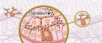
- Melanoma of the skin - arises from its pigment cells - melanocytes.
There is another type - skin adenocarcinoma (glandular skin cancer), which arises from the sweat glands. A rather rare type of skin cancer.
There are a number of rules, following which, you can identify the disease yourself. To do this, you need to know the signs of skin cancer in the early stages of the disease.
What should you be wary of?
- If you notice that the nevus has become asymmetrical, for example, one half of the mole is different from the second;
- the edges of the nevus have become uneven, swelling or indentations have appeared;
- a change in color has occurred, the mole has acquired a bluish tint, has become significantly darker, or its pigmentation is not uniform;
- if the mole begins to grow rapidly or its size is more than six millimeters;
- when there is a scar on the skin and it does not heal for a long time or liquid begins to ooze from it;
- the unreasonable appearance on the skin of a spot or bump in the form of a nodule with a glossy surface with an unusual pigment (red, pink, black).
Moreover, over the past ten years, the incidence has increased by more than 30%. Skin cancer (without melanoma, which is usually treated separately) is not as life-threatening as other malignancies. Only 0.53% of all cancer deaths are caused by skin cancer (1.26% by melanoma).
There are three main types of skin cancers: basal cell carcinoma, squamous cell carcinoma, and melanoma. They develop from different types of cells and have significant differences in both symptoms and the course of the disease.
Basalioma is the most common type of skin cancer and the least dangerous. Most often, basal cell carcinoma forms on the face or neck. It develops very slowly, over years or even decades, and, as a rule, does not spread within the body (that is, it does not metastasize). In 90% of cases, basal cell carcinoma can be completely cured.
Squamous cell carcinoma progresses faster, not years, but months. Develops on exposed areas of the skin exposed to sunlight. In its advanced form, when the tumor grows to a significant size, squamous cell carcinoma is capable of forming metastases.
Melanoma is the most dangerous skin cancer. Although it is less common than other types, it is the one that claims the most lives. Melanoma develops rapidly and metastasizes easily. Sometimes it develops from moles, which is not typical for other types of skin cancer, but often occurs as a primary tumor. In patients with melanoma, even with timely treatment, relapses are likely - re-growth of the tumor after a few years.
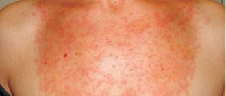
Skin cancer can go unnoticed for a long time, especially if the tumor forms in a hard-to-reach place, such as under the hair, on the back, behind the ear or on the skin of the feet. The first serious symptoms that you should pay attention to are: an observed increase in the size of the pigment spot, a change in its color, inflammation, itching and pain, and possible bleeding.
The main cause of all types of skin cancer is ultraviolet radiation. And not only the sun: staying in a solarium poses the same risk as traditional sunbathing. Those at risk are those who spend a lot of time outdoors, residents of southern countries and, especially, those who moved south from less sunny latitudes. People with fair skin ( phototypes I and II on the Fitzpatrick scale ) are more susceptible to cancer.
For the development of mutation, the total accumulated dose of ultraviolet radiation is important. Therefore, the second risk factor is age: skin cancer most often develops after 50 years.
Malignant skin neoplasms can also be caused by long-term exposure to carcinogens. For example, squamous cell carcinoma was first described in 1775 as an occupational disease of London chimney sweeps. Thus, stove soot became the first carcinogen identified.
A risk factor for melanoma is a large number of moles, especially large ones. Heredity also plays a large role in the development of melanoma.
The main measure for preventing skin cancer is avoiding direct sunlight and other sources of ultraviolet radiation and using sunscreen.
TNM classification is necessary for a more accurate assessment of the prevalence of skin cancer
T - primary tumor:
- TX - it is impossible to evaluate the tumor due to lack of data;
- TO—the tumor is not detected;
- Tis—cancer in situ;
- TI—tumor size up to 2 cm;
- T2 - cancer tumor size up to 5 cm;
- TZ - the size of the formation is more than 5 cm;
- T4 - skin cancer grows into the underlying deep tissues: muscle, cartilage or bone.
N—state of lymph nodes:
- NX - the condition of regional lymph nodes cannot be assessed due to lack of data;
- N0 - there are no signs of metastases in the lymph nodes;
- N1 - there is metastatic damage to regional lymph nodes.
M - presence of metastasis
- MX - lack of data regarding the presence of distant metastases;
- MO - distant metastases not detected;
- M1 - distant metastasis is present.
The degree of differentiation of tumor cells is assessed within the histopathological classification of skin cancer.
- GX - it is not possible to determine the degree of differentiation;
- G1 - high differentiation of tumor cells;
- G2—average differentiation of tumor cells;
- G3 - low differentiation of tumor cells;
- G4 - undifferentiated skin cancer.
Types and signs of malignant skin tumors
areas affected by certain types of tumors
The classification of malignant skin tumors is based on their histological structure, depending on which the following types of cancer are distinguished:
- Basal cell (basalioma);
- Squamous cell carcinoma.
In recent years, melanoma growing from melanin-forming tissue and therefore not associated with the surface epithelium and glands of the skin is often excluded from malignant tumors of the integumentary epithelium. Melanoma will be mentioned a little later.
In rare cases, the growth of glandular tumors (adenocarcinomas) is possible, the source of which is the skin appendages - sweat and sebaceous glands.
Oncologists use the TNM classification of tumors, based on which the stages of the disease can be distinguished:
- If the tumor is no more than 2 cm in size and there are no metastases, then they speak of the first stage.
- In the case when the tumor reaches 5 or more centimeters, destroys the surface layers, is located under the skin, but still does not metastasize, we can talk about the second stage of the disease.
- In the third, it is possible that deep structures outside the skin may be involved without metastasis, or the tumor itself is small, but regional lymph nodes affected by cancer cells are detected.
- The fourth stage of cancer is established when distant metastases are detected, regardless of the volume of the primary tumor and the condition of the regional lymph nodes.
The initial stage of skin cancer often does not manifest itself with any pronounced or specific signs, so patients are in no hurry to see a doctor, and the appearance of skin changes is attributed to various inflammatory processes, infectious lesions, etc. Meanwhile, the presence of even a spot or small nodule should be a reason for visiting a dermatologist and excluding a malignant tumor.
Signs, combinations of which may cause concern:
healthy moles (top) and tumor/preneoplastic processes (bottom) (primarily melanoma)
The first symptoms of skin cancer may be a local change in the color of the skin, tissue hardening, the presence of nodules, plaques that are prone to ulceration, often bleeding. Such changes progress quickly and cannot be treated, especially at home. In the absence of timely qualified assistance, metastases may damage regional lymph nodes, deeper tissues, including cartilage, muscles, and ligaments.
Basal cell carcinoma
The most common type of skin cancer is considered to be basal cell carcinoma, accounting for about two-thirds of all detected neoplasms in this localization. This tumor, as a rule, is detected in elderly people, is often located on the face, does not metastasize, but is capable of germinating tissue, causing its destruction. The course of basal cell carcinoma is quite favorable, and slow growth along with the absence of metastases allows one to achieve good treatment results.
Basal cell skin cancer is made up of cells that resemble the basal layer of the epidermis, hence its name. Externally, the tumor resembles a node (this is the most common type of growth) of dense consistency, with a smooth surface, and pink color. Metastasis is not typical, on the basis of which there has been a long debate about the degree of malignancy of the tumor, however, its histological structure and the nature of the cellular elements still speak in favor of cancer.
In addition to nodular, there are also superficial types of basal cell carcinoma, ulcerative, pigmentary and others.
In most cases, the first signs of basal cell carcinoma are the presence of a convex pink neoplasm with a pearlescent tint or a nodule with ulceration. The size of the tumor increases slowly, so patients are in no hurry to see a doctor, however, damage to deeper tissues, ulceration or bleeding still force them to seek help.
Basalioma is the most common type of facial skin cancer. This tumor can be located in the area of the eyes and temples, growing into nearby tissues, which can lead to death in a fairly short time.
Squamous cell carcinoma
Squamous cell carcinoma is diagnosed in approximately a quarter of all patients. If basal cell carcinoma is more common for older people and reflects age-related degenerative changes, decreased immunity, impaired regenerative capabilities of the skin, then squamous cell carcinoma, as a rule, is a consequence of long-existing dermatoses, inflammatory processes, scars, etc. Due to the direct participation of external factors in the process tumor formation, most often such neoplasms are found in open areas of the body - on the hands, face.
Skin cancer looks different in patients, and therefore its main types have been identified: infiltrative-ulcerative and papillary. This division is based not only on the appearance of the tumor, but also takes into account the nature of tissue damage as a whole.
The infiltrative-ulcerative form is manifested by the appearance of a deep ulcer, growing deep into the tissue, the bottom of which is covered with crusts and bloody discharge. Over time, the size of the lesion increases both in width and depth.
The papillary form of skin cancer is similar in appearance to cauliflower; the formation has a dense consistency and a lumpy surface. In the early stages, this type of growth can be mistaken for a papilloma (benign tumor), however, the presence of a previous skin lesion, rather intensive growth with a tendency to ulceration and bleeding indicate a malignant process.
Squamous cell carcinoma, unlike basal cell carcinoma, may be accompanied by the presence of metastases. As a rule, cancer cells reach regional lymph nodes and settle in them. The spread of tumor cells through blood vessels can lead to the appearance of secondary tumor nodes in the liver and lungs, but this route of metastasis is extremely rare.
Video: doctor about identifying skin cancer
Skin cancer - the first symptoms of the disease:
- pain syndromes in the affected area of the skin as the tumor spreads, the pain intensifies;
- open ulcers and wounds on the body that do not heal for a long time, the appearance of ulcers on a mole;
- hair loss from the surface of the nevus;
- change in color (darkening, lightening, uneven coloring);
- bleeding;
- active growth, doubling in six months;
- the size of the mole is more than 7 mm, with asymmetrical uneven edges and unclear boundaries;
- the appearance of nodes.
In later stages of the disease, skin cancer is characterized by symptoms such as:
- weight loss;
- loss of appetite;
- weakness;
- fast fatiguability;
- apathy;
- general malaise;
- nausea;
- vomit;
- increase in body temperature, etc.
With complete metastasis, deterioration of vision, hearing, and headaches may occur. Without appropriate treatment, death is quite possible.
Symptoms of the disease
The tumor in this disease is a small nodule. Its surface is characterized by unevenness, sometimes it is covered with somewhat keratinized epithelium of a rough appearance. The disease can manifest itself in areas such as:
- oral mucosa;
- language;
- lips;
- nostrils (rarely);
- eyes;
- senile keratomas;
- papillomas, etc.
As a rule, a person pays attention to it only when it begins to ulcerate, deepen and expand.
A rather characteristic sign appears, which indicates that squamous cell skin cancer is present - the edges and bottom of the resulting ulcer have a bright red color, a dense consistency, the edges seem to turn outward.
Clinical picture
It has pronounced symptoms that can be independently detected on the body. The disease usually begins to develop from a node, strange ulcer, scar or plaque. Such skin neoplasms in squamous cell carcinoma can grow excessively quickly. Sometimes pain even begins due to infection.
Basal cell skin cancer results from abnormal growth in the top layer of epidermal cells. It accounts for 75% of skin cancer cases, making it the most common form.
Squamous cell skin cancer involves changes in the skin cells in the middle layer of the epidermis and appears as a growing bump with a rough, scaly surface and flat, reddish spots.
Basalioma is a superficial form, characterized by an abundance of blood vessels. A semi-malignant skin tumor that does not metastasize.
- Symptoms of the disease in ulcerative form. On the body, roller-like, clearly raised edges are visible that surround the ulcer. It itself resembles a crater in shape. As a rule, a terrible bloody discharge comes out of the outbreak, which smells very unpleasant. Such a neoplasm can progress quickly, increasing not only in width, but even in depth.
- Symptoms when a node forms. In this form, skin cancer resembles cauliflower in appearance. The base of the formation is quite wide, and the surface is characterized by tuberosity. The color of the formation usually varies from brown to deep red. The knot itself feels very dense to the touch. Ulcers and various erosions sometimes appear on top. Nodular cancer also progresses rapidly.
- Symptoms of a malignant tumor developing in the form of a plaque. It is characterized by a red color, systematic bleeding and the presence of small tubercles on the entire surface. As a rule, plaques have a very dense consistency. The tumor grows excessively quickly, first affecting the skin, and then going deeper into the tissue.
- When squamous cell skin cancer affects the scar, its thickening immediately begins, and the surface of the scar becomes covered with cracks.
- In areas of metastasis (regional), that is, in the armpit, directly on the neck or in the groin area, painless, but mobile and dense lymph nodes may appear. Subsequently, they lose their mobility, begin to hurt, and grow together with the skin.
Diagnosis of skin cancer
To diagnose skin cancer, a number of studies are required:
Attention! If you notice any strange formation in the form of a spot, ulcer, nodule, or an existing mole has changed color or begins to increase in size, you should immediately consult a dermatologist.
- Independent research. At least once every six months you need to independently examine your skin.
- Examination by a doctor. At the appointment, the dermatologist will carefully examine the suspicious formation using a magnifying glass or microscope. If it is suspicious, the doctor will prescribe tests for skin cancer.
- Dermatoscopy is a visual examination of skin formations without the use of surgical intervention, which makes it possible to significantly clarify the diagnosis of the early stages of a malignant skin tumor.
- Biochemical research. A blood test for skin cancer shows an elevated level of lactate dehydrogenase, but it is detected in the later stages of the disease, when there are already metastases. But a high level of this enzyme does not always indicate the presence of cancer; it may indicate other diseases.
- Biopsy. This method is considered the main one for detecting oncology; the procedure is carried out in several ways, after first numbing the puncture site.
A biopsy can be taken using:
- scalpel, cutting off a segment of the tumor;
- with a blade, completely cutting off the existing growth;
- using a special needle, separating a piece of tissue from the affected area;
- completely removing the source of inflammation along with the surrounding tissues.
After the procedure, the resulting material is sent for cytological and histological examination.
- Cytological analysis. This study examines the structure and shape of cells, which makes it possible to determine whether a tumor is malignant or benign. Also, this examination of skin cancer determines its type, which allows you to prescribe the right treatment by finding out which type of therapy the tumor is more sensitive to. The examination result, as a rule, comes 5-6 days after the biopsy is taken.
- The following factors indicate the malignancy of the formation. The cells look atypical, namely their nuclei are larger and darker in color, they do not perform their function and have signs of active division.
- Histological analysis. The tissue obtained during the biopsy is combined with paraffin, which makes it harder, after which it is cut into thin sections, placed under a microscope, and stained with a special preparation. This procedure allows you to judge the malignancy of the tumor, determine how aggressive its course is and help you choose the right therapy. The suspicion of the presence of a malignant tumor is confirmed by the accumulation of atypical cells, their large nuclei and their surrounding by cytoplasm.
- Radioisotropic research. Positron emission tomography is a new type of hardware examination that determines the accumulation of cancer cells and allows us to identify the presence of microtumors and distant single metastases. The procedure is considered expensive, and the necessary equipment is not available in every clinic.
If all the examinations and tests performed for skin cancer have confirmed the diagnosis, in the later stages (3-4) additional methods may be prescribed:
Treatment methods
Most often, basal cell and squamous cell skin cancers are diagnosed in the early stages. In such cases, the radical treatment method is surgery. There are three types of operation:
- Curettage and electrodesiccation can be used in the early stages, for small tumors. The tumor is cut off and the wound is cauterized with electric current.
- Larger tumors are excised, including some surrounding healthy tissue. The removed specimen is sent to the laboratory for microscopic examination and evaluation of the resection margin. No tumor cells should be found near the edge of the incision.
- Mohs surgery is indicated for tumors with a diameter of 2 cm or more, with a high risk of relapse, when the tumor is localized in the face, genital area, when there is perineural invasion. This is a highly specialized and labor-intensive surgical procedure. After the tumor is removed, it is frozen, sections are prepared and examined under a microscope. This helps to figure out how deep the cancer has penetrated, how much tissue and in what places still needs to be removed. Thus, high precision, effective removal of pathologically altered tissues and maximum preservation of healthy ones are achieved.
All these types of surgical interventions are performed under local anesthesia; general anesthesia is not required.
Radiation therapy for skin cancer can be used when surgery is contraindicated if the tumor is in places that are inconvenient for excision, for example, in the nose, eyelids, ears. Radiation may also be prescribed after surgery if the surgeon is not sure that all the tumor tissue has been removed, in cases where the risk of recurrence is increased.
Sometimes in the early stages, cryosurgery can be used - the destruction of tumor tissue using low temperature.
In some cases, with squamous cell carcinoma, removal of regional lymph nodes is indicated. This is necessary when a malignant tumor has grown heavily into the skin, if the lymph nodes are enlarged and hardened.
In the later stages, when the cancer spreads to various organs and is inoperable, chemotherapy, targeted therapy, and immunotherapy are used. For basal cell carcinoma, targeted drugs vismodegib and sonidegib are used in global oncology practice. For squamous cell carcinoma:
- The immunodrug cemiplimab (Libtayo).
- Various types of chemotherapy drugs and targeted drugs, including their combinations.
Additional studies and laboratory tests
Additional studies are necessary after making an accurate diagnosis and before prescribing treatment, as well as after undergoing a course of radiation or chemotherapy, surgery:
- Ultrasound of lymph nodes and abdominal cavity (places of frequent diagnosis of metastases);
- CT, MRI;
- chest x-ray;
- ECG;
- biochemical coagulogram;
- general blood and urine analysis;
- serum biochemistry;
- test for the absence of diabetes mellitus;
- blood test for Rh factor and group;
- Wasserman reaction, as well as determination of antibodies to HIV
Initial, zero (0) stage of skin cancer TX(Tis)N0M0
In the provoked area of the skin, malignant cells appeared and began to actively divide. So far the process is developing quickly, but not rapidly. The first divisions of modified cells are always slower than subsequent ones. This is due to the fact that the cells have not yet completely lost the characteristics of normal cells of the tissue from which they were formed. At this stage, superficial skin formations begin to change shape and color, but do not yet bleed or ulcerate.
If you visit an oncologist during this period and get rid of a mole, nevus or keratinized protrusion, the probability of avoiding the development of skin cancer is almost complete. The patient learns that the patient has developed skin cancer after the cellular composition of the removed lesion is analyzed.
Skin cancer and its treatment
The choice of treatment method depends on many factors:
- tumor localization;
- types of skin cancer;
- stages of pathology;
- histological and cytological structure (its type).
The main type of treatment is surgery.
Indications for surgery are:
- deep tissue damage;
- large size neoplasm;
- relapse of the disease;
- tumor on the scar.
To prevent the tumor from growing again, radiation therapy is often used together with surgery, the purpose of which is to completely destroy possibly remaining microscopic cancer cells.
Surgical intervention has many advantages over other methods:
- allows you to remove all atypical cells in one procedure;
- even large skin cancer can be excised;
- possibility of control of remaining tissues;
- low relapse threshold.
Radiation therapy, as mentioned earlier, works better with surgical treatment.
As an independent method, it is prescribed if:
- due to health reasons, the patient cannot be given anesthesia for surgery;
- the tumor size is too large, late stage of the disease requiring palliative treatment;
- hard-to-reach place of education;
- relapse treatment;
- for cosmetic purposes.
Chemotherapy as an independent method is not highly effective for skin cancer; in combination with radiation therapy and surgery it gives better results. Treatment with chemicals has many contraindications, and the course of therapy is long.
Most often used if:
- the patient is categorically determined to undergo surgery;
- in the treatment of recurrent basal cell carcinoma;
- stage 1 tumor with possible treatment with chemical-based ointments;
- presence of metastases.
Additional, gentle methods in the early stages of the disease are:
- laser destruction;
- cryotherapy;
- drug treatment.
How does melanoma manifest?
Melanoma differs from basal cell and squamous cell skin cancer primarily in that, unlike previously described types of cancer that do not penetrate into the deeper layers of tissue, this tumor has the potential to grow and spread. The deeper it penetrates the skin, the higher the risk of its further spread throughout the body. Metastatic melanoma is an extremely dangerous disease. However, melanoma can be completely cured if it is detected at an early stage, before it has time to grow deeper into the skin and form metastases in the internal organs.
Melanoma not only penetrates deeper into the skin, but individual cells spread around the primary tumor. Therefore, when resection of melanoma, the surgeon necessarily removes a fragment of normal tissue. For example, if the tumor is comparable in size to a pencil cut, you need to additionally remove about a centimeter of healthy skin around the entire circumference of the melanoma - and possibly more if the cancer has penetrated into the deeper layers of the tissue.
When melanoma grows deep into the skin, there is a risk of it spreading to other organs. Typically, in cases of metastases, secondary lesions form in the lymphatic system, namely in the lymph nodes. Lymph nodes in the armpits, groin or neck begin to enlarge and can reach considerable sizes. From there, cancer cells can spread to internal organs. Therefore, in cases of deep germination of melanoma, the doctor not only removes the tumor itself, but also checks the lymph nodes. They should not show any signs of cancer spreading.
Check the price with a specialist
Preventing skin cancer
Prevention of skin cancer involves protecting the skin from adverse chemical, radiation, ultraviolet, traumatic, thermal and other influences. It is necessary to avoid open sunlight, especially during the daytime solstice. Use sunscreens and ointments that protect the skin from direct sunlight. Those people who work in hazardous industries must adhere to safety rules with hazardous substances and use protective equipment.
It is also necessary to undergo medical examinations and visit a dermatologist more often. If there are precancerous diseases, you should immediately begin their treatment. Preventing the transformation of melanoma-dangerous nevi into skin cancer lies in the correct choice of treatment tactics and method of their removal.
How to fight cancer?
Traditional treatments for skin cancer include surgical removal, radiation therapy, and chemotherapy. The choice of a specific method of combating the disease is determined by the form of the neoplasm, location, stage, and the nature of damage to surrounding tissues.
It is important to remember that any tumor and skin cancer, in particular, is much easier to treat in the early stages, so a timely visit to a dermatologist can be the key to successful and effective therapy in the future.
tumor cryodestruction
The main and, perhaps, the most effective method of treatment is surgery to remove the tumor. For small and shallow tumors without metastasis, preference is given to gentle techniques, and it is also possible to use low temperatures (cryodestruction using liquid nitrogen), laser and electric current (electrocoagulation). However, the use of these procedures is unacceptable in case of a significant volume of lesions and the presence of tumor metastases.
Surgeons may experience particular difficulty when treating facial skin cancer, since removal of such a tumor inevitably entails a cosmetic defect in the future. Whenever possible, the most minimally traumatic methods are used, but they should not be at odds with the radical nature of the operation.
In the case of a tumor location that makes it difficult to remove (temples, eye area), local radiation treatment is resorted to, the effectiveness of which may be no less than surgical intervention.
Chemotherapy has no independent significance for skin cancer and is used in combination with other methods.
In the presence of metastatic damage to the lymph nodes and advanced stages of the disease, it is necessary to remove all affected tissues and lymph nodes, followed by radiation and chemotherapy.
Treatment with folk remedies in case of skin cancer is unacceptable, since it will not lead to even a slight improvement, and instead of timely assistance from a specialist, the patient will lose time. In addition, when using locally various herbal preparations prepared at home, an inflammatory reaction, infection, necrosis, and bleeding of the tumor may occur.
Skin cancer prevention is extremely important, but is often ignored by the public and even by medical professionals. In order to prevent cancer, you must follow the simplest rules:
- Avoid excess ultraviolet radiation, including in solariums;
- If possible, limit skin contact with household and industrial carcinogens;
- Lead a healthy lifestyle that does not include smoking;
- Regularly visit a dermatologist or oncologist if you have close relatives who are sick.
If it is difficult to give up sun tanning, then various sunscreens that contain filter substances against ultraviolet rays will help. Particular attention should be paid to children exposed to direct sunlight, as their skin is more sensitive to radiation. Pregnant women, due to hormonal changes, may also be more susceptible to the negative effects of ultraviolet radiation, therefore, when sunbathing, they should be very careful.
The prognosis for skin cancer is favorable for most patients, and survival rate is about 95%. Recurrence of the tumor is possible, however, even such cases can be treated quite effectively.
Skin cancer prognosis
The mortality rate for skin cancer is the lowest compared to other cancers. The prognosis depends on the type of skin cancer and the degree of differentiation of cancer cells. Skin basal cell carcinoma has a more benign course of metastasis. With timely and correct treatment, the five-year survival rate is 95%. As for skin melanoma, its prognosis is unfortunately disappointing. The five-year survival rate is only 50%.
Skin cancer is one of those cancers that is easiest to detect at an early stage, due to visualization of the source of inflammation. To make timely diagnosis and adequate treatment, you just need to be attentive to your body and do not delay visiting a doctor if suspicious tumors are detected.
Treatment of early stage skin cancer
Treatment of early-stage skin cancer allows you to get rid of the tumor without relapses or complications. Since at the first stage of development there are no symptoms of metastasis, it is sufficient to perform a surgical operation to remove the malignant focus.
Squamous cell carcinoma and basal cell carcinoma can be removed using minimally invasive methods - cryodestruction, laser cauterization, electrocoagulation. These types of operations are performed without anesthesia with minimal damage to surrounding tissue, which allows for rapid recovery.
If it is not certain that all cancer cells have been removed, radiation or chemotherapy is used as a preventive measure. Sometimes treatment for early-stage skin cancer begins with radiotherapy before surgery to help the tumor shrink in size or disappear completely.
Melanoma always requires surgical excision, often with lymph node removal, to prevent recurrence. Minimally invasive methods are not used, as they can provoke the spread of metastases. After surgery, biochemotherapy is recommended - a combination of cytostatics and immunomodulators.