What is oral cancer
Regular examination by a doctor - prevention
Oncological diseases of the oral cavity are caused by malignant cell growth in various tissues of this anatomical region. Since almost the entire oral cavity is lined with epithelial tissue, the likelihood of developing cancer is quite high.
This does not mean that anyone is susceptible to this disease. For tumor growth to occur, the influence of negative factors is necessary.
Malignant growth can affect the following parts of the oral cavity:
- Lips.
- Gums.
- Language.
- Buccal mucosa.
- Sky.
- Floor of the mouth.
The most common tumors are the tongue, the inner lining of the cheeks and the floor of the mouth. The clinical picture and the rate of spread of tumor cells depend on the location of the disease. When a tumor grows in the buccal area, the salivary glands may be affected.
general information
Cancer of the oral mucosa is diagnosed quite rarely, in approximately 3% of cases of all cancers. But, according to research results, the number of cases is increasing every year.
Pathologists call the main causes of development negative factors that affect the mucous membranes, such as smoking, poor diet or living in areas with poor ecology.
It has been established that men are more likely to suffer from the disease, which is due to the fact that they smoke and drink alcohol more often than women. Experts have also found that this disease occurs mainly in elderly patients.
On this topic
- Oncostomatology
Consequences of surgery on the salivary glands
- Natalya Gennadievna Butsyk
- December 6, 2020
Pathology poses a great danger to life and health. This is due to the fact that, against the background of the spread of mutated cells, the blood supply to the tissues of the oral cavity deteriorates.
The pathological process can also affect the lymph nodes, brain, and respiratory tract. If complications develop, death occurs.
Causes
Oral cancer
The mechanism of development of oral cancer is generally similar to the stages of formation of any malignant tumor. Healthy epithelial cells, after dividing, perform their functions and are gradually destroyed.
Cell division is regulated by genetic information and special intracellular mechanisms. Under certain conditions, the division process can be disrupted, resulting in the formation of an abnormal cell mass called a malignant tumor.
Scientists are still trying to identify all possible causes of disruption of the cell division cycle. This may be a random genetic mutation as a result of exposure to negative factors and a response to trauma. At the same time, the growing tumor damages healthy cells and cannot be corrected by the immune system.
The most dangerous form of cancer is considered to be the undifferentiated form of cancer, which is characterized by active spread to other tissues.
Possible causes of oral tumors:
- Congenital defects in the development of the oral cavity. Cleft lip, cleft gums, and other defects may increase the risk of malignant growth.
- Use of tobacco in any form. Chewing tobacco is especially dangerous, since when consumed, harmful substances actively affect the mucous membrane.
- Alcoholism.
- Excessive exposure of lips to sunlight.
- Sexually transmitted viruses. The human papillomavirus significantly increases the risk of cancer.
- Mechanical, chemical and other irritating effects on the oral cavity. Even regularly eating too spicy foods can be a risk factor.
- Pathology of the immune system.
- Large amounts of red and processed meat in the diet.
- Gastroesophageal reflux (stomach contents can even enter the oral cavity).
Read: Intestinal oncology: symptoms and treatment features
The occurrence of malignant growth in the oral cavity is quite unpredictable. Sometimes the disease occurs in those patients who have never been exposed to negative factors.
It is assumed that the presence of pathological genes in DNA plays a key role in the development of the disease.
Precancerous conditions
Precancerous processes in the oral cavity include leukoplakia, papilloma, warts, ulcers, and fissures. Timely contact with the dentist allows you to identify these processes and prevent their transition to malignant cancer. Chronic injuries, poor hygiene and the addition of a secondary infection, all this can provoke malignancy.
Bowen's disease is a slowly growing, patchy nodule. They can merge, forming hyperemic plaques with a smooth surface. Subsequently, atrophy may develop. Based on current understanding, Bowen's disease is an intraepithelial cancer. It is excised surgically; if this is not possible, close-focus radiotherapy is used.
Leukoplakia is increased keratinization of the oral mucosa. The disease looks like flat whitish areas of mucous membrane. There are verrucous and erosive leukoplakia. Treatment consists of timely sanitation of the mouth, treatment of gastrointestinal diseases, taking vitamin complexes, and glucocorticosteroid ointment applications for rapid healing.
Papillomatosis is a papillary proliferation of connective tissue on a stalk. They are prone to keratinization and have a whitish color. They are treated surgically.
Malignancy of formations in the mouth is preceded by the following conditions:
- Bowen's disease is characterized by the appearance of a spot on the oral mucosa, the size of which does not exceed 5 cm. The spot is smooth or velvety, the edges are uneven. When sunken, erosion occurs.
- Leukoplakia. With leukoplakia, the upper layers of the epithelium become denser and coarser. This is due to inflammatory processes in the oral mucosa. Leukoplakia is divided into three subtypes: flat or simple, verrucous, also called leukokeratosis or warty, and erosive.
- Papilloma is a benign epithelial formation, which is a papillary growth of connective tissue. The color of papillomas is light, their size reaches two centimeters. They have a thin stem and a wide base. In addition to papilloma, benign tumors of the oral cavity include cysts and angiomas.
- Simple and erosive ulcers are caused by poor-quality dentures or tooth fragments that injure the oral mucosa.
- Rhomboid glossitis usually has a chronic course. The tongue is hard, the patient suffers from excessive salivation, and the tongue hurts. The formation is shaped like a diamond on the back of the tongue.
- Dysplasia is an intermediate condition between precancer and cancer. Damaged cells become malignant.
Symptoms and signs
Poorly healing ulcers are a warning sign
Symptoms of any cancer can be variable. Often patients do not have any complaints until the tumor becomes large enough.
Symptoms depend on the location of the malignant growth and the individual body's response to the disease.
The most common symptoms include:
- The appearance of a lump, thickening, rough patch, or slight swelling in any area of the oral mucosa.
- Unexplained bleeding in the mouth.
- Numbness and loss of sensation in the face or neck.
- Constant appearance of sores in the mouth area.
- Painful sensations while eating food.
- Difficulties with chewing and swallowing. Speech dysfunction.
- Partial numbness of the tongue.
- Pain in the ear area.
- Gradual decrease in body weight.
The listed signs are not specific to oncology and may indicate a variety of diseases of the oral cavity. However, if such symptoms are detected, you should consult a doctor for a detailed diagnosis.
Squamous cell carcinoma of the esophagus
The risk of this type of malignancy increases with progressive gastroesophageal reflux disease. Against the backdrop of gastric juice reflux into the esophagus, a small tumor first forms on its walls, which gradually reaches an impressive size. Due to nonspecific symptoms, treatment for squamous cell carcinoma often begins in advanced stages. Common symptoms:
- pain in the chest area of a compressive nature;
- dysphagia (difficulty swallowing);
- blood vomiting;
- unpleasant odor when exhaling, especially if the tumor has become necrotic or has a bacterial infection;
- belching with pieces of food;
- heartburn;
- bloody spots in the stool.
Stages of the disease
Oral cancer is treatable!
As the pathology progresses, the malignant tumor will grow deeper into the mucous membrane and spread to neighboring tissues.
Late stages are dangerous because tumor cells enter the lymphatic and circulatory system, as a result of which metastases can occur in any organs and tissues of the body.
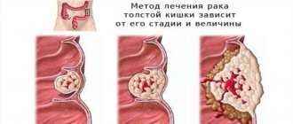
The following stages are distinguished:
- First stage. The malignant mass has a superficial structure.
- Second stage. The size of the tumor does not exceed 2 centimeters in diameter. Internal structures are not affected.
- Third stage. The size of the tumor can reach 4 centimeters. Possible spread to one lymph node in the cervical region.
- Fourth stage. The malignant tissue spreads to adjacent areas of the mouth and penetrates the lymph nodes. Metastasis to distant organs.
Read: Diagnosis of liver cancer: a biopsy is often not needed
The staged process is not always clearly expressed. Depending on the type of disease, a malignant tumor can grow and spread at different rates.
Classification
According to its microscopic structure, cancer of the oral mucosa is of the squamous cell type. There are several of its forms:
- Keratinizing squamous cell carcinoma. It looks like a cluster of keratinized epithelium (“cancer pearls”). Accounts for up to 95% of cases of development of pathology of this localization.
- Non-keratinizing squamous cell. It is manifested by the proliferation of epithelial cancer cells without areas of keratinization.
- Poorly differentiated (carcinoma). This is the most malignant and difficult to diagnose form.
- Cancer of the oral mucosa in situ. The rarest form.
Depending on the characteristics of tumor growth, the following forms are distinguished:
- An ulcer is one or more ulcers that gradually grow and tend to grow and merge. Usually the bottom of the ulcers is covered with an unpleasant-looking coating.
- Nodular - characterized by the appearance on the mucous membrane of a dense growth in the form of a node, covered with whitish spots.
- Papillary - manifests itself as fast-growing, dense growths resembling warts. The growths are usually accompanied by swelling of the underlying tissues.
Certain forms of cancer of the oral mucosa
- Tongue cancer. The typical location of the pathology is the lateral surface of the tongue; less often, the tumor is found on the root of the tongue, the back or the lower surface. A malignant tumor already in the early stages leads to chewing and swallowing disorders, which facilitates diagnosis.
- Photo: initial stage of cheek cancer
Cancer of the buccal mucosa. This tumor often masquerades as aphthous ulcers located along the line of the mouth on the cheeks. An increase in the diameter of the ulcer and its growth into the masticatory muscles leads to restrictions in opening the mouth, which is a typical symptom of cheek cancer.
- Cancer of the palate and gums of the upper jaw. A rapidly growing nodule with a tendency to ulceration forms on the palate. Early onset of pain may occur, especially in situations where deeper tissues are affected.
- Cancer of the mucous membrane of the floor of the mouth. The first signs of the disease almost always go unnoticed. The tumor grows early into the surrounding tissues, including bone. The oncological process is often accompanied by increased salivation (salivation), and the development of bleeding is dangerous. The growth of a tumor affecting the bone tissue of the lower jaw is accompanied by facial deformation.
- Cancer of the mucous membrane of the alveolar processes. At an early stage, it is accompanied by problems with teeth - patients are bothered by toothache, teeth become loose and fall out, and gums become swollen. A typical phenomenon is bleeding from the socket of a fallen tooth.
Let's consider the stages of development of oral cancer and the manifestations of the disease at different stages:
- tx - there is practically no data available to evaluate the primary tumor;
- then - the tumor is not diagnosed;
- tis — preinvasive stage of carcinoma;
- t 1 - formation within 2 cm;
- t 2 - up to 4 cm;
- t 3 - more than 4 cm in its largest part;
- t 4 - pathology grows in neighboring sections;
- nx - insufficient information to evaluate regional lymph nodes;
- no—no metastatic effects detected;
- n 1 - metastases in one node;
- n 2 - in a single lymph node on the affected side up to 4 cm;
- n 2a - in one lymph node up to 6 cm in maximum dimension;
- n 2b - in multiple lymph nodes in the affected area up to 6 cm;
- n 2c - too, but on both sides;
- n 3 - metastases more than 6 cm;
- mx - there is no data to establish distant metastasis;
- mo - completely absent signs of distant metastasis;
- m1—separate lesions were identified.
The disease can manifest itself in various forms depending on the form of development. Thus, doctors have identified three main types of cancer:
- ulcerative – manifests itself in the form of an ulcer, which takes a long time to heal and can increase in volume;
- nodular - seals form in different parts of the oral cavity, progress quickly, have clear outlines and shapes, sometimes covered with white spots;
- papillary - outgrowths of a dense formation that hang in the mouth and bring significant discomfort to the patient.
The tumor can also be localized in different places. Based on location, doctors have identified several forms of cancer:
- cheeks;
- floor of the mouth;
- language;
- in the area of the alveolar processes;
- palate.
The disease develops with varying intensity depending on the etiological factor. However, in each patient the pathology develops in 5 stages:
- zero – the tumor does not extend beyond the mucosa, the size of the tumor is relatively small;
- first – the tumor is no more than 2 cm in volume, does not grow further along the cavity;
- second – the neoplasm reaches 4 cm in diameter, the progressive disease has not yet affected the lymph nodes;
- third – the tumor is more than four centimeters, the lymph nodes are damaged;
- fourth - metastases spread to internal organs, a pathological process develops in the lungs, spreading to the bones of the face and nasal sinuses.
Diagnostic methods
If you suspect a disease, you should contact an oncologist. During your appointment, your doctor will ask about your complaints, take a medical history, and examine your mouth to look for signs of malignant growth.
Any abnormality of the epithelial mucosa may be suspected, including areas of irritation, ulcers and white spots. To exclude other diseases and confirm oncology, instrumental and laboratory research methods will be required.
Special research methods:
- Sampling of a section of the oral mucosa followed by histological examination. A biopsy is the most accurate method for diagnosing cancer and identifying the type of malignant tumor. Also, the results of the method may indicate precancerous changes in the epithelium, which increase the risk of cancer.
- Endoscopic examination. During the procedure, a small flexible tube equipped with a camera and a light source is placed down the patient's throat. The nasal cavity is examined in the same way. Endoscopy is necessary to detect the primary area of tumor spread if oral cancer is a secondary formation.
- Visualization. To obtain high-precision images of certain tissues and organs, doctors prescribe radiography, as well as computer or magnetic resonance imaging. These methods are useful for assessing tumor size and finding the primary site of malignant tissue spread.
Screening diagnostic methods aimed at detecting the early stages of oncology are also important. If a patient has certain risk factors, regular examinations are necessary.
Methods for detecting cheek cancer
To determine pathology at the initial stage of its formation, it is necessary to independently examine the oral cavity. This procedure is recommended to be carried out at least once a month. Self-diagnosis will allow you to promptly identify changes on the inside of your cheek and seek qualified medical help.
If any pathologies are detected, you should consult a dentist, who, if a cancerous tumor is suspected, will send the patient to an oncologist.
In turn, the oncologist will use palpation to determine the extent of the growth’s spread. Further, to clarify the diagnosis, it is necessary to undergo a cytological analysis (biopsy), which allows you to determine the affected area and the stage of the tumor.
Also, other additional diagnostic methods are used to identify, clarify and prescribe effective treatment for malignant tumors.
Ultrasound diagnostics
This method allows you to evaluate the structure and size of the cancer node.
Radiography
Determines possible damage to bone tissue (spread of metastases). Since the bones of the skull are located in close proximity to the site of the disease, they are primarily affected by cancer cells.
CT scan
With its help, the nature of the neoplasm is determined (benign or oncological). According to the results of such a study, a treatment method is prescribed.
Treatment
Depending on the degree of the disease and the stage of its development, appropriate treatment methods are selected.
Radiation exposure
Radiation has a negative effect on cancer cells, leading to their death. It is carried out as independent therapy or as an adjunct after surgery. It all depends on the size of the tumor.
Chemotherapy
Refers to a medicinal method of treatment and is used in combination with surgery or radiation. It is prescribed to patients who have no contraindications to the chemotherapy drugs used.
Surgery (tumor removal)
This method is considered the most effective. However, it is worth considering that after removal of a cancerous tumor on the cheek, the patient often needs to undergo additional plastic procedures to restore his appearance.
Treatment and prevention
Oral cancer can masquerade as non-serious illnesses
Treatment of oral cancer should be aimed at removing the tumor, preventing the spread of malignant growth and correcting possible complications.
The method of cancer therapy for a particular patient is determined by the attending physician based on the stage of the disease, site of origin, type of tumor and other factors.
Read: Complications and symptoms of liver cancer
Main methods of treatment:
- Surgical removal of the tumor. During the operation, the doctor removes not only the malignant mass itself, but also part of the healthy tissue adjacent to it to prevent relapse. Removing a large tumor may require intervention in the area of the skull bones. The affected lymph nodes are also sometimes removed.
- Further surgical treatment should be aimed at restoring the natural anatomy of the mouth.
- Radiation therapy is the use of high-energy X-rays to destroy cancer cells and prevent metastasis. In some cases, this is the only treatment available.
- Chemotherapy is treatment using special chemicals that specifically target tumor cells. Often this method of therapy is combined with surgical treatment.
- Immunotherapy to prevent new malignant growth.
Disease prevention measures include:
- Quitting smoking and alcohol.
- Drawing up a balanced diet.
- Limit sun exposure.
- Undergo regular oral examinations.
- To undergo examinations, you must visit a dentist and otolaryngologist.
Forecast
The survival period directly depends on the type of tumor and the stage of spread. During the first stage, when malignant cells remain on the surface of the mucous membrane, surgery helps to completely get rid of the problem.
A poor prognosis is typical for late stages, when tumor cells have spread to the deep cervical lymph nodes. In this case, survival rarely reaches two years.
Thus, oral cancer, whose symptoms may be nonspecific, is best treated in its early stages of growth. Screening diagnostics help detect precancerous changes and prevent tumor growth.
Informative video about oral cancer - in the video:
Read along with this article:
- Rectal cancer: survival, risk factors and treatments
- Temperature with stomatitis in children: symptoms and signs...
- Candidiasis on the lips: information, etiology, possible symptoms,…
- The first signs of lip cancer, a description of symptoms by stage...
- Tubular adenoma of the rectum: symptoms, treatment and prognosis
- Symptoms of tongue cancer, its treatment and causes
- Important topic: the structure of the human oral cavity
- Signs of lip cancer, risk factors and treatment options
- What diseases cause a yellow coating on a child’s tongue?