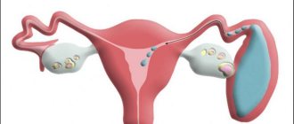
Endometrial cancer is an oncological disease that affects the genital organs of women and is characterized by the formation of a tumor on the body of the uterus. The pathology rarely leads to death because it responds well to treatment.
The pathology is accompanied by characteristic symptoms even in the early stages, so advanced stages are rare. In most cases, the pathology is detected at the initial stage, so this disease becomes a cause of mortality in extreme situations.
Causes
Endometrial cancer of the uterus is a disease that develops due to an increase in the amount of estrogen produced by the ovaries at the beginning of menstruation. During this period, convoluted glands appear, which are necessary in the process of implantation of a fertilized egg.
When there is an excess of hormones, the endometrium begins to grow, called hyperplasia. The growth of endometrial gland cells also increases. All this contributes to disruption of cell division and the formation of a cancerous tumor.
Older women often face a similar diagnosis. But pathology can occur at an earlier age.
Endometrial growth, or hyperplasia, has several types:
- simple not atypical,
- adenomatous complex not atypical,
- simple atypical,
- adenomatous atypical complex.
Two types of non-atypical adenomatous or simple hyperplasia are characterized by increased growth of the uterine lining and an increase in the number of glands in the uterus.
Atypical simple and adenomatous atypical complex type of hyperplasia mean the appearance of a pathogenic genetic malignancy - endometriosis.
How are tumor types classified?
In oncology, there are different types of endometrial classifications. Among them:
- classification according to TNM,
- stages of the disease according to the FIGO system.
The first system takes into account the overall assessment of the cancerous tumor (T), lymph nodes (N), and the presence of metastases (M). It looks like this:
- T 0 - the tumor was removed during the curettage procedure and is no longer detectable,
- T 1 - a tumor is found in the uterus,
- T 2 - the neoplasm has grown into the uterine cervix,
- T 3 - the tissue around the uterus and the lower part of the vagina is affected,
- T 4 - the tumor has spread beyond the pelvic area, affecting the rectum and bladder.
In addition, this type of cancer is classified according to the WHO system. There are epithelial (endometrioid carcinoma, adenocarcinoma, etc.), nonepithelial (stromal sarcoma, etc.), epithelial and nonepithelial (adenosarcoma, carcinosarcoma, etc.), other types (lymphoma, stromal cell, germ cell neoplasms).
Stages
There are the following degrees of uterine tumors:
- initial stage - the tumor is localized only in the uterus,
- the second is cancer in the mucous membrane and cervix,
- third - cancer in the pelvis with possible spread to the abdominal cavity,
- the fourth is a neoplasm in the pelvis with penetration into the rectum and bladder with possible metastases to the lymph nodes and other organs.
All stages of the disease can be cured after surgery and examination of the removed tumor, tissue, lymph nodes and other tissues. A medical examination will allow you to accurately identify the extent of tissue damage, the structure of the cancer tumor and the degree of differentiation. These examinations make it possible to draw up the correct treatment regimen and give a subsequent prognosis.
Classification of malignant tumors of the cervix
The second place among oncological pathologies in women is cervical cancer. Stage 1 uterine cancer usually progresses asymptomatically, which leads to its untimely detection and treatment. Every two minutes in the world a woman dies from a malignant tumor located in the vaginal area. Every year, specialists register 600 thousand new cases of cancer.
Squamous cell carcinoma most often progresses in women over 40 years of age, which accounts for 65% of the total number of cases. A favorable prognosis is only possible with timely treatment, for example, chemotherapy for cervical cancer.
According to morphological structure
The nature of tumor progression depends on its morphological structure. The following types of cancer are called:
- low-grade;
- squamous non-keratinizing and keratinizing;
- glandular – adenocarcinoma.
Poorly differentiated cervical cancer
In 85% of cases, squamous forms are detected. The prevalence of adenocarcinoma localized in the cervical canal is only 15%. The keratinizing variant occurs in 25% of women with a history of pathology and has a favorable prognosis. A pronounced degree of malignancy is observed in rare, poorly differentiated neoplasms.
According to the nature of progression
The tumor has different growth directions:
- endophytic - progression inward, followed by transition to the uterine cavity, tubes, ovaries and vaginal wall;
- exophytic – development into the vaginal lumen.
Types of cervical cancer progression
The neoplasm may have a mixed growth pattern.
Stages
The progression of carcinoma is determined by stages. They describe the prevalence of the formation, the degree of involvement of distant organs and lymph nodes according to the FIGO, TNM classifications (tnm - tumor, lymph nodes, metastases). Gynecologists call 4 stages of pathology development, which differ in the degree of tissue damage, symptoms, treatment and prognosis.
Stage 1 cervical cancer has several types, classified depending on the prevalence of pathological cells.
Stage IA (T1a) is determined microscopically:
- IA1 (T1a1) – the depth of the lesion is no more than 3 mm;
- IA2 (T1a2) – the tumor penetrates the epithelium by 5 mm.
Stage IB (T1b) is detected macroscopically:
- IB1 (T1b1) – the neoplasm measures 4 cm;
- IB2 (T1b2) – carcinoma size exceeds 40 mm.
Detection of the first stage of cervical cancer, for example, T1b, occurs during the examination process, including laboratory and instrumental techniques. Stage 1 cervical cancer is more often diagnosed in women under 40 years of age.
Symptoms
Symptoms and signs of uterine cancer can manifest as simple menstrual irregularities, infection, or premenopausal hormonal changes. During the development of endometrial cancer of the uterus, symptoms may manifest themselves in the form of small or, conversely, heavy bleeding. Symptoms of endometrial cancer may include discharge (purulent, watery, thin, clear).
Women of childbearing age can independently identify the process of tumor formation by such signs of endometrial cancer of the uterine body as bleeding between periods or heavy bleeding during menstruation. It is worth noting that cancer is often completely asymptomatic, so it is worth regularly visiting a gynecologist for a preventive examination.
Late signs of endometrial cancer are manifested by pain in the lower abdominal cavity, lower back, and sacrum. Painful sensations appear due to compression of nerve cells by the parametric infiltrate and the entry of the serous membrane of the uterus into the development of the oncological process.
Discharge in the final stages of the disease is characterized by a putrid odor and a color similar to meat waste. If the cancer spreads to the cervix, there may be an accumulation of pus inside the uterus, as well as cervical stenosis, narrowing and even closure.
If the cancer begins to put pressure on other organs, for example, the rectum, then the sick woman may experience constipation and spotting in the stool. If the ureter is compressed, hydronephrosis begins with simultaneous uremia and pain in the lumbar region. If the cancer has spread to the bladder tissue, then blood appears in the urine and urination becomes more frequent or, on the contrary, difficult.
Signs of spread of metastases may be:
- in the last stage of cancer, bone pain appears, fractures are possible,
- if the liver is affected, the skin becomes jaundiced, and the patient suddenly loses weight,
- if the cancer has reached the pelvic organs, ascites may begin.
Symptoms
Endometrial cancer can cause symptoms such as unexplained pain, fatigue, and heaviness in the pelvic area. Photo: Vk.
Early signs and symptoms include unusual bleeding, such as after menopause or between periods.
Pain may occur in the pelvic area or during sexual intercourse. Some women also experience pain when urinating or have difficulty emptying their bladder.
As the disease progresses there may be:
- feeling of excess weight or heaviness in the pelvic area;
- unintentional weight loss;
- fatigue;
- nausea;
- pain in several parts of the body, including the legs, back and pelvic area.
Other noncancerous health problems have similar symptoms, such as fibroids, endometriosis, endometrial hyperplasia, or polyps in the lining of the uterus.
difference between normal endometrium and hyperplasia. Photo: Pinterest.
polyps in the uterine cavity. Photo: Pinterest.
It is important to rule out endometrial cancer if other conditions cause similar symptoms.
Diagnostics
If a woman has complaints about any problems in the genital organs, she should immediately consult a gynecologist and also undergo an ultrasound examination.
Among the diagnostic measures that need to be done are the following:
- ultrasound examination of the pelvis,
- chest x-ray,
- curettage or biopsy of the uterine cavity,
- examination by a gynecologist,
- urine and blood tests, hemostasis studies.
These diagnostic measures will help confirm or refute the fact of the development of a cancerous tumor and clarify its size. It will not be difficult for a specialist to recognize endometrial cancer on an ultrasound. Tests will determine the exact location and nature of possible damage to other organs.
How to cure a tumor of the uterine body
There are many treatment options for uterine cancer. But they have the greatest effect if the tumor was detected at the first or second stage of the disease. This period is considered the most favorable. At this time, even hospitalization is excluded. Surgery will be required, no matter what stage of the lesion is detected. The operation will be short and quite easy: extirpation of the uterus and uterine appendages is performed.
Among the non-drug methods of treating endometrial cancer, radiation therapy is used (in rare cases). It all depends on the individual characteristics of the disease.
After examination by a doctor, hormone therapy can be performed, but it has many contraindications.
As for chemotherapy, this method has not been used for a long time due to many negative side effects.
How is endometrial cancer treated?
The goal of therapeutic measures is to eliminate malignant tumors localized in the body of the uterus, to prevent metastasis and relapse of the disease.
At the moment, when endometrial cancer is detected in women, doctors recommend complex therapy, including surgery, radiation exposure, and taking appropriate medications.
The order of measures, as well as their intensity, is selected according to the stage of development of the cancer process. Treatment is carried out on an individual basis and is prescribed exclusively by an oncologist.
Prognosis and prevention
The main factors influencing the prognosis of uterine cancer are the level of differentiation and the spread of the tumor to neighboring organs or tissues. In the first stages, the tumor can be completely cured. At the third stage of uterine cancer, according to statistics, approximately a third of patients survive; at the last, fourth stage, only 5% of patients survive.
Therefore, preventing endometrial cancer and detecting the disease in the early stages is extremely important.
The prognosis can be worsened by other present diseases in patients, as well as advanced age. For older people, it is difficult to choose a full range of treatment, which becomes an additional problem for recovery.
Any woman should be regularly examined by a gynecologist to avoid the risk of tumor formation. And if a woman has already had endometrial cancer, she should undergo regular tests to exclude the development of a relapse.
Risk factors
Although the direct causes of endometrial cancer are unknown, there are several factors that can increase the risk of the disease.
A major factor in endometrial cancer is increased exposure to high levels of estrogen.
They are usually elevated in those who:
- never been pregnant;
- menstruation began before age 12;
- Menopause occurred after age 55.
Estrogen-only contraceptive hormone therapy also contributes to the risk of endometrial cancer.
Polycystic ovary syndrome (PCOS) can increase estrogen levels and is therefore also a risk factor.
Diabetes may increase your risk because estrogen levels tend to rise with insulin levels. Long-term high levels of estrogen increase the likelihood of developing uterine cancer.
Other factors include:
- endometrial hyperplasia, or abnormal growth or thickening of the lining of the uterus;
- obesity;
- hypertension;
- using tamoxifen to prevent or treat breast cancer;
- radiation therapy to the pelvis;
- family history of uterine cancer;
- previous diagnosis of ovarian or breast cancer.
Some reports link acrylamide, a cancer-causing compound found in charred, carbohydrate-rich foods, to endometrial and ovarian cancer in postmenopausal women.
However, other reports disputed this.


