Content
The temporomandibular joint (TMJ) is considered one of the most used and mobile joints in the human body. It is responsible for opening and closing the jaw, so it almost always works when people talk, chew, yawn, etc.
Diseases associated with damage to the jaw joint are very common. A huge number of patients suffer from pain when moving their jaw; according to statistics, about 40% of the population have at least once experienced discomfort in the TMJ area . However, not everyone seeks medical help; people do not attach serious importance to pain in the temporomandibular joint and usually self-medicate.
Pain in the jaw joint is considered the most common dental pathology after caries and pulpitis.
Temporal bone
Position of the temporal bone in the skull.
The temporal bone is part of the skull and is connected to the surrounding bones through sutures. It is a stationary component of the joint - all movements are carried out relative to its surface.
Main characteristics of the temporal bone:
- On its upper part there is a flat and durable plate called scales. On the sides it forms the cranial vault and connects to the occipital, parietal and sphenoid bones.
- The tympanic part is connected to the lower jaw. Its feature is a large number of holes and channels.
- All these openings and canals contain vessels and nerves emerging from the cranial cavity, as well as a number of formations of the auditory apparatus.
- The formation of the temporomandibular joint includes only a small depression in the tympanic part, called the articular surface.
- It is located slightly anterior to the external auditory canal, located between it and the temporal tubercle.
- As a result, a rounded depression is created, which almost completely corresponds to the condylar process of the lower jaw.
Important! Due to the presence of an articular disc in the joint cavity, movements can become block-like in nature and are carried out only along one axis.
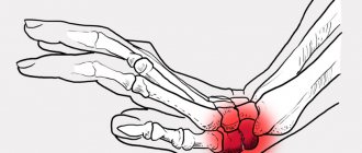
Why does the jaw joint hurt?
There are many reasons for joint pain, the most common are:
- jaw injuries - fractures, bruises, muscle strains, etc.;
- inflammatory and infectious diseases (purulent mumps, boil, abscess, arthritis, osteomyelitis, arthrosis of the jaw, purulent otitis media, etc.);
- wearing dentures;
- malocclusion;
- endocrine diseases;
- pathologies of peripheral nerves and blood vessels;
- tumors;
- bad habits (gritting your teeth, biting your nails, opening a bottle with your teeth, etc.).
If your temporomandibular joint hurts, you should consult a doctor immediately. There are many causes of pain, and pain can be a signal of the development of a serious disease, such as osteomyelitis or arthritis.
Types of TMJ diseases and main symptoms
When the maxillofacial joint hurts, depending on the cause of the disease, the following symptoms may additionally be observed:
- feeling of fullness in the ears;
- enlarged lymph nodes under the jaw;
- dizziness and migraine;
- crunching sound when moving the jaw;
- spasms in the facial muscles, etc.
Important! When the jaw joint hurts near the ear, this may be a signal of the development of a complication of a dental disease (the development of arthritis, arthrosis, etc.).
With TMJ diseases, patients are concerned not only with pain in the jaw; a common symptom of inflammation is ear pain
Pain in the temporomandibular joint indicates the development of some disease or pathology, the most common cases:
- Osteomyelitis is a purulent inflammation of the jaw joint. The main symptoms are severe jaw pain, swelling, fever, and inflammation of the lymph nodes. The appearance of osteomyelitis in most cases is caused by caries or pulpitis, so when examining the jaw, patients often complain of toothache. Osteomyelitis is a serious disease that requires immediate medical attention.
- Dysfunction of the temporomandibular joint is a consequence of endocrine diseases and psycho-emotional shocks, stress, trauma, malocclusion, prosthetics, etc. Sometimes TMJ dysfunction can be caused by congenital underdevelopment of the articular head. Signs of the disease: facial asymmetry, pain when chewing in the ear and temple area, inability to open the mouth smoothly, swelling, a characteristic crunch when moving the jaw, fatigue when chewing, etc.
- Arthritis. Symptoms: swelling and pain of the joint, a clicking sound when moving the jaw near the ear, limitation of motor activity, etc.
- Arthrosis. Acute pain occurs with strong opening of the mouth, clenching of teeth and chewing. Often the disease is accompanied by a crunching sound in the jaw joint.
- Dislocation of the joint. The pathology develops as a result of muscle disorders, manifested by displacement of the head and disc of the jaw bone, inability to close the mouth, a characteristic sound when moving the jaw, etc.
Important! Sometimes the temporomandibular joint hurts after having dentures installed. Pain in this condition is considered normal, since the joint is simply not accustomed to the presence of a “foreign” object in the mouth. If discomfort does not go away for a long time, you should consult a doctor.
Patients with pain in the jaw joint are examined by several doctors to determine the cause of the pathology. Usually the patient goes to see a family doctor, who gives a referral to an appropriate specialist.
Soft fabrics
The main muscles of the anatomical region in question.
The temporomandibular joint, the structure of which is complex, consists of an articular cavity and a capsule, which also has its own characteristics.
They are associated with the division into 2 anatomical floors through a cartilaginous disc:
- The first, or upper, half consists of the articular surface located on the temporal bone and the articular tubercle of the lower jaw. The shells are adjacent to the edge of the fossa in its outer and posterior parts, expanding anteriorly. In this place the capsule is attached very widely and captures a significant part of the head into the joint cavity. This feature is associated with the need to perform a number of movements: lateral displacement of the jaw and its rotation.
- The lower part is narrower than the upper, so the joint cavity has the shape of a cone, the apex of which faces down. Moving from the borders of the disc, in the area of the head the capsule forms an extension, which is strengthened from the outside by ligaments. Further, in the region of the neck of the condylar process, the capsule decreases, and its shells are attached to it and complete the articulation cavity.
Important! The articular cavity of the joint is not significant in size, since the disc occupies its main part.
Joint ligaments
Ligamentous apparatus.
The temporomandibular joint is small, so its tendons are not large.
But, despite this, they are divided into large and small entities:
- The most powerful ligament, which is called the lateral ligament, is directly connected to the shell. It is located on the outer half of the capsule. Anatomically, it cannot be distinguished separately, because the ligament is a thickening of the articular membrane. Additionally, it is divided into the external oblique and internal transverse ligaments.
- There are two small tendons that are located separately: the sphenomandibular and stylomandibular ligaments. They are not independent, but are parts of the inner fascia of the neck that form a loop. Their function is to limit the mobility of the head of the lower jaw, preventing it from making movements with a significant displacement.
- The disco-mandibular ligament is an intra-articular ligament. It stabilizes the lower half of the joint and provides additional connection between the disc and the temporal process of the mandible.
- The smallest structure is the malleusomandibular ligament. It is responsible for connecting the bones of the middle ear to the lining of the joint.
Important! A large number of tendons are practically not involved in supporting the articular joint. This function is performed by the muscles that set it in motion.
Intra-articular disc
Location of the intraarticular disc.
The cartilaginous plate is located inside the joint cavity, so it is difficult to judge its structure for sure.
In its structure and function, the disc resembles the menisci of the knee joint, although there are still some features:
- The disc is formed by cartilage tissue of a fibrous nature. It is distinguished from a similar structure covering the surface of the joint by greater strength and flexibility.
- It differs from the menisci of the knee joints in the lack of shock absorption during movement. The role of the articular disc in the temporomandibular joint is to provide additional support and support.
- The disk itself is not homogeneous. In the outer sections it is thickest, and in the central part it is thinner.
- The disc is fixed to the joint shell, so it is relatively immobile. During movements, only its lateral displacement is possible.
How to treat?
If your jaw joint hurts, you should seek medical help. In the process of diagnosing and treating the disease, depending on its cause, various specialists can participate - dentists, orthopedists, rheumatologists, infectious disease specialists, neurologists, otolaryngologists, etc.
If there is acute pain in the jaw joint, you need to apply a cold compress
To determine the cause of pain in the temporomandibular joint, the patient is interviewed, his condition is assessed, and anamnesis is collected. Diagnostics includes the following studies:
- determination of the amount of uric acid in blood serum;
- UAC;
- radiography of the temporomandibular joint;
- analysis of synovial fluid samples;
- CT;
- MRI, etc.
If the pain is a complication of a dental disease, the patient is treated by a dentist. Caries is a common cause of infection and inflammatory diseases.
Often, diseases of the jaw joint occur in an acute form, so providing the patient with emergency medical care is the first step to recovery. Patients with acute pain in the TMJ are advised to rest completely, immobilize the affected joint, and apply cold.
Drug treatment depends on the cause of the pain, for example, for inflammation of the jaw joint, the patient is prescribed non-steroidal anti-inflammatory drugs and painkillers (Nimesil, Diclofenac, Ibuprofen, etc.). In case of an infectious lesion of the joint, antibiotics are prescribed, psychological relaxation drugs are prescribed to relieve tension in the muscles, and special dentures and acrylic devices are prescribed to correct the bite.
Physiotherapy effectively combats diseases of the jaw joints; patients are prescribed a course of electrophoresis, UHF, diadynamic currents, etc.
Important! Patients with TMJ inflammation and other joint diseases should not open their mouths wide and eat solid foods. To immobilize the damaged joint, doctors recommend wearing a chin sling or parietal-mental bandage, using dental splints, etc.
If the cause of the pain cannot be determined after laboratory tests, the patient is referred to a CT and MRI of the temporomandibular joint
If conservative therapy does not help, surgical treatment is resorted to. Most often, operations are performed after serious joint injuries or for purulent mandibular arthritis.
Whatever the cause of pain in the TMJ, be sure to see a doctor. Self-medication for this pathology is ineffective and threatens the development of complications.
Diagnosis of muscle dysfunction
Treatment of temporomandibular joint dysfunction is prescribed by a doctor after a comprehensive examination. He examines the patient. A survey about associated symptoms is also conducted. An examination can confirm the presence of stiffness. The face also looks asymmetrical.
When the teeth are compressed, creaking may occur. The doctor performs palpation. In this case, pain most often occurs. Muscles become hyperactive. Sometimes subluxations of the head of the jaw or its disc occur. In some cases, examination reveals hypertrophy of the masticatory muscles.
During the diagnostic process, the functional state of occlusion in the oral cavity is analyzed. The position of all elements of the joint is also determined using special diagnostic methods. Based on the data obtained, the doctor prescribes comprehensive treatment. It depends on the degree of neglect of the disease, the characteristics of its course and provoking factors.
What is arthrosis of the maxillofacial joint and how to treat it?
Just a few decades ago it was believed that arthrosis was a disease of old age. If the disease occurred in young people, it was only as a result of previous injuries. Today, more and more patients under 50 years of age suffer from this disease. By this age, some form of arthrosis is observed in almost 50% of people. By the age of 70, the number increases to 90% and this is only according to official statistics.
Read also: Diseases of the temporomandibular joint
- Content:
- What is arthrosis of the jaw joint
- Why is arthrosis of the jaw dangerous?
- Symptoms and treatment
- How to cure arthrosis of the jaw
- Treatment with folk remedies
Although arthrosis of the maxillofacial joint is not as common as compared to gonarthrosis and coxarthrosis, it is still quite common and often begins in patients between the ages of 30 and 50 years.
Ankylosis
Considering diseases of the temporomandibular joint, a few words should be said about such an ailment as ankylosis. When it occurs, the surfaces of the joints grow together due to fibrous or bone changes. In this case, partial or complete loss of mobility may be observed in the joint.
The cause of the development of ankylosis can be chronic or acute diseases in the joint area. Most often, osteomyelitis, purulent arthritis, and fractures in this area lead to similar consequences. Children are more susceptible to ankylosis. At this age, the surfaces of the joint, covered with periosteum without cartilaginous cover, quickly grow together. In adults, fibrous ankylosis occurs more often.
One of the main symptoms is persistent difficulty or lack of movement in the affected joint. In most cases, it is determined when moving in a horizontal plane. If the ankylosis is unilateral, the face will be asymmetrical. If the disease develops on both sides, the chin will be pushed back.
With the fibrotic form of the disease, pain is more common. In this case, the muscles quickly get tired when chewing, and it is difficult to open the mouth, especially in the morning.
On x-ray, there is no joint space in some areas. This is a characteristic sign of the disease. In this case, the head and neck of the joint thicken.
What is arthrosis of the jaw joint
The intermaxillary joint provides mobility to the jaw, allowing it to make movements necessary for chewing food and producing sounds. With arthrosis, the cartilage becomes thinner. At the slightest movement the patient experiences acute pain. As the pathology develops, the pain syndrome shifts to the nasal area and can lead to ear congestion.
The main reasons contributing to the development of pathology are the following:
- Age and heredity. In women, pathology is especially common during menopause. Hormonal changes lead to metabolic disorders. Lack of nutrients leads to thinning of cartilage tissue and its cracking.
- Surgical intervention.
- Consequences of injuries.
- Malocclusion associated with missing teeth.
- Inflammatory and infectious processes.
- Heredity.
- The presence of other types of arthrosis.
Degenerative changes in the initial stages go unnoticed and are quite difficult to determine.
Symptoms of arthrosis of the jaw joint are as follows:
- Pain syndrome - manifests itself when there is a load on the joint. The patient experiences pain when chewing food. In this case, drug treatment is almost always necessary; arthrosis of the temporomandibular joint has progressed to stage 2-3.
- Extraneous sounds - when the jaw opens, a characteristic crunching and clicking sound appears.
- Facial asymmetry. Even with a cursory examination of the face, deviations become noticeable.
- Numbness - the jaw may be difficult to open after sleep, and limited movement may continue throughout the day.
- Hearing loss occurs in the later stages of the disease.
- Growths - upon palpation, compactions can be detected. Pressure causes pain.
Treatment of arthrosis of the maxillofacial joint begins only after a general diagnosis of the patient. It includes:
- X-ray diagnosis of arthrosis.
- Computed tomography.
- MRI.
Surgical treatment of arthrosis of the mandibular joint is prescribed extremely rarely, only in cases where conservative and restorative therapy has not brought results.
Arthrosis
Arthrosis of the temporomandibular joint is a very common disease. It refers to dystrophic pathologies. Arthrosis is a consequence of various injuries, inflammation, disorders of the endocrine system or metabolic processes. When certain deviations appear, muscles begin to adapt to them. This leads to compression in some areas and stretching in others.
In this case, the head of the lower jaw suffers from excessive load. It is taken over by cartilage. They do not become unable to withstand stress over time. As a result, the nerve endings are pinched. Bone tissue is gradually destroyed. If no measures are taken to treat the disease, the degenerative processes will be irreversible. This will lead to disability, the inability to open your mouth or speak. Therefore, the disease cannot be started.
Arthrosis of the temporomandibular joint is accompanied by pain. This is caused by injury to the nerve endings during movement. Here blood circulation is disrupted. The joint does not move correctly. When the mouth opens, the lower jaw moves to one side.
Such a disease can be caused by poor-quality prosthetics and tooth loss. The jaw takes an incorrect position when moving or chewing. If you need to perform prosthetics, you need to contact a trusted specialist. Otherwise, over time you will have to face a problem such as arthrosis.
Why is arthrosis of the jaw dangerous?
Initially, the disease has virtually no symptoms and is often discovered by chance when visiting a dentist. Deforming arthrosis of the jaw joint is observed in later stages as degenerative processes develop.
Ultimately, the disease leads to immobilization of the jaw, and eating becomes problematic. Headaches caused by inflammatory processes are debilitating. A displacement of the jaw is often observed. Inflammation can spread to other organs.
It becomes extremely difficult to reduce pain and achieve improvements in well-being during this period; conventional anesthetics do not have the desired effect. This leads to migraines, high blood pressure, stress, etc.
Surprisingly, arthrosis can be cured quite simply at the beginning of the development of degenerative processes. The disease, which is practically untreatable at stage 3 of development, is well treated in the early stages.
Symptoms and treatment of temporomandibular arthrosis
For almost all types of arthrosis, the same treatment is provided, aimed at combating clinical manifestations. Osteoarthritis of the facial joint has its own ICD (International Classification of Diseases) code, which allows you to compare treatment methods and select those that bring the best results.
The first signs of the disease are usually discovered during a dental examination. The patient himself will be able to seek help if the motor functions of the jaw decrease.
Symptoms and causes of arthrosis of the temporomandibular joint may influence the prescription of drugs. The essence of drug therapy is as follows:
- Reduce inflammation and swelling - for this purpose, the patient may be recommended one of the NSAID drugs. Vasodilators are prescribed in combination. Quite often a drug blockade is prescribed. The inflammatory process and swelling can affect pinching of the facial and trigeminal nerve. If inflammation of the periosteum is observed and diagnostic results show the presence of infection, antibiotics are prescribed.
- Establish normal metabolism. Removal of salt deposits and removal of ossifications is possible, subject to normal synthesis in the body. Lack of blood flow to the affected area only makes the situation worse. Osteoarthritis of the jaw is treated with vasodilators.
- Restore cartilage tissue. If radiological signs show only initial degenerative changes, the course of therapy includes chondroprotectors. Long-term use of chondroprotectors (at least six months) helps restore cartilage tissue. Chondroprotectors are most effective during non-exacerbation periods.
- Provide nutrients to the affected tissue. Vitamins and minerals are prescribed in combination with other drugs. It is especially important to take medications with increased calcium content.
How to cure arthrosis of the jaw
Degenerative changes in cartilage tissue are an almost irreversible process. Therefore, all methods of therapy, from traditional to treatment of arthrosis of the jaw joint at home, are aimed at eliminating and combating the symptoms of the disease.
In the early stages, it is possible to stop the destruction process and at least partially restore the cartilage. Moderately severe arthrosis can be stopped and stable remission achieved.
Early diagnosis is extremely important to achieve positive treatment results. Based on the results of the examination, therapy is prescribed.
The following contributes to recovery:
- Prescribing a gentle diet. It is extremely important to reduce the stress on the joint. It is necessary to limit the diet to soft food in grated or pureed form. The load during this period should be kept to a minimum.
- Provide protection from stress and nervous tension. The consequence of this is bursitis or teeth grinding during sleep. Chewing gum and long conversations, especially on the phone, are highly undesirable.
- Attendance at physiotherapy treatments. The arthrologist will recommend visiting ultrasound, laser and magnetic therapy. Massage and special exercises for the jaw can help.
- Eliminating the causes of the problem. Lumps near the ear and under the jaw due to arthrosis can be caused by malocclusions. Therefore, the arthrologist will issue a referral to the dental clinic. If the reason lies in this area, bite plates, mouth opening limiters, braces will be installed - implantation for arthrosis of the temporomandibular joint, prosthetics and dental treatment will be performed.
- Drug therapy is prescribed if all of the above measures have not brought results and the progression of the pathology is obvious. Tinnitus, headaches, swelling - require the use of NSAIDs. At the same time, anti-inflammatory ointments and injections are prescribed.
- Surgical intervention. During the operation, the head of the lower jaw is removed and prosthetics are performed. The surgical measure is effective and often helps to completely restore all functions of the damaged joint in full.
Read also: Inflammation of the maxillary joint
Treatment of jaw arthrosis with folk remedies
Traditional methods of treatment are aimed at the same thing as traditional therapy: eliminating the symptoms and causes of the disease. They use tinctures, compresses, and homemade warming ointments. You can find many such recipes.
The most popular are the following:
- Ingredients with garlic - 200 gr. peeled heads are added to 500 g. cranberries Grind thoroughly in a meat grinder or blender. Add 1 kg of honey to the resulting pulp. Take 1 teaspoon before each meal.
- Apple cider vinegar - two teaspoons of solution diluted in half a glass of water. Drink diluted vinegar before meals for 1-2 months. The course of treatment is repeated after 3-4 months. Apple cider vinegar flushes salt deposits from the body and helps normalize blood circulation in the muscle tissue around the joints.
- Bee venom - apitherapy or treatment with bee stings, is somewhat reminiscent of acupuncture, but in its beneficial effects it is in many ways superior to it. Bee venom is a powerful antibiotic, causes a rush of blood and activates the immune system.
It is impossible to list all the folk recipes, and everyone probably has their own “special” compositions that help in 100% of cases. And if they help, then it’s really nice. The main thing is that folk remedies do not interfere with basic drug therapy.
Increased pain, swelling of the skin, urticaria - all these are clear signs indicating the need to discontinue the folk remedy.
Arthritis of the maxillofacial joint: symptoms and treatment
Along with other types of arthritis, arthritis of the maxillofacial joint occurs, the symptoms and treatment of which have some similarities with them.
In medical terminology, this disease is abbreviated as TMJ arthritis, or arthritis of the temporomandibular joint. This joint ensures the mobility of the lower jaw; movement in it should be carried out in several directions - the ability to open and close the mouth, moving the jaw forward, shifting the jaw to the side.
Both older people and children are susceptible to the disease. If in children the occurrence of such a pathology is caused by the growth of teeth and bones and injuries, then in older people it can occur as a result of infectious or inflammatory diseases. If we talk about adults, the disease occurs equally often in both men and women. Temporomandibular arthritis is characterized by increased pain when opening the mouth.
Dislocation and subluxation
Another common pathology is dislocation and subluxation of the temporomandibular joint. Such disorders can be caused by injury or certain diseases, such as muscle dysfunction. In this case, the elements of the joint are displaced relative to each other.
This condition is characterized by pain. It becomes difficult to open your mouth. The head and disk change their position relative to each other. With muscle dysfunction, subluxation or dislocation is directed posteriorly.
With functional disorders, the capsule and ligaments are stretched. Subluxations occur periodically. In this case, jaw mobility at some point becomes impossible. The patient eliminates this situation on his own. This condition requires timely treatment. If a dislocation occurs, the head takes a fixed, incorrect position. At the same time, it becomes impossible to close the mouth.
What causes inflammation of the maxillary facial joint?
Inflammation of the jaw joint occurs for a number of reasons, which are grouped into 3 groups - infection, trauma and inflammatory diseases. Injury can occur from a blow or fall, resulting in the formation of a focus of inflammation. This process causes swelling, impairing the mobility of the joint.
As for the infectious causes of arthritis, the root cause is pathogenic microorganisms that, when they enter the joint, cause an inflammatory process. Penetration of infectious microorganisms occurs through direct, contact and hematogenous routes. The direct route is when the infection occurs when a joint is injured. This may be a fracture of the lower jaw as a result of a blow, knife or gunshot wound. The result of direct infection is specific and nonspecific inflammation. Specific causes tuberculous and syphilitic arthritis of the TMJ, nonspecific causes staphylococcal and streptococcal arthritis.
Hematogenous infection occurs with infectious diseases of the ear, nose, throat, and lower jaw. In this case, the infection is spread by the bloodstream. The contact route involves the spread of pathogenic bacteria from nearby tissues that have already been infected. Infection by contact is a consequence of:
- furunculosis;
- purulent mumps;
- purulent inflammation of the soft tissues of the face;
- purulent otitis;
- abscess;
- infectious diseases in the oral cavity;
- osteomyelitis.
The third group of causes of maxillofacial arthritis - inflammatory diseases - includes mainly rheumatic diseases, which are characterized by systemic inflammation of the organs and tissues of the body. Such systemic diseases that provoke inflammation include:
- Reactive arthritis.
- Lupus erythematosus.
- Rheumatoid arthritis.
- Gout.
Types of disease and forms of its manifestation
Arthritis of the maxillofacial joint is classified according to the form of the disease and the reasons that caused it. Thus, according to the form of arthritis of the temporomandibular joint, it can be acute or chronic. By the nature of occurrence - infectious and traumatic. Infectious arthritis is divided into specific and nonspecific. A separate type of disease is rheumatoid and purulent arthritis of the temporomandibular joint.
The acute form of arthritis is usually inflammatory-infectious or traumatic in nature, and the chronic form is rheumatoid in nature. Common symptoms of facial arthritis:
- pain that may radiate to other parts of the face;
- characteristic crunching and clicking sound;
- soreness when palpated;
- shifting the chin to the side;
- swelling and redness;
- hearing impairment;
- formation of infiltrate;
- chills and fever;
- dizziness, insomnia.
Each type of arthritis of the mandibular joint has its own distinctive features. Thus, with infectious arthritis, severe increasing pain is observed in the joint affected by the deformity. The pain usually increases with jaw movements. The soft tissue and skin around the joints also become inflamed. This form of the disease is characterized by the inability to open the mouth.
A distinctive feature of purulent arthritis is the formation of infiltrate. When palpating the joints, the pain increases significantly, general intoxication of the body occurs, and body temperature rises. A characteristic symptom of this form is hearing loss, which is the result of narrowing of the ear canal under the influence of infiltrate.
Traumatic arthritis is accompanied by sharp pain when opening the mouth and swelling of nearby tissues. Palpation is also accompanied by sharp pain. There is a displacement of the chin to the side.
general information
The interaction of all components of the joint is aimed at carrying out physiologically significant movements - opening and closing the mouth. Thanks to this, a person can perform a large number of actions: from chewing to vocal functions. Therefore, the temporomandibular joint, the anatomy of which is highly complex, has such a strong impact on a person’s quality of life.
This joint is located at the base of the skull, where a large number of other anatomical structures are located. Therefore, its structures are quite compact and do not interfere with the functioning of numerous vessels, nerves and the organ of hearing. The joint itself cannot be called simple, since it is surrounded by soft tissues responsible for certain functions.
General diagram of the structure of the joint.
The structure of the temporomandibular joint is always considered from both sides, since the joints work simultaneously.
Their main characteristics:
- Based on its location, the joint is classified as a joint of the skull . They form direct contact with the facial region and the base. Although the temporal bone has its own characteristics: it participates in the formation of both the base and vault of the skull.
- The shape of the joint is ellipse . This means that the convex process of the mandible is in contact with the concave and rounded articular surface. Their interaction is not limited in any way, which leads to a wide variety of movements.
- Based on the nature of their structure, the compounds are classified as complex . This means that the bone structures do not have common points of contact. Between them there is a cavity that is filled with a fibrous cartilage disc. It allows you to further increase the range of movements.
- At the same time, the joint is classified as combined . When muscles of the same name contract, their work is observed to be the same. Unilateral movements are possible only with pathologies, for example, with fractures or dislocations.
Summary table of the main parameters of the temporomandibular joint:
| Joint name | Articular surfaces | Type of joint | Axes of motion | Type of joint movements |
| Temporomandibular joint |
|
|
|
|
Important! The anatomy of the temporomandibular joint is externally hidden due to the powerful fibers of the masticatory muscle, which carries out the main joint movements - opening and closing the mouth.
Symptoms of acute and chronic form
Symptoms of arthritis of the maxillofacial joint, which occurs in an acute form, are characterized by the presence of a pronounced inflammatory process. The sensitivity of the nerve endings increases, which causes a feeling of pain, swelling of the soft tissues occurs.
Acute inflammation manifests itself in the form of sharp pain, which increases as the jaw moves. This situation significantly reduces the patient’s quality of life, because he cannot open his mouth due to the resulting pain, which spreads throughout the head and face.
Acute inflammatory arthritis of the TMJ is accompanied by redness and swelling of the soft tissues of the joint. This symptom is usually a sign of purulent arthritis. Actively reproducing pathogenic microorganisms cause dilation of blood vessels in the inflamed cavity and increased blood flow to the site of inflammation. This leads to swelling and redness of the soft tissues.
Another symptom is a feeling of fullness in the area of the affected joint. This sensation is caused by tissue swelling and accumulation of exudate. The patient may experience hearing loss. This occurs due to the fact that the inflammatory process spreads to the ear canal and causes it to narrow. The patient experiences a feeling of ear congestion.
Read also: Arthritis of the maxillary joint
Acute inflammation is often accompanied by an increase in temperature in the joint area. The dilation of blood vessels and the influx of warm blood to the inflamed area leads to an increase in temperature by several degrees. If the temperature rises above 38 degrees, a fever occurs. The headache intensifies, general weakness appears and fatigue increases. These kinds of symptoms, as a rule, indicate purulent inflammation in the jaw joint.
Symptoms of maxillofacial arthritis in the chronic form of the disease are as follows:
- restrictions on the functioning of the joint;
- painful sensations of aching and pulling nature;
- stiffness in the jaw;
- crunching and clicking;
- hearing impairment;
- slight increase in temperature;
- feeling of weakness and excessive fatigue.
Symptoms of TMJ arthritis
The inflammatory process of the jaw joint is characterized by the occurrence of pulsating pain, which intensifies significantly when opening the mouth and any attempts to move the jaw.
When pressing on the joint and on the chin, the pain becomes more intense. The area around the TMJ may become swollen. If nearby soft tissues are involved in the process, hyperemia and swelling of the skin in the ear area may be observed. It is impossible to fold the skin in this area.
The ability to fully open your mouth is sharply limited. The patient cannot open it more than 1 centimeter. The development of an acute inflammatory process is accompanied by the following symptoms:
- high body temperature;
- dizziness;
- chills and others;
- symptoms of general intoxication.
As a result of severe swelling, a narrowing of the external auditory canal occurs; the patient feels as if his ear is blocked.
Such symptoms may appear on only one side, for example, with jaw arthritis caused by osteomyelitis of the mandible.
Bilateral inflammation of the TMJ is characteristic of hematogenous infections, sepsis and autoimmune diseases.
Read also: Joint inflammation symptoms
Maxillofacial arthrosis - features of the disease
Osteoarthritis of the maxillofacial joint, unlike arthritis, occurs as a result of joint deformation, and it mainly affects older people. The affected joint becomes thinner, which causes limited mobility of the lower jaw and pain that occurs when moving the lower jaw. This form of arthrosis can be asymptomatic for a long time and then becomes chronic.
The risk group for the likelihood of developing maxillofacial arthrosis includes people over 50 years of age, as well as women during menopause, when the body undergoes significant hormonal changes. In addition, individuals who have undergone temporomandibular joint and facial surgery and suffer from osteoarthritis are also at risk of having a history of arthrosis of the maxillofacial joint in the future. This deformation is a consequence of improper distribution of the load on the jaws and occurs for the following reasons:
- excessive stress on the joint;
- bruxism, or the habit of grinding your teeth;
- prolonged stress;
- various joint injuries;
- malocclusion;
- deformation of the dentition.
Arthrosis of the temporomandibular joint often occurs without symptoms. Some time after the onset of the disease, as the articular cartilage undergoes degenerative changes, the patient may experience pain when putting stress on the jaw, and then discomfort even when talking or chewing non-solid food. Here are the most common symptoms:
- asymmetrical facial change;
- painful sensations in the jaw joint when chewing;
- tension in the chewing muscles;
- crunching and clicking when opening the mouth;
- limitation of motor function of the lower jaw;
- inability to open your mouth wide;
- pain radiating to the ears or eyes;
- in rare cases - hearing loss and headaches.
Diagnosis of the disease has some difficulties due to its asymptomatic course. Therefore, visiting a doctor often occurs in advanced forms. Arthrosis of the mandibular joint can be diagnosed using instrumental methods - conventional and contrast radiography, as well as computed tomography. X-rays can only reveal gross changes in cartilage tissue in the joint, while computed tomography can detect pathology in the early stages of its development.
Examination methods
Of the variety of examination methods, it is worth highlighting the most significant and relevant today.
These methods include:
- X-ray diagnostics of the temporomandibular joint;
- use of nuclear magnetic resonance technology;
- ultrasound examination of joints.
X-ray of the TMJ
Using radiography you can determine:
- condition of the bone structures of the joint;
- the correct relationship in space of the individual elements of the joint;
- size and configuration of the joint space;
- signs of arthrosis;
- deformation of articular surfaces.
Table 1. Distinctive signs of arthrosis and arthritis of the temporomandibular joint:
| Radiological criteria | Arthritis | Arthrosis |
| Configuration of articular surfaces | May be no change | Flattening of the head of the lower jaw, its uneven edges |
| Joint space dimensions | May be normal or reduced | Significant size reduction |
| Osteosclerosis | No | Eat |
| The ratio of the components of the joint | May not be changed | May be broken |
| Subluxation of the articular head | Likely | Likely |
An orthopantomogram shows both joints at once, this is its advantage.
Computed tomogram of the TMJ
On a computed tomogram, it is possible to identify structural changes in the bones in more detail, layer by layer and in detail. The possibilities of MRI of the temporomandibular joint are quite wide. You can see the correct implementation of this method in the photo below.
TMJ MRI technique
The reasons for conducting magnetic resonance imaging may be signs that were not identified using previously indicated methods, as well as if it is necessary to see the condition of the soft tissues in this area.
Contraindications to MRI are:
- the presence of metal implants;
- pacemakers;
- severe neuroses, in particular hysteria;
- fear of closed spaces;
- early childhood.
The advantage of using this method is that this method avoids radiation exposure to the body and makes it possible to evaluate:
- bone structures;
- soft tissues;
- disk;
- the entire periarticular area.
Using ultrasound of the temporomandibular joint, you can visualize the head, disc, ligaments, and muscles of the joint. The relative echogenicity of tissues is determined, similar signs of a pair of joints are compared, and the performance of functions is observed.
Of course, the choice of diagnostic method and technique remains with your doctor, because only he is competent in what signs and by what criteria he needs to evaluate in order to identify or exclude joint pathology.
Treatment of facial arthritis
Diagnosis of the noted pathology is carried out by various methods - CT, MRI and radiography, as well as palpation. Laboratory methods for diagnosing such arthritis include a general blood test, a blood test with determination of ESR and C-reactive protein, and uric acid.
If instrumental methods revealed a narrowing of the joint space, the formation of marginal patterns on the head of the joint or tubercle, then this indicates chronic arthritis of the TMJ. If the joint space is widened, this is a sign of acute arthritis. Depending on the causes of the disease, its treatment is carried out by doctors of various specialties - dentists, orthopedists, traumatologists, phthisiatricians, rheumatologists, neurologists, infectious disease specialists. Treatment of maxillofacial arthritis is carried out according to the symptoms of manifestation.
Regardless of the type of maxillofacial arthritis, treatment includes mandatory rest and exclusion of any stress. For several days, a special bandage is applied and an interdental plate is inserted, so during this period the patient can only take food in liquid form.
Treatment of acute jaw joint arthritis includes the following therapeutic measures:
- Drug therapy - the use of non-steroidal anti-inflammatory drugs, antibiotics, hormone-containing drugs, chondroprotectors and intra-articular injections.
- Physiotherapeutic treatment – laser and magnetic therapy, ultraphonophoresis, paraffin therapy, acupuncture.
Treatment of arthritis of the mandibular joint caused by injury is carried out with the use of painkillers, and ice is applied to the site of inflammation for several days. In addition, physiotherapy measures are used. In case of purulent arthritis, emergency surgery is performed. The source of inflammation with infiltrate is opened and drained. In the postoperative period, physiotherapy procedures are indicated.
Treatment of arthritis of the maxillofacial joint in chronic form is carried out with the help of massage, physiotherapy, and physical therapy. Sanitation of the oral cavity and nasopharynx, as well as restoration of the integrity of teeth, are also provided.