Cardiac rupture (HR) is a pathological process in which the walls between the ventricles or the walls of the atria are deformed. In most cases, the process occurs in the initial 7 days after a heart attack.
With any MS, almost immediate death occurs, or severe heart failure occurs.
If a re-infarction occurs, a second rupture is rarely present. This happens because the scar that forms during the first heart attack steadily tolerates oxygen deficiency.
In most cases, this disease affects older people, mostly females. In order for myocardial rupture to occur, serious changes are needed, because the heart is the strongest muscle of the body.
Heart rupture cannot occur due to unexpected fright.
It is possible to cure a ruptured heart only through surgery performed by cardiac surgeons.
Classification
Cardiac ruptures are divided into internal (structures inside the heart are damaged) and external (a defect forms on its outer walls), caused by external MS.
Also, cardiac ruptures are divided into three types:
- 1st type. An unexpected small gap in the form of a gap, which is characterized by dangling ends. Mostly appears one day after a heart attack, but can be provoked by a form of severe atherosclerosis, as well as an ischemic attack;
- 2nd type. The deformation in this case proceeds slowly, and is provoked by the destruction of the surface of the heart when blood circulation fails. Appears over longer periods of time after ischemia or myocardial infarction;
- 3rd type. This type is formed when a vessel rapidly expands due to its damage, which leads to subsequent rupture. Usually progresses in the early stages of an ischemic attack.
The most dangerous rupture is deformation of the ventricular wall, as it leads to death almost every minute.
Various types of cardiac ruptures
Also distinguished are complete cardiac ruptures, which are caused by deformation of the muscle over its entire area. Whereas incomplete rupture is not characterized by complete damage to the myocardium, but with progression of expansion and deformation.
There is also a rupture of the aorta in medicine, this occurs due to hemorrhage between its walls. Quite often, heart valve rupture is also recorded, which is caused by illness or traumatic situations.
Also, gaps can be:
- Early. Deformations that occur within 3 days from the moment of tissue death are classified;
- Late. Ruptures that occur after three days with excessive physical activity. Large areas of tissue death can lead to instantaneous ruptures, which lead to immediate death.
External breaks
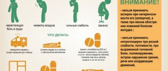
Symptoms:
- loss of consciousness,
- heavy breathing until it stops,
- swelling,
- enlargement of the neck veins,
- unpalpable pulse
- low pressure,
- a state of shock leads to loss of blood pressure.
Increasing hemotamponade with cardiogenic shock can cause death if immediate medical attention is not provided. Up to 90% of external ruptures occur unexpectedly and are fatal.
Elderly people, especially women, as well as patients suffering from diabetes, hypertension, and chronic cardiac ischemia are more susceptible to heart rupture
Why does deformation occur?
Factors that provoke rupture of the heart wall belong to structural changes, due to the fact that healthy heart muscle is very strong and elastic. Therefore, it cannot become deformed in a healthy state. What tissue the heart muscle is made of can be read here .
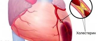
Heart rupture due to a heart attack
The most common cause of myocardial rupture is infarction (tissue death); with tissue necrosis, deformation occurs in the first 24 hours and is recorded in 3 percent of patients.
Occurs regardless of gender. But statistics say that in women, cardiac rupture is recorded in greater numbers.
The reasons that will lead to further myocardial rupture may be:
- Heart attack. Death of heart muscle tissue;
- Diseases that attack the myocardium;
- Hemorrhage of the aorta between its walls, caused by its dissection;
- Tumor formations in the heart;
- Striking, diseases of infectious origin;
- Traumatic situations of the chest, which are both blunt and penetrating in nature;
- Implantation of special medical devices;
- Open heart surgery.
There are a number of factors that in one way or another influence an increase or decrease in the chances of getting a heart rupture:
| Risk zone after a heart attack | Reduced chances of getting a rupture |
| Persistent high blood pressure | The presence of chronic cardiac ischemia, in which bypass pathways of coronary blood circulation are well developed |
| For mental and physical stress during acute myocardial infarction | Timely surgical intervention on the coronary artery |
| Age category over sixty years old | Early use of beta blockers after a heart attack |
| Females are more likely to be targeted | Heart failure |
| The use of steroid and non-steroidal drugs against inflammation during the healing period after a heart attack | Previous heart attacks |
| Small size of the first death of myocardial tissue | Increased thickness of the walls of the left ventricle |
| Resolving blood clots (later than eleven hours) after the onset of tissue death | |
| Chest pain after myocardial infarction |
Also, deformation of the heart muscles is possible during surgical interventions.
These include the following:
- Open heart surgery;
- An operation to connect electrodes from pacemakers to the heart;
- Balloon valvuloplasty (a method of treating narrowing of the heart valves);
- Pericardiocentesis (surgery to remove fluid in the pericardium);
- Cardiac catheterization for diagnosis, as well as cardiac muscle biopsy;
- Transcatheter aortic valve implantation (a modern minimally invasive transcatheter method for replacing the aortic valve).
A completely healthy heart muscle cannot rupture due to severe fear. During severe stressful situations, there is a significant increase in heart rate and increased pressure in the heart. As a result, tissue deformation may occur in the affected heart.
Hemorrhage due to a myocardial defect leads to almost instant death. Since blood flows inside the heart move under high pressure, when holes are formed in the myocardium, blood quickly fills the pericardium, which leads to disruption of the heartbeat and its complete stop.
After which all blood flow stops, and the patient dies from shock.
On the left - post-infarction rupture of the myocardium (heart muscle), on the right - external rupture of the heart with hemotamponade
It is noted that ruptures occurring inside the heart are a little easier. During the period of such deformities, the patient can live up to 14 days, but immediate surgical intervention is required.
Manifestation
The period before the rupture is characterized by intense cardiac pain, radiating to a similar spinal region (narcotic painkillers do not cope). Due to severe cardiac tamponade and shock, general health will deteriorate.
Clinical picture: low blood pressure, high venous pressure, swollen veins. Sinus heart rhythm disturbance develops.
Rapid cardiac contraction provokes sudden rupture of the myocardium:
- dizziness to the point of complete loss of consciousness,
- the skin of the upper body becomes blue,
- thickening of the neck,
- low blood pressure/unpalpable pulse.
After one minute, breathing stops. An electrocardiogram records an abnormal rhythm with gradual cardiac arrest. The manifestation of slow destruction - from several hours to several days. The pain increases and decreases (sometimes stronger/weaker).
As the symptoms of acute heart failure progress, the pulse becomes thready, blood pressure drops, and a change in consciousness is possible
Patient's condition:
- the skin becomes grayish,
- sticky sweat is released
- fast heart rate,
- diastolic pressure drops to zero, arterial pressure is reduced.
When blood clots form, the patient's general condition stabilizes. Urgent puncture of the pericardium and surgical intervention are necessary.
Diagnostics:
- The patient's medical history/complaints are analyzed (thoracic injuries, previous myocardial infarction, coronary artery disease).
- They identify diseases of the patient, close relatives (heredity), took hormonal or anti-inflammatory drugs, and check for the presence of tumors.
- The skin condition is examined for the presence of cyanosis/swelling, and blood pressure is measured. Listen to the heart for characteristic murmurs.
- Urine and blood analysis.
- Analysis for the presence of troponin T/I (intracellular substance of the heart muscles).
- Blood test for sugar, cholesterol, uric acid, urea (to detect damage or pathologies of other organs).
In the blood, high levels of norepinephrine, serotonin, magnesium, creatine phosphokinase and lactate dehydrogenase are determined
- Coagulogram - determination of the level of blood clotting. Check for the presence of blood clots and blood clot breakdown products.
- Determine heart rate.
- Echocardiography is performed (location of the rupture, presence of fluid in the pericardium, strength of heart contractions).
- A catheter is inserted into the right side of the heart to reduce the oxygen concentration.
How to identify signs of MS?
Progression of cardiac deformation can appear within 72 days after myocardial infarction. In the vast majority of cases, destruction occurs in the first five days.
The symptoms that appear depend on the extent of damage to the heart muscle, the degree of circulatory failure, and the location of the damage.
With a small hole, when blood does not enter the heart sac, symptoms are recorded from several minutes to increasing pain over several hours.
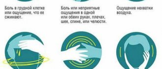
The following signs of cardiac rupture clearly appear:
- Sharp pain in the chest and heart area, which can radiate to the area between the shoulder blades. Such pains are not relieved by Nitroglycerin and Morphine;
- Feeling of strong anxiety, possible fear of death;
- Hard breath;
- Blue tint of the skin;
- Swelling throughout the body;
- Weak pulse;
- Decline in blood pressure;
- Loss of consciousness;
- Pain in the liver area associated with blood stagnation;
- Cold sweat;
- Cardiac tamponade;
- Swelling of veins;
- Cardiogenic shock;
- Pulmonary edema;
- Expectoration with “foamy” sputum, pale red in color. Occurs when the papillary muscle is torn.
Symptoms, with the progression of rupture of the heart muscle or components of the heart, are expressed quite clearly. Accompanied by severe pain.
If one of the above symptoms is detected, you should immediately call an ambulance and hospitalize the patient.
Left ventricular aneurysm
Kinds
The following types of pathology exist:
- External gap. The integrity of the ventricular wall is completely disrupted, leading to the accumulation of blood in the pericardial sac.
- With an internal rupture, the structure of the heart muscle is disrupted (the interventricular septum is torn/the papillary muscles are damaged).
- Slow flowing.
Heart ruptures occur during myocardial infarction in 3% of cases, more often in the first week (in 50% of cases) of the first, usually anterior extensive transmural “Q-wave MI”
- Subacute. Lasts several hours and is characterized by an unstable heart rhythm.
- Acute (acute hemotamponade). Most often, elderly people with transmural myocardial infarction and hypertension (during the thrombolysis procedure) suffer. After severe tamponade, instant death occurs.
- Late. The necrotic area becomes thinner.
- Early (80%). Occurs on the third or fifth day after MI.
Diagnostics
When cardiac deformation develops, it is necessary to diagnose it as quickly as possible and apply immediate surgical intervention. During the initial examination, the doctor listens to the patient’s complaints and performs an initial examination to identify obvious symptoms, and also examines the medical history.
In case of an internal rupture, with which a person can live for up to 14 days, the doctor, suspecting a diagnosis, can send the patient for the following hardware tests:
- Chest X-ray . This study helps to detect large heart sizes and pulmonary edema. These indicators may give rise to the diagnosis of cardiac rupture and the appointment of surgical intervention;
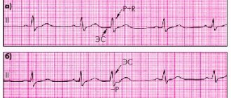
- ECG (electrocardiography). In this study, an electrical method is used to study the activity of the heart muscle. In a state close to heart rupture, the electrocardiogram shows signs of a heart attack;
- Echocardiogram (Echo-CG). In this case, the examination is carried out using ultrasound. The structure of the heart and its performance are studied. The presence of blood in the pericardium, blood transfusion through the hole in the septum between the ventricles, and mitral valve insufficiency are recorded. Used for urgent diagnosis of all types of heart muscle ruptures;
- Cardiac catheterization. In this study, a contrast agent is injected into a vein, after which an x-ray is taken. The use of such an examination is indicated only for patients who are in a stable condition and are preparing for surgery.
Intra-aortic balloon counterpulsation
Types of heart rupture
Depending on the location of the damage, it can be internal or external. Internal ruptures include ruptures of the interventricular septum, which separates the left and right ventricles.
This leads to a rapid disruption of blood flow, a drop in pressure and death. Also included in the group of internal ruptures are damage to the papillary muscles of the heart, which move the valves.
Death in this case develops due to pulmonary edema against the background of congestion. It is these patients who can be saved through emergency surgical treatment, since they are able to survive several days before death.
External ruptures cause blood to leak into the pericardium (the sac around the heart), which puts the heart under compression and it stops working.
According to the timing of the appearance of the pathology, it is as follows:
- early rupture - occurs 72 hours after a heart attack or other disease;
- late rupture - observed 72 hours or later after a heart attack.
The duration of the pathology may vary. Simultaneous ruptures lead to instant death due to cardiac tamponade; slowly flowing over several hours or days cause circulatory disorders and human death.
A complete rupture damages the muscle to its full depth, an incomplete rupture damages it partially, followed by the formation of a bulge (aneurysm) of the heart.
This pathology is divided into external and internal.
A rupture of the organ walls is diagnosed as external. Blood begins to rapidly flow into the area of the pericardium, which performs a barrier function. If the space fills with blood, the heart may “choke” and eventually stop.
External damage has the following features:
- The risk of deformation increases significantly if the injured myocardial area occupies more than one fifth of the entire muscle.
- The chambers of the heart react differently to tissue separation. The left ventricle of the heart (side wall) often ruptures; its right “colleague” is subject to this pathology much less frequently. Atrial damage is also rare.
Internal rupture occurs due to disruption of the integrity of the papillary muscle or the septum separating the ventricles.
This leads to mixing of venous and arterial blood. Complete separation of the papillary muscle is quite rare and can be fatal.
A partial tear or stretch of the heart muscle leaves doctors a chance to save the patient. In both cases, a favorable outcome directly depends on the promptness of the medical care provided.
Additionally, gaps are classified as:
- Long-lasting and instant. With a simultaneous rupture, a sharp disruption of the cardiac tissue occurs. The organ fills with blood, which, entering the pericardial sac, begins to compress the myocardium. In cases where the pathology slowly spreads across the affected area, the blood flows less abundantly. Small blood discharge can thrombose and, as a result, stop further discharge. In such situations, the patient needs urgent hospitalization and prompt assistance from specialists.
- Early and late. Late ruptures are considered to occur between 4 and 7 days after a heart attack. Tissue deformation occurs at the border of healthy and damaged areas. An early rupture is one that occurs one day after a heart attack.
The heart is the most important organ of our internal system, which has a complex structure and a lot of features that must be taken into account by specialists when treating patients.
Doctors identify several forms of such pathology as rupture of the heart muscle, which allow them to more accurately diagnose and diagnose the cause that led to such a situation.
Depending on where the damaged areas are located, they speak of external and internal rupture of the heart. In the first case, a defect forms directly in the wall of the heart, due to which blood begins to leak into the pericardial sac, which leads to its filling.
In the second case, the septum, valves and other structural components of the organ suffer. It is believed that internal tears are not as painful as external ones.
The patient can even live with them for up to two weeks before he suspects something is wrong and consults a doctor.
According to the part of the heart muscle where the rupture occurred, there is a rupture of the left, right ventricle, rupture of vascular structures, and rupture of the atria. The latter is very rare.
During a heart attack, the heart can rupture, and the reasons for this are not only tissue necrosis, but also a number of factors that can lead to the fact that the muscle tissue of the heart can rupture:
- age over 50, when regeneration processes are slowed down, in addition, many older people have heart problems such as coronary disease or wear and tear of the heart muscle;
- Increased blood pressure can exert a powerful load on the myocardium muscle;
- treatment of a heart attack not started on time;
- excessive physical activity of the patient during a heart attack, as well as heavy lifting;
- low body weight, exhaustion;
- taking hormonal and non-steroidal drugs.
How is heart rupture treated?
In the vast majority of cases, cardiac rupture requires immediate surgical intervention. Sometimes medications are also used, but only to maintain the patient’s condition.
Sometimes the use of medications and surgical intervention cannot be done on time, since the progression of the pathology is very rapid, and in many cases leads to instant death.
With heart defects, the time for hospitalization and preparation for surgery is very limited.
Among the surgical interventions used in modern surgery for cardiac ruptures are the following:
- Coronary artery bypass surgery . Restore blood flow in the arteries of the heart by bypassing narrow vessels using vascular prostheses (shunts);
- Valve replacement . During this operation, an artificial valve is implanted in the patient's heart. The replaced valve will completely replace the damaged one;
- Suturing the defect and installing special patches . An open heart operation is performed, in which the defect in the myocardium is sutured and a special synthetic patch is sewn on top;
- A special patch is used to repair the hole between the atria;
- Heart transplant . Implantation is performed if the patient’s heart cannot be saved, the area of damage is too extensive, or further complications are possible that will lead to death.
Patch to eliminate ventricular septal defect
All patients with heart damage require urgent cardiac surgery. After which, transfer to the cardiac intensive care unit and long-term therapy.
To temporarily stabilize the patient's condition, balloon counterpulsation may be introduced into the aorta.
It will improve the body’s blood circulation due to the fact that a specialized balloon is inserted into the lumen of the aorta through the femoral artery, which inflates with each contraction of the heart muscle.
Treatment and surgery
Treatment begins with normalization of blood circulation and coronary insufficiency. Surgical treatment involves pericardiocentesis (normalizes blood flow/closes the gap).
Coronography is performed in an equipped laboratory or directly in the operating room. To restore the functioning of the ventricle, mechanical blood supply is used. To remove accumulated fluid, the needle is inserted into the pericardial sac.
A mitral valve prosthesis is surgically installed and the transformed interventricular septum is closed. Open surgery involves suturing the fracture site using linings. Install the patch.
Surgical treatment consists of suturing the myocardial defect during open surgery, possibly strengthening the site of damage using a special “patch”
Depending on the damage to the heart muscle, cardiac surgery is distinguished:
- mitral valve replacement,
- remove the damaged area,
- carry out bypass surgery,
- remove the aneurysm area,
- perform a complete heart transplant.
Moderate impairment is amenable to medical treatment with cardiac medications. Mitral regurgitation requires the use of vasodilator medications. Timely cardiac surgery does not guarantee that a person will live (50% of patients die after the intervention from self-cutting sutures).
What therapy is prescribed for MS?
The use of medications can be used at the stage when the patient is still being examined and prepared for surgery, in order to maintain the patient's condition.
The most commonly used drugs are:
- Vasodilator medications (Nitroprusside, etc.). They are used when the papillary muscle is torn off, in order to reduce the load on the heart. In some cases, drugs are used intravenously;
- Inotropic drugs . Taking these medications increases heart contractions, dilates blood vessels, and has diuretic properties. The indication for use is a hole in the septum between the ventricles;
- Infusion and inotropic drugs are prescribed for rapid intravenous administration, during transport of the patient to the operating room, in case of rupture of the myocardial wall.
How to prevent heart rupture?
Due to the fact that the majority of heart ruptures occur as a result of myocardial infarction, preventive actions are aimed at preventing the provoking disease.
Doctors recommend following these steps:
- Get rid of excess weight (if any);
- Do not use cigarettes;
- Stick to a healthy diet;
- Set aside an hour of time for walking and also play sports;
- Stick to the prescribed treatment.
To reduce the risk of heart muscle rupture after a heart attack, you should also:
- Remove or minimize the use of non-steroidal and corticosteroid drugs against inflammation;
- Monitor blood pressure levels, and proactively use beta blockers (Metoprolol, etc.) in patients with heart attack;
- Use surgical interventions for heart lesions as early as possible;
- Reduce the risk of blunt chest trauma. When in cars, wear a seat belt.
Prevention
Timely preventative treatment and doctor's instructions can avoid serious heart damage.
How to prevent heart rupture:
- Get tested regularly to check your cholesterol levels,
- check your blood pressure daily,
- switch to proper nutrition,
- quit drinking/smoking,
- If you experience pain in the heart area, consult your doctor,
- patients with coronary artery disease must be under medical supervision/follow the instructions,
- in case of a heart attack, perform first aid and call an ambulance,
- do not abuse physical activity after a heart attack (bed rest with minimal physical activity).
Heart rupture is a serious pathology requiring immediate surgical intervention. However, even after surgery, the chances of death are high. It is better to prevent the disease, regularly see a doctor, and follow his recommendations.