© Author: Z. Nelly Vladimirovna, laboratory diagnostics doctor, qualification in immunodiagnostics, especially for SosudInfo.ru (about the authors)
Such a biochemical study as the determination of rheumatoid factor in blood serum is well known to many patients, especially those with joint problems, because the very name of the test is associated with a specific disease - rheumatoid arthritis (RA). Indeed, rheumatoid factor (RF) is one of the main laboratory tests that determine this disease, but, in addition to rheumatoid arthritis, rheumatoid factor can be used to identify other pathological conditions, in particular, acute inflammatory diseases in the body and some systemic diseases.
By its nature, rheumatoid factor is antibodies (mainly class M - up to 90%, the remaining 10% are immunoglobulins of classes A, E, G) against other antibodies (class G) and Fc fragments.
The norm for rheumatoid factor is the same for everyone: in women, men and children it is absent (qualitative test) or does not exceed 14 IU/ml (quantitative analysis), if everything is fine in the body in this regard. However, there are cases when RF is not detected, but the symptoms are obvious (the main reason for the increase is rheumatoid arthritis), or it is present, but the person is healthy. You can read about this below.
The essence and types of analysis
The essence of the analysis is to detect autoantibodies, in most cases belonging to class M immunoglobulins (IgM). Antibodies (IgM up to 90%) under certain pathological conditions under the influence of an infectious agent change their characteristics and begin to act as an autoantigen capable of interacting with other self-antibodies - immunoglobulins class G (IgG).
Currently, the following types of laboratory techniques are mainly used to determine rheumatoid factor:
- The latex test with human immunoglobulins of class G aggregated on the latex surface, agglutinating in the presence of a rheumatic factor, is a qualitative (not quantitative) analysis) that determines the presence or absence of RF, but does not indicate its concentration. The latex test is very quick, inexpensive, does not require special equipment or special labor costs, but it is used primarily for screening studies. Express analysis often gives false-positive answers, and therefore cannot be the basis for establishing a final diagnosis. Normally, the rheumatic factor in such a study is negative;
- The classical Waaler-Rose test (passive agglutination with sheep red blood cells treated with anti-erythrocyte rabbit serum) is being used less and less, This study is still more specific than the latex test;
- It agrees well with the latex test, but is superior in accuracy and reliability - nephelometric and turbidimetric determination of rheumatoid factor. The method is standardized, the concentration of antigen-antibody complexes (AG-AT) is measured in lU/ml (IU/ml), that is, this is a quantitative analysis that indicates not only the presence of rheumatoid factor, but also its quantity. Rheumatologists consider the result elevated if the concentration values exceed the limit of 20 IU/ml, but in approximately 2-3% of healthy people and up to 15% of elderly people (over 65 years of age), this indicator also sometimes gives elevated values. In persons suffering from rheumatoid arthritis, especially with a rapidly developing and severe form, it can be quite high (RF titers exceed 40 lU/ml, in other cases - quite significantly).
- The ELISA method (enzyme-linked immunosorbent assay), which is capable of detecting, in addition to IgM, autoantibodies of classes A, E, G, which are not captured by other methods, making up 10% of a specific protein, which we call rheumatic factor. This test has become widespread and has been implemented almost everywhere (except perhaps in rural outpatient clinics), since it is recognized as the most accurate and reliable. It has been noted that the concomitance of vasculitis in rheumatoid arthritis gives an increased concentration of class G immunoglobulins, and the appearance of class A autoantibodies is characteristic of a rapidly progressive and severe course of the disease (RA).
Until recently, the above laboratory tests were taken as the basis for establishing a diagnosis (RA). Currently, diagnostic measures, in addition to mandatory immunological studies, have been supplemented by other laboratory methods, which include: A-CCP (antibodies to cyclic citrullinated peptide - anti CCP), acute phase markers - CRP (C-reactive protein), ASL-O. They make it possible to quickly and more accurately differentiate rheumatoid arthritis from another pathology that is symptomatically similar, or from diseases in which the clinical picture is different from RA, but the RF also tends to increase.
Blood test for rheumatoid factor - what is it?
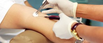
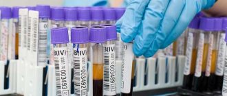
To test blood for rheumatoid factor, blood is taken from a vein. Before donating blood, you must avoid drinking alcohol, smoking, and exercising 12 hours before the test. During this period, you should not drink tea, coffee and sweet drinks, but clean water will only be beneficial. It is advisable to stop taking any medications for a while. If this is not possible, you should tell your doctor what medications you have taken recently. The test is taken on an empty stomach; it is advisable to rest for 10–15 minutes before taking blood.
Venous blood is taken to test for rheumatoid factor.
As a rule, RF is studied in combination with two other indicators - CRP (C-reactive protein) and ASL-O (antistreptolysin-O). The determination of these indicators is called rheumatoid tests, or rheumatoid tests.
A referral for a study of rheumatoid factor in the blood is usually given by a traumatologist, rheumatologist or immunologist.
In addition to rheumatoid tests, the following additional studies may be prescribed to diagnose systemic diseases and other immunological pathologies:
- a general blood test with a detailed leukocyte formula - allows you to identify the inflammatory process in the body and tumors of the hematopoietic system;
- ESR (erythrocyte sedimentation rate) - its increase is also a marker of inflammation;
- biochemical blood test - in particular, the level of uric acid, the amount of total protein and the ratio of its fractions are important;
- anti-CCP test (antibodies against cyclic citrullinated peptide) - allows you to confirm the diagnosis of rheumatoid arthritis;
- determination of antibodies to cellular organelles.
High RF level and low factor values
Rheumatoid factor is most often used to diagnose rheumatoid arthritis; its elevation is observed in approximately 80% of patients with the most common form of the disease (synovitis).
From this we can conclude that there are two forms of the disease: seropositive, when RF is detected in the blood serum, and seronegative, when rheumatic factor is absent, but the symptoms clearly indicate the presence of an inflammatory process.
A high level of RF may indicate a progressive course of the disease. It should also be noted that, while having high sensitivity, rheumatoid factor does not show such high specificity (every 4th result is false positive), because its nature has not been fully studied, however, it is known that autoantibodies are actively produced in many chronic inflammatory processes.
In addition, RF may not be detected in the presence of signs of the disease in rheumatoid arthritis at the beginning of the development of the pathological process in 20-25% of patients, so a single negative result cannot be reassuring if symptoms of the disease occur. In suspicious cases, the analysis should be repeated after six months and a year (to give time to update the pool of plasma cells that produce autoantibodies).
It is inappropriate to rely on this analysis to control the course of the process and the effectiveness of therapy - the medications received by the patient can influence the results of studies, which no longer reflect the real picture and thereby mislead the patient (he begins to prematurely rejoice in the cure, attributing the merits to some some folk remedies).
How to prepare for the test
To perform a laboratory test, blood is drawn from a vein from the patient. Today, there are various methods for determining the concentration of a specific protein. The most informative is a study called ELISA. Thanks to it, you can determine not just the presence of rheumatic factor in the blood, but also a type of autoantibodies.
Before taking the test, the patient must prepare. In this case, he must adhere to certain rules and recommendations of specialists:
- Blood from a vein is taken in the first half of the day, ideally before 9 am. Before the procedure, it is recommended to refrain from eating for 12 hours.
- For two hours before the procedure, it is necessary to avoid various types of physical activity. Smoking and drinking alcohol are also not allowed.
- You should not eat salty or fried foods for 24 hours before the procedure.
- You should refrain from taking medications for 24 hours before the procedure. If it is not possible to avoid taking a particular drug, you must notify your doctor and the laboratory that is conducting the test.
Rheumatoid factor in children does not predict the diagnosis of RA
If in adults (women, men - it doesn’t matter) rheumatoid factor is quite closely associated with rheumatoid arthritis, then in children the situation is somewhat different. Juvenile RA, which develops before the age of 16, even with the rapid development of the inflammatory process, gives an increase in RF titers (mainly due to IgM) only in 20% of cases - at the onset of the disease in children under 5 years of age. The beginning of the development of the process in children under 10 years of age is manifested by an increase in this indicator in only 10% of cases.
Meanwhile, children who are often and long-term ill have elevated RF even without obvious signs of any disease. This suggests that autoantibodies (IgM) can be produced in them due to prolonged immunostimulation (chronic infections, recent viral diseases and inflammatory processes, helminthic infestations), and the reason does not lie in the development of rheumatoid arthritis.
Given these characteristics of rheumatoid factor, pediatricians do not attach any special diagnostic value to this laboratory test.
Diseases in which the indicator is increased
If the level of this immunoglobulin is increased, this may indicate the presence of such pathological conditions in the human body:
- presence of rheumatoid arthritis. If, during the development of such a disease, a high level of rheumatoid factor is noted, this indicates the development of a severe form of the disease;
- pathological processes in connective tissues, for example, the development of lupus erythematosus, systemic scleroderma, sarcaidosis, dermatomyositis;
- development of pericarditis, chronic myocarditis and heart diseases.
Regardless of what disease is detected, it is important to undergo timely treatment as prescribed by a doctor.
Other causes of elevated rheumatic factor values
The cause of an increase in the concentration of rheumatoid factor in the blood, in addition to the classic version of rheumatoid arthritis, can be many other pathological conditions:
- Acute inflammatory diseases (influenza, syphilis, infectious mononucleosis, bacterial endocarditis, tuberculosis, viral hepatitis);
- A wide range of chronic inflammatory processes localized in the liver, lungs, musculoskeletal system, kidneys;
- Sjögren's syndrome is an autoimmune disease that affects connective tissue and involves the exocrine glands (tear, salivary glands - primarily) into the process. Sjögren's syndrome is also characterized by corresponding symptoms: dryness of the mucous membranes of the eyes, mouth, external genitalia, suffering from the respiratory system, cardiovascular system, and kidneys;
- Felty syndrome, which is a special form of RA, characterized by an acute onset with a decrease in the number of white blood cells in the blood - leukocytes (leukopenia);
- Still syndrome (Still syndrome) is a form of juvenile (childhood) rheumatoid arthritis, the symptoms of which coincide with those of Felty syndrome, but differ in the indicators of a general blood test - the number of leukocytes is increased (leukocytosis);
- Scleroderma;
- Hyperglobulinemia of various origins;
- B-cell lymphoproliferative diseases (myeloma, Waldenström's macroglobulinemia, heavy chain diseases);
- SLE (systemic lupus erythematosus);
- Sarcoidosis;
- Dermatomyositis;
- Surgical interventions;
- Oncological processes.
Obviously, the list of conditions that can cause an increase in the concentration of rheumatoid factor is not limited to rheumatoid arthritis alone.
In addition, it should be borne in mind that this indicator naturally increases in older people (60-70 years old), as well as when using certain medications (methyldopa, anticonvulsants and contraceptives), therefore it should be considered specific and especially significant for diagnosis inappropriate.
However, the treating doctor will figure it out, and our article is intended for people trying to independently interpret the results of biochemical studies. After all, it happens that upon hearing information about high numbers of some kind of analysis, especially suspicious citizens fall into panic or (even worse) begin to show initiative and be treated with various dubious means.
Types of rheumatic tests
As noted above, there is no unambiguous interpretation of this term in classical medicine. Therefore, the question invariably arises, what tests should be taken for rheumatic tests? To diagnose any systemic disease, the following indicators are most often determined:
- Rheumatoid factor is a special antibody that is produced by immune cells (leukocytes) and affects connective tissue. Despite their name, their increase in the blood indicates not only the presence of rheumatoid arthritis, but also a number of other diseases;
- AntiDNA is another substance that leukocytes produce in the presence of autoimmune inflammation. It has a specific target, unlike rheumatoid factor - a strand of DNA in cells where most of the genetic information is contained. That is why the presence of AntiDNA in the blood leads to a large number of disorders in various organs;
- HLAB27 is a specific gene that is responsible for predisposition to a number of autoimmune pathologies, including rheumatoid arthritis, ankylosing spondylitis, urethro-oculosynovial syndrome and others. Its detection in a patient with characteristic symptoms of one of the above diseases allows it to be confirmed in 90% of cases;
- Antinuclear factor is a group of antibodies that appear in the blood during autoimmune pathologies. Their main target is the nuclei of cells, mainly connective tissue organs.
About 10-20 years ago, markers were often added to the list of examinations that helped detect a past streptococcal infection: Antistreptolysin (ASL-O), Antistreptokinase and Antistreptohyaluronidase. This helped to detect or exclude the presence of acute rheumatic fever (rheumatism) in the patient. However, at present, this disease has practically disappeared in the Russian Federation, thanks to adequate treatment of upper respiratory tract infections.
What is the Russian Federation
Rheumatoid factor is nothing more than an antibody to the cells of one’s own body.
Appears in the blood if there is a malfunction in the human immune system. Rheumatoid factor appears in the blood when the immune system fails. It is an antibody that reacts as a self-antigen with its own IgG class immunoglobulins. Most often, RF refers to IgM, much less often to IgA, IgD, IgG.
Autoantigens that react with one’s own antibodies are extremely dangerous. RF forms a stable circulating complex with immunoglobulin, which has a cytotoxic effect. He:
- damages the synovial membrane of the joints;
- causes inflammation;
- has a destructive effect on the vascular wall.
Accordingly, due to its occurrence, the patient experiences joint pain. And for an accurate diagnosis, the doctor needs to know not only the presence, but also the concentration of RF in the blood. Directed by:
- if rheumatoid arthritis is suspected;
- to monitor the treatment of the disease;
- for the diagnosis of autoimmune pathologies;
- for chronic inflammatory diseases.
To determine its concentration, the ability of RF to agglutinate (glue together) red blood cells in the presence of immunoglobulins is used. This is one of the manifestations of the reaction between it and ordinary antibodies.
Rheumatoid factor is detected using various methods:
- latex agglutination;
- Waaler-Rose reaction;
- nephelometry;
- enzyme immunoassay (ELISA).
Most often, they are used to determine RF related to IgM. But identifying autoantibodies of classes G, A and D is much more difficult. That is why, in case of a seronegative (negative) reaction in the presence of clinical symptoms of the disease, other clarifying diagnostic methods are recommended.
The reaction is considered positive if agglutination occurs at a dilution of 1:40 or 1:20 (modified Speransky method). Due to the use of different methods for determining RF in clinical laboratories, repeated tests must be carried out in the same one where the analysis was initially taken.
Which doctor should I contact?
Most people associate the concept of “rheumatoid factor” with a pathology such as rheumatoid arthritis.
However, this substance appears in the blood of people with other autoimmune diseases. Rheumatoid factor, which is an autoantibody, when reacting with immunoglobulins, has a destructive effect on the joints. And its appearance in the blood indicates that the patient has rheumatoid arthritis, another autoimmune or infectious disease. A very high RF titer signals an extremely severe course of the disease. Its presence in the blood is determined in clinical laboratories. A rheumatologist refers him for research. An orthopedist, neurologist or neurosurgeon can prescribe such a study if the patient comes to them with complaints of pain in the spine, joints, and limited movement.
Rating: (votes - 3 , average: 1.67 out of 5)