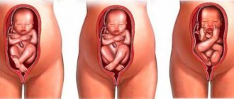
Ultrasound diagnostics is considered the most informative and safe procedure aimed at monitoring the development of pregnancy. Ultrasound in the first trimester of pregnancy helps diagnose conception that has occurred already at 5 weeks, when a specialist can detect a fertilized egg, and at 6 weeks a formed embryo is detected in the uterus.
The most exciting moment is seeing your baby for the first time.
Indications and limitations of the first ultrasound screening
An ultrasound is performed in the first trimester to confirm intrauterine pregnancy, determine the number of fetuses, their viability, anatomy, and also clarify the timing of gestation. An abnormal structure increases the likelihood of having a child with a disability, Patau, Edwards, Turner, Down syndrome. The absence of movement with a heartbeat indicates the freezing of the embryo. In all cases, the woman is taken under observation and a repeat ultrasound is performed.
Indications for an unscheduled examination include a woman’s history of miscarriages, ectopic embryo implantation, or cesarean section.
In the 1st trimester, emergency sonography is done for:
- vaginal bleeding of any intensity;
- abdominal pain;
- suspected fetal death;
- other complications of gestation.
Contraindications for sonography in the 1st trimester include early pregnancy up to the 6th week of gestation. The device does not see the embryo, there is a risk of failure of the fertilized egg. Transvaginal examination is not performed if there are signs of miscarriage.
Transabdominal ultrasound cannot be done if:
- skin damage in the lower abdomen;
- obesity;
- urinary incontinence.
In the 1st trimester, according to the plan, an ultrasound is performed between the 77th and 97th days of pregnancy: , , 13 weeks of gestation inclusive. This is approximately 3 months from the first day of the last menstruation (the calculation takes into account the phase of the cycle before ovulation). At this time, it is possible to determine the size and anatomical structure of the child’s body, brain, internal organs, bones, heart functioning, and motor ability.
The ISUOG Society Committee has classified sonography as a mandatory type of examination in the 1st trimester. It allows you to identify conditions dangerous to the child or mother from the 6th week of gestation. A woman’s refusal to have an ultrasound scan relieves doctors of all responsibility in the event of the birth of a baby with a defect in the brain, internal organs or skeleton.
We recommend watching a video on the topic:
Interpretation of additional ultrasound parameters
The presence of chromosomal abnormalities in fetal development is determined by the thickness of the nuchal translucency. This indicator allows you to determine only screening of the 1st trimester. You can find out how an ultrasound is done on the Internet or ask your doctor.
Analysis of the structure and location of the chorion allows us to determine the future placement of the placenta and determine how the pregnancy develops. It is important to note that if the chorion is attached near the internal os of the uterus, then there is a possibility of developing placenta previa.
The yolk sac is almost completely neutralized by week 12, since by this time the placenta begins to mature, which will perform all the same functions and provide the fetus with nutrients and all the factors necessary for normal development.
Analysis of the condition of the woman’s genital organs is also very important. Since the non-standard shape of the uterus (saddle-shaped, bicornuate) can cause miscarriage or fetal death. The appendages are also examined for the presence of cysts. In some cases, during pregnancy there is a need for surgical intervention to eliminate the pathology.
To describe the pathologies found, the ultrasound specialist writes a comment at the end of the protocol. Ultrasound screening of the 1st trimester is a very important examination that allows you to fully identify all possible pathologies and abnormalities in the development of the fetus and genital organs of the pregnant woman.
Preparing for the first ultrasound screening
Three days before a transabdominal ultrasound, it is necessary to exclude from the diet foods that contribute to gas formation in the intestines. You can create a menu according to Pevzner’s recommendations for treatment table No. 1b.
Be sure to exclude from the diet:
- kefir, yogurt;
- salty/canned/pickled food;
- Rye bread;
- sparkling water, drinks;
- coffee, strong tea;
- legume dishes;
- sauerkraut;
- raw vegetables;
- fresh fruits, berries, tropical fruits.
For people with flatulence, the doctor prescribes carminatives that are safe for the fetus. On the day of sonography, take a shower without soap/gel. It is not advisable to apply cosmetic or medicinal creams to the skin of the abdomen. They take a diaper, a towel, shoe covers with them to the antenatal clinic, and a condom for TVUS. Before a transabdominal ultrasound, the bladder should be full. A pregnant woman should drink 3-4 glasses of still water half an hour before the procedure and not go to the toilet.
No preparation is required for a transvaginal ultrasound. But before the examination you need to empty your bladder.
Algorithm for first pregnancy screening
In the 1st trimester, it is allowed to do an ultrasound through the skin of the lower abdomen (transabdominal) or using a vaginal sensor from the side of the cervix. The last method is more reliable. But this method requires caution from the diagnostician so as not to provoke pregnancy failure.
Algorithm for conducting transabdominal ultrasound in the first trimester:
- The couch near the sonographer is covered with a disposable diaper.
- The woman lies on her back, exposing her stomach.
- The diagnostician smears the suprapubic area with acoustic gel and places the sensor perpendicular to the skin.
- The lower abdomen is scanned along longitudinal, vertical lines. During the examination, the size and height, structure of all fetal structures are calculated, and photographs are taken.
- During the procedure, the diagnostician may require you to change your position: bend your knees, place your palms under your lower back, and roll over onto your side.
- After the procedure, the gel is washed off with a napkin/towel. The doctor prints out the protocol.
For a transvaginal ultrasound, the woman undresses as for a gynecological examination. Then he lies down on the couch with his stomach up, bends his legs, places his heels closer to his buttocks, shoulder-width apart. The diagnostic sensor is protected with a condom, lubricated with gel, and carefully inserted into the vagina. The fetus is examined from several angles. Then the woman gets dressed, the doctor prepares a protocol.
The examination is carried out for 2–7 minutes with the minimum frequency of ultrasound waves in mode “B”, “M”. This ultrasound technique is reliable and excludes damage to the embryo or fetal failure in the 1st trimester.
Interpretation of the results of the first ultrasound screening
For interpretation, the ultrasound protocol is given to the gynecologist-obstetrician managing the pregnancy. To obtain a second medical opinion, the document is provided to a qualified doctor of an independent medical institution.
Normal fetal development in the 1st trimester:
| What is being examined? | Medical standards by week of gestation | ||
| 11 | 12 | 13 | |
| Yolk sac (mm) | From 7 | No rating possible | Can't examine |
| Collar space (thickness in mm) | From 1.5 | Up to 2.5 | 2.6―3 |
| Nasal bone (mm) | Visible on ultrasound | From 2 | 3―4.2 |
| Heartbeat (beats per minute) | 140―170 | 140―170 | 140―170 |
| KTR (size of the fetus from the crown to the coccyx in mm) | From 42 | From 51 | 73―88 |
| BDP (biparietal diameter of the head, size between the temples in mm) | From 13 | 18―24 | 20―27 |
The site of egg implantation, the development of the placenta, the closure of the cervix, the size of the corpus luteum in the ovary, and the structure of the uterus are compared with the norm. Before the 14th week of gestation, a woman may be diagnosed with a cyst, fibroids, polyps, tumor, and other gynecological diseases.
Interpretation of ultrasound of the first trimester:
- hypertonicity of the uterus - the myometrial layer is thickened;
- underdevelopment of the amnion - small diameter of the aqueous amniotic membrane;
- intrauterine infection - large amnion diameter;
- risk of pregnancy failure - chorionic villi abnormalities, changes in heart rate, arrhythmia;
- spinal cord herniation - a dark-colored protrusion in 1 part of the ridge;
- pregnancy fading - no heartbeat, fetus does not move;
- hyperechogenicity of the intestine - the intestinal tract of the fetus is white;
- malformations - the structure of the brain, organs, bones or body differs from the anatomical norms of the fetus corresponding to 11–13 weeks of gestation.
In the 1st trimester, deviation from the norm only indicates the possibility of a chromosomal disorder or developmental defects. To confirm the anomaly, a follow-up examination is performed at least 1 week later.
To watch a video review from a specialist on the topic:
The results are normal: how the fetus develops
The first ultrasound is performed to diagnose the development of fetal organs
The interpretation of the results is carried out depending on the stage of pregnancy, and the indicators will differ.
The results record the number of fetuses, coccygeal-parietal size, thickness of the nuchal zone, location and structure of the chorion:
Normal ultrasound indicators:
- The thickness of the nuchal translucency at 10-11 weeks of gestation should be in the range of 0.8-2.2 mm, at 12-13 weeks - 0.7-2.7 mm.
- The coccyx-parietal size, the indicator from the coccyx to the crown, should be 53 mm. If the value ranges from 42 to 58 mm, then this is not a reason to worry, since the value will differ depending on the gestational age. If the indicator is lower, this may indicate genetic abnormalities, missed abortion, or developmental delays associated with genetic or infectious abnormalities in the mother.
- The length of the nose bone is also an important indicator. Normally, at 12-13 weeks its size should be in the range of 2.0-4.2 mm.
- The normal heart rate is 161-179 beats/min at 10 weeks, 153-177 beats/min at 11 weeks, 150-174 beats/min at 12 weeks of pregnancy.
- The amount of amniotic fluid is about 50 ml. They are updated daily.
- The yolk sac should be visible by 12 weeks. In the future it is reduced. The yolk sac should be round in shape and its size should be within 4-6 mm. If the shape and structure of the sac changes, a frozen pregnancy or congenital pathologies are diagnosed.
- At the 9th week of pregnancy, the embryo should normally have an interhemispheric fissure, the lateral ventricles and choroid plexuses of the brain should be clearly visible. The size of the choroid plexuses increases by the end of the first trimester and reaches 5 mm in width.
- If the embryo develops normally, then the chest size is 24 mm, the biparental head size is 21 mm, and the thigh length is 9 mm.
Up to 12 weeks, these parameters can be used to identify chromosomal diseases, for example, Down's disease. In the first trimester, the diagnosis is established with an accuracy of up to 98%. In the second trimester, diagnosis is problematic and is carried out after blood sampling. In addition, the correct location of the internal organs is assessed, the placenta insertion can be examined and the uterine tone can be assessed.
Ultrasound in the first trimester can reveal isthmic-cervical insufficiency, which can cause termination of pregnancy.
It should be remembered that deviations from the average values do not always indicate congenital pathologies. In each case, they are individual in nature and can return to normal during the formation of fetal organs.
Fetal pathologies
Ultrasound can detect intrauterine defects in the fetus and chromosomal diseases
In case of a genetic disorder, the following pathologies can be identified at the first screening:
- Down syndrome. In Down syndrome, the indicators differ significantly from the norm. The disease is diagnosed if the thickness of the collar zone is greater than normal, if the nasal bone cannot be detected on ultrasound, and at 15-21 weeks it is smaller than the permissible size.
- De Lange syndrome. Diagnose developmental defects and mental retardation. After birth, the child may notice craniofacial anomalies, a short nose, long eyelashes, and fused eyebrows. This syndrome is characterized by abnormalities of the musculoskeletal system: shortening of the limbs, absence of one or more fingers, etc.
- Patau syndrome. The newborn has pathologies of the central nervous system, mental retardation, body weight below normal, and a sunken bridge of the nose. Life expectancy does not exceed a year.
- Edwards syndrome. The newborn has not only low weight, but also abnormalities in skeletal development. Defects of the cardiovascular system are also observed. Children with the syndrome live for no more than 3 months.
- Smith-Opitz syndrome. The development of the syndrome is associated with impaired cholesterol synthesis. The child develops encephalopathy; external signs include low-set ears, a protruding forehead, and a small and flattened nose.
- An umbilical hernia and neural tube pathologies can also be detected. Omphalocele or umbilical hernia is a congenital pathology, which is characterized by a defect in the development of the abdominal muscles, as a result of which internal organs protrude from the abdominal cavity. Neural tube defects include hydrocephalus, herniation of the spinal cord and brain, and prolapse of the meninges.
Read: When to do the first ultrasound and fetal development in the first trimester of pregnancy
If an ultrasound reveals genetic abnormalities, the woman should seek advice from a geneticist. A qualified specialist will prescribe a chorionic villus biopsy. The procedure involves examining chorionic villi, which makes it possible to accurately diagnose chromosomal pathologies.
Cost of the first ultrasound pregnancy screening
In antenatal clinics with state status, ultrasounds are performed free of charge upon referral from a gynecologist-obstetrician. In private medical centers, the cost of screening in the 1st trimester varies between 600–6500 rubles. In case of multiple pregnancy, the price increases by 100–1000 rubles.
In the 1st trimester, ultrasound is absolutely safe and is performed in conjunction with blood tests. After transvaginal sonography, a slight discharge of ichor is possible from the vagina within 24 hours. In the absence of pain or pulling sensations in the lower abdomen, this is considered normal.
Comment on the article, spread the information on social networks, and share your experience of examination in the 1st trimester. Have a safe pregnancy and childbirth.
What is an ultrasound scan in the first trimester?
Ultrasound examinations take place in specially equipped private clinics or antenatal clinics, which have appropriate professionals who can carry out the necessary diagnostics.
Ultrasound screening of the 1st trimester will help to conduct a full examination at a short stage of pregnancy. The attending physician will explain how the study is carried out, and if necessary, he will also tell you how to prepare for the diagnosis.
The screening study is carried out transabdominally (through the abdominal wall) using an ultrasound diagnostic device. The final ultrasound protocol indicates the following indicators:
- structural features of the uterus and appendages;
- visualization of the fetus and yolk sac;
- location and structure of the chorion;
- fetal heart rate;
- fruit size from crown to tailbone;
- thickness of the neck fold.
An ultrasound specialist will be able to determine the exact duration of pregnancy, exclude any genetic pathologies and malformations of the fetus, and also see whether there are pathologies in the woman’s reproductive system that can complicate the course of pregnancy or cause its termination. Ultrasound screening of the 1st trimester provides a complete examination of the pregnant woman and fetus in all necessary parameters.