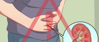
Kidney stones are an insidious disease. Developing virtually asymptomatically, kidney stones can suddenly begin to move. People are afraid of treatment, but when the stones begin to move, they experience unbearable pain.
A particularly difficult stage of self-removal of stones is their passage through the ureters. These narrow tubes, up to 30 cm long, are capable of passing through only small dense formations. But even in this case, severe pain is guaranteed. In addition, the risk of blockage of the ureter increases, which leads to acute renal failure, rupture of the ureter and death.
Stones in the ureters: what are they?
A stone in the ureter is a dense formation consisting of salts, uric acid and proteins. The diameter of the ureter is 4-15 mm. But even if the stone is smaller in diameter but has an uneven surface, its movement injures the mucous membrane, causing severe pain. The most difficult process is to remove oxalate stones, the surface of which is strewn with thorns.
Women most often develop struvite stones. Formed as a result of the processing of urea by bacteria, they are rectangular in shape and very dense. With this type of stones, folk remedies and drug therapy are ineffective; stone removal from the ureter is carried out using ultrasound or surgery.
If a stone is stuck in the ureter, in most cases this indicates the presence of stones in the kidneys and their unstable position. It is extremely rare (for example, with ureteral diverticulum) that dense conglomerates form directly in narrow urinary tubes.
This may be caused by chronic inflammation of the bladder, diseases of the nervous system with impaired innervation of the urinary tract (paralysis), or renal pathology that occurs with stagnation of urine. In women, stones in the ureters often form when the bladder prolapses. This situation occurs after a difficult birth, multiple pregnancy, in women who have given birth many times and is caused by incompetence (weakness, overstretching) of the pelvic muscles.
Often stones begin to form after surgery. For example, when performing mini-traumatic loop uroplexy for stress urinary incontinence, the likelihood of stones appearing in the ureter increases several times.
Stone formation in the urinary tract is promoted by an addiction to spicy, salty foods, women's restriction of drinking regime and regular use of laxatives for weight loss. The risk of kidney stones is high in women with a sedentary lifestyle (sedentary work).
Treatment options
The treatment process for this disease depends on the severity of the disease and the size of the stones and can develop in two directions: conservatively or actively. The first type of therapy (conservative-expectant) is used if the size of the stones does not exceed 2-3 mm.
The patient has been taking special medications for some time that have a destructive effect on the stones and also relieve inflammation and pain.
Conservative expectant therapy includes:
urolitic drugs; antispasmodics; painkillers; physiotherapy; diet; physiotherapy.
This treatment is used if the calculus does not completely block the ureteral canal and does not interfere with urination. It may well be that in this case the stone will come out on its own.
Also, in some cases, the so-called endovesical treatment method is used, when drugs are injected into the ureteral canal to stimulate the passage of the stone. To speed up the passage of a stone, the urinary system may be stimulated using an electrical current.
Active methods for treating ureteral stones are as follows:
lithotripsy; ureteroscopy; ureterolithotomy.
Lithotripsy is a method of remotely crushing a stone into small pieces using various waves (for example, electromagnetic). The crumbled calculus is excreted along with urine. The procedure lasts about an hour and is often performed without anesthesia. This is one of the most effective and low-traumatic ways to deal with stones.
Ureteroscopy is the forced removal of a stone using a special instrument - a ureteroscope, which is inserted through the entire genitourinary system directly into the ureter. The doctor removes the stone using forceps. If necessary, the stone is crushed using laser fiber. The patient is under partial or general anesthesia.
Ureterolithotomy is a surgical method that is used for large stones. The stone is removed from the ureter directly through a dissection of its wall in the part of the canal where the stone is located. The patient is under general anesthesia.
Ureterolithotomy - the essence of the procedure
As a rule, surgical intervention cannot be avoided if the calculus has blocked the lumen of the ureter or is very large in size, and the disease is accompanied by serious complications.
Stones in the ureter are not a case when you should postpone a visit to the hospital. The disease is serious and can end in disaster. But if an attack of pain when a stone passes takes a person by surprise far from medical institutions, then you can try to expel the stone yourself.
You should take an antispasmodic, which will relax the muscles, and it will be easier for the stone to move towards the exit. To relieve an attack of pain, the patient is given an analgesic. Then you can take a warm bath and try to drink a lot of water or a diuretic herbal decoction. However, even if the stone passed on its own, you should consult a doctor as soon as possible and undergo the necessary examinations - it may well be that there was more than one stone, and the attack will soon happen again.
Symptoms of stones in the ureters in women
When sand (tiny dense particles) passes through the ureters, a woman feels a dull pain in the lower back, and sometimes a tingling sensation occurs in the lower abdomen (from the side of the damaged kidney). The female urethral canal is wide and short, so the passage of stones through it does not cause severe pain. In the future, grains of sand can be seen in the urine. This condition lasts up to several days and, usually, the woman does not seek medical help.
Movement of larger stones always causes renal colic, an acute condition with unbearable pain. Women who have experienced colic prefer to give birth twice. This comparison gives an idea of the intensity of pain when stones move through the ureters.
Characteristic symptoms of stones in the ureters in women:
- Pain
It appears suddenly. The cramping pain is immediately very intense and is initially localized in the lumbar region on the side of the affected ureter. Stones stuck in the upper and middle sections of the ureter cause incredibly severe pain in the side, spreading throughout the abdomen with possible irradiation under the scapula.
Obstruction of the ureter in the upper sections is characterized by irradiation into the umbilical region. A dense conglomerate moving in the lower part of the ureter provokes pain in the lower abdomen with irradiation to the leg, perineum and rectum.
The patient clearly understands that the epicenter of pain is localized in the lower back, and pain in the abdomen, leg, etc. is just an echo. The pain weakens or completely disappears when the stone stops. Changing position for renal colic does not bring relief.
- General state
An attack of colic is accompanied by severe sweating. Often a woman notices nausea and vomiting, which does not bring relief. Colic is accompanied by a periodic increase in temperature and a false urge to have a bowel movement.
During an attack, the temperature periodically rises, and pressure may rise. The pressure surge is not controlled by antihypertensive drugs. If it lasts for a long time, bloating, constipation or loose stools may occur.
- Urinary dysfunction
During an attack, the patient notes an increased urge to urinate. In this case, little urine is released, and the pain at the time of emptying the bladder intensifies. The urine may become cloudy and mixed with blood. Large blood clots indicate deep damage to the ureteral mucosa.
The main question is how long does it take for a stone to come out of the ureter? An attack of painful colic lasts 3-18 hours. If the stone does not leave the ureter, acute urinary retention occurs, and renal failure provokes shock. The pain decreases, the pressure drops, the heart sounds become dull and rare, the patient loses consciousness.
Differences between colic and other diseases
Although the pain syndrome during colic is quite pronounced and specific, one should clearly distinguish between pain associated with the passage of stones through the ureters and those caused by other diseases.
- With intestinal obstruction, the pain increases gradually and is much weaker. Excretory urography and functional chromocystoscopy allow differentiation of the diagnosis.
- With an ectopic pregnancy (an acute condition occurs at 3-4 weeks), the pain is constant and localized in the lower abdomen. The woman lies with her legs brought to her chest. The doctor records all signs of peritoneal irritation - board-shaped abdomen, etc.
- A perforated gastric ulcer is characterized by “dagger” pain in the epigastrium. Symptoms of an acute abdomen are combined with signs of intra-abdominal bleeding. An x-ray shows a crescent-shaped accumulation of gases under the diaphragm.
- A stone in the right ureter in the lower section produces symptoms similar to appendicitis. An attack of appendicitis begins with fever, nausea/vomiting. With renal colic, these symptoms appear a little later. Pain with appendicitis forces a woman to adopt a “fetal” position - on the right side with her legs brought to her stomach. Symptoms of an acute abdomen are accompanied by a rapid increase in leukocytes in the blood and ESR.
- Pancreatitis causes girdle pain, however, unlike renal colic, the temperature remains normal, although the patient's condition is quite serious.
- An attack of acute cholecystitis is always preceded by a violation of the diet (feast, alcohol, intake of fatty foods, etc.). Pain that occurs in the right hypochondrium can radiate to the side and abdomen. Palpation of the right hypochondrium increases pain. The temperature is steadily elevated, and slight yellowing of the sclera is possible.
Principles of treatment - stone removal and surgery
stone from the ureter, photo
The method of stone evacuation is chosen by the doctor depending on the composition of the stone. The following methods for removing stones from the ureters are currently practiced:
Remote (non-surgical) destruction of stones using ultrasonic waves or laser. Crushing stones in the ureter with ultrasound or laser is painless; fragments come out on their own with less pain. Lithotripsy is effective only for small stones, the diameter of which does not exceed 0.5-2 cm. The procedure is carried out under visual control, so this treatment option is not suitable for crushing urate stones. Several procedures are required to achieve the effect.
Non-invasive technique for examining the bladder and ureters using an endoscope. In this case, the doctor determines not only the level of blockage of the ureter, but also uses special mini-instruments to capture and evacuate the stone.
Destruction of dense formations using an ultrasound or laser emitter passed through the urethra and bladder. Unlike the remote technique, contact lithotripsy gives results comparable to surgical intervention already in the first procedure. In most cases, a hospital stay is not required.
Radical surgical intervention is advisable only in severe cases: diagnosis of multiple kidney stones and complete blockage of the ureter with a large stone. Surgery is difficult to tolerate. Its only advantage is a wide overview, which makes it possible to eliminate all associated pathologies (suturing a diverticulum, removing large kidney stones, etc.).
Skin punctures provide access to the site of injury. The stone is removed through an incision in the ureter. The recovery period after such an operation is easier than from open surgery. The patient is discharged home within 2-3 days.
Most often, evacuation of a stone from the ureter is carried out non-invasively (lithotripsy, cystoscopy), surgical intervention is a last resort in especially severe cases. Removing a stone from the ureter does not mean recovery. Kidney stones are the result of a violation of the water-salt balance. Therefore, in order to destroy kidney stones that have already formed in the kidneys and prevent the formation of new ones, treatment should be continued, including long-term use of drugs (Urolesan, Phytolysin, Blemaren, etc.), diet - nutrition is adjusted in accordance with the composition of the stones.
source: zdorova-krasiva.com
Stones can be anywhere in the urinary system. But stones in the ureter, the symptoms of which we will consider in this article, are the most common.
The sensations in this case, as a rule, are the most unpleasant. Moreover, they are as unpleasant as you can imagine. Especially if the stone is not a single one, but a whole group of them, following each other (and there are such things).
Finding stones in the ureter can cause dangerous complications and the most severe consequences compared to other locations of stones.
This is due to the characteristic features of the ureter as an organ of the human urinary system, the main of which is a narrow passage.
about where stones are located in the ureter , as well as what kind of pain a person feels in this article.
Stone in the ureter - what to do?
It is impossible to quickly remove a stone from the ureter. The main task for renal colic is to relax the ureter as much as possible to reduce pain and unhindered movement of the stone to the bladder.
Stone in the ureter: how to remove it at home:
- Taking painkillers (Ketorolac, Ketanov, Dexalgin) and antispasmodics (No-spa). The best effect is achieved when these drugs are combined and administered intramuscularly. Pills will not bring immediate pain relief. An excellent combination drug for relieving renal colic is Baralgin. Subcutaneous injections of anticholinergics Atropine and Platyphylline.
- Bath with water temperature 37-39ºС for 15-30 minutes. will enhance the antispasmodic effect of drugs.
- Physical exercises - jumping, running up stairs - help move stones.
It is advisable to urinate in a separate container. It is recommended to submit the released stone for analysis. The composition of the stone can significantly affect further treatment tactics:
- Phosphate stones can be crushed using hardware;
- Urate stones often occur against the background of gout and constant dehydration. They are not detected on x-rays, but they can be eliminated with the help of diet and adequate drinking regimen;
- Struvite stones are easily crushed under the influence of ultrasound;
- Cystine (protein) stones are in most cases removed with the help of drug therapy.
- Oxalate stones are the hardest, in such cases radical treatment is resorted to.
How are stones removed from the ureter? Methods and preparations
Treatment methods for urethral stone disease are selected depending on the complexity of the process, determined by the size and location of the formations. Only a doctor can determine how it is possible to remove stones from the ureter. Treatment methods are based on two directions - conservative expectant therapy and surgical intervention.
Expectant therapy is justified for small stone sizes (up to 3 mm), when under the influence of medications it is possible to provoke spontaneous passage of stones. For this purpose, drugs are prescribed, the basis of which is natural raw materials - terpenes. Their properties are due to different actions:
- enhance blood circulation processes, causing hyperemia;
- increase the volume of urine excreted;
- eliminate spasm of the pelvic and urethral muscles;
- promote increased peristalsis, thereby increasing the likelihood of tumors emerging;
- have a bactericidal effect on infectious flora.
Preference is given to the drugs “Enatina”, “Olymetina”, “Spasmacistenal”, “Rovatinex”, “Canephron”, “Cyston”, “Fitolysin” or “Palina”.
The final choice of drug is based on the indications of the chemical type of stones, their size and location. These indicators determine how long it takes for the stone to leave the ureter. Typically, this happens after a two-week course of full therapy, including:
- Painkillers – “Ketanov”, “Baralgin” and antispasmodics – “Papaverina”, “Drotaverina”, which promote painless passage of stones through the urethral canal, which is relaxed by antispasmodics and has an increased diameter.
- NSAIDs - Ibuprofen or Naproxen.
- When an infection occurs, antibiotics are prescribed.
- As emergency measures to relieve renal colic, narcotic analgesics, antispasmodics and blockade drugs are prescribed.
- Long-term course use of herbal preparations that can dissolve stones of any composition, alleviate symptoms and restore impaired urination - “Curly silkworm”, a collection of 17 medicinal herbs - “Litolysin”.
- Exercise therapy techniques.
- Physiotherapy in the form of diathermy, diadynamic currents and subaqueous baths.
Diet and drinking regimen. In addition to drug treatment, patients are advised to balance their drinking and nutritional diet. Patients need to drink at least 2 liters of fluid per day.
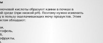
When choosing a diet, the doctor is guided by the chemical nature of the stones. For example, when diagnosing oxalate stones, dishes from cabbage, spinach, sorrel, parsley, currants, legumes, nuts, etc. should be excluded from the patient’s diet. The menu must include products that help dissolve stones and dilute urine. A watermelon diet, foods, drinks and decoctions that have diuretic properties are useful.
The same criteria are used to select a diet for phosphate, urate, struvite, carbonate, cystine and coral stones.
As endovesical techniques , to enhance peristalsis and facilitate the passage of stones, solutions of Papaverine, glycerin or Novocaine are injected into the lumen of the urethra. Electrical stimulation of the canal with urethral catheters is possible. Sometimes, to remove stones from the ureter, an endourological method is used - ureterolithoextraction.
- Removal of stones is carried out using special loop devices (traps) inserted into the urethra using a urethroscope.
When removing strangulated stone formations at the mouth of the urethra, or large stones, a procedure is performed for non-invasive laser crushing of stones in the ureter, using an ultrasound or electrohydraulic method (lithotripsia), followed by stenting of the urethra, which ensures the complete passage of urine, sand and broken fragments.
In the active treatment of stone disease in the urethra, methods are used - remote, contact and endoscopic crushing. Laparoscopic or open ureterolithotomy is indicated in exceptional conditions:
- the presence in the ureter of formations larger than 1 cm.
- in case of failure of treatment of infectious processes;
- in severe cases of intractable pain;
- immovable stone;
- in cases of obstructive processes, especially if there is only one kidney;
- failure of other methods.
Removing stones from the ureter at home
Patients often ask questions about how to remove stones from the ureter at home. Despite the many supposedly proven methods advertised on the Internet, you should not self-medicate. Since with stones in the ureter, delay in drug treatment is very often complicated by an infection of the urethra.
The ascending spread of infection very quickly invades the kidney tissue. As a rule, lesions in the kidneys are noted within a day, in the form of varying severity of inflammatory processes, and after two weeks, you can lose your kidneys due to the development of an abscess and sepsis. Therefore, in principle, home treatment methods cannot be an alternative to adequate drug treatment.
What's the prognosis?
The prognosis is not alarming if stones are removed from the urethra in a timely manner and subsequent treatment is adequate. Multiple neoplasms complicated by chronic renal pathologies may cause concern.
To avoid relapses of the disease, patients need periodic follow-up examinations to identify the possible activity of inflammatory processes and the degree of urodynamic disorders.
source: zdrav-lab.com
Kidney stones are an insidious disease. Developing virtually asymptomatically, kidney stones can suddenly begin to move. People are afraid of treatment, but when the stones begin to move, they experience unbearable pain.
A particularly difficult stage of self-removal of stones is their passage through the ureters. These narrow tubes, up to 30 cm long, are capable of passing through only small dense formations. But even in this case, severe pain is guaranteed. In addition, the risk of blockage of the ureter increases, which leads to acute renal failure, rupture of the ureter and death.
Main causes of violation
Urinary stones can affect not only the kidneys and bladder, they also form in the ureter. Urolithiasis develops in the internal organ independently or is a consequence of migrating stones from other urinary organs. Often the source of abnormalities in the ureter is a genetic predisposition or trauma to organs of varying complexity localized in the pelvis . There are also external causes of the disorder: an unbalanced diet or impaired fluid intake. The most common factors that cause stones to form in the ureter in men and women:
- kidney diseases associated with infection;
- not completely empty bladder;
- deviations in the functioning of the gastrointestinal tract;
- endocrine pathologies;
- disturbed structure of the pyelocaliceal system;
- lack of water in the body;
- excess spicy and salty foods in the daily diet;
- heredity.
People with impaired phosphoric acid or oxalic acid balance are at risk of developing stones in the ureter. Men with gout or prostate adenoma are also often exposed to urolithiasis. The likelihood of getting sick increases with stricture of the urinary canal or with existing signs of diverticulosis.
Factors contributing to stone formation
- Genetic predisposition, which is manifested by the presence of urolithiasis in family members and relatives.
- Chronic diseases of the urinary system, especially of infectious origin, for example chronic pyelonephritis.
- Disturbances in the outflow and stagnation of urine caused by congenital anomalies of the urinary system: strictures (narrowing) of the ureter, its kinks, underdevelopment, abnormalities of the bladder.
- Metabolic disorders, both acquired and congenital, accompanied by the entry of lithogenic (promoting the formation of stones) substances into the urine. For example, calcium for hyperparathyroidism, oxalates and cystine for congenital fermentopathy in children, urate for gout, etc.
The formation of uroliths is promoted by living in a hot, dry climate, high-calorie diet with a high content of animal protein, taking certain medications, for example, uroseptics from the nitrofuran group, diseases of the gastrointestinal tract with impaired absorption (for example, prolonged diarrhea). Very often there is not one, but several factors that provoke the onset of the disease.
Men are more likely to suffer from urolithiasis than women.
For quite a long time, urolithiasis does not manifest itself in any way, and the person does not know about its existence. Often, stones (see photo) in the kidney are found by chance during an ultrasound examination during an examination for another reason. Manifestations of the disease begin when an infection attaches or when the stone “moves” from its place.
In the photo - ultrasound of the kidneys
How do stones get into the ureter?
A stone formed in the renal pelvis can move down into the ureter through the flow of urine and through peristalsis of the urinary tract. This movement can be provoked by prolonged driving on a bumpy road, horse riding, lifting and carrying heavy objects, eating too much food and especially drinking. The advancement of the calculus is accompanied by striking clinical manifestations, primarily pain in the abdomen or back - this is called renal colic.
Very rarely, uroliths form in the ureter itself. This happens, first of all, with its congenital anomalies (expansion, bending). This situation occurs more often in children.
In older people, especially men suffering from prostate adenoma, stones in the bladder formed as a result of constant stagnation of urine: the causes of pain in the lower abdomen, difficulties when urinating, for example, when the stream of urine suddenly stops, although a person feels a full bladder.
How does renal colic manifest?
- Pain, quite strong, intense, in the side or lumbar region with irradiation down (along the ureter) to the iliac, suprapubic, and inguinal regions. In nature, it can be constant, cramping, causing a lot of anxiety, patients “rush around” and cannot find a place for themselves.
- Frequent urge to urinate, sometimes painful.
- Blood in urine.
- There may also be nausea, vomiting, impaired intestinal motility, and dizziness.
The duration of renal colic varies greatly, from a few minutes to 12 hours, and depends on the size of the urolith, their number, and motility of the urinary tract.
Diagnosis of the disease
A test analysis is being carried out.
In order to understand what to do and how to expel a stone from the ureter, you should undergo diagnostics. After a complete diagnosis, a specialist will be able to help remove the stone. The diagnostic process will include a number of procedures: - general blood test; — biochemical analysis; — determination of PH level; — sowing tank; All these tests will show the doctor a certain clinical picture and suggest what to do next. Additionally, you need to undergo a kidney ultrasound, CT scan, urography, urethroscopy and other medical procedures.