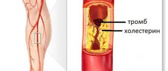
One of the manifestations of urolithiasis is stones in the bladder, the so-called cystolithiasis. Salt or calcified stones form in the bladder cavity when minerals coalesce into small, hard structures. A prerequisite for such a process is a violation of the urine output, which, as a result of stagnant processes, becomes concentrated and leads to the crystallization of individual elements.
The formed stones can become fixed on the wall of the bladder or urethra, causing unpleasant consequences or growing asymptomatically. While they are small in size, they are able to come out with urine, causing pain, urination disorder, hematuria and other disorders.
According to the international classification ICD-10, the formation of stones in the lower urinary tract is coded N21. This disease is diagnosed using ultrasound of the genitourinary system, general urine tests, radiography, cytoscopy and other methods. Therapy is carried out conservatively if the sand or stones in the bladder are not too large and can come out on their own. Surgery is performed for large formations and complications of the condition.
Classification of bladder cameas
In medicine, stones in the bladder are classified according to several criteria.
Age category. The older the patient, the more uric acid the stones contain; in children, stones contain predominantly uric acid in crystals, calcium oxalates and phosphates. Quantity. You can diagnose one stone (single) or several (multiple). Size. There are stones that are very small and also reach the size of the bladder. Structure. Stones can be soft or hard. Surface. There are stones that are smooth, similar in shape to pebbles, and more dangerous - with sharp spikes.
Types of stones
Stones and sand in the bladder are particles that vary in color, size, shape, density and composition.
Particles measuring 1–3 millimeters are considered small. They are called "sand". Medium - from 4 to 9 mm, large - from 10 mm or more.
The shape can be round, oval, triangular, prickly, with uneven edges, or needle-shaped.
The density and color of urinary stones depend on the organic acids and minerals from which they are composed. Find out what they are in chemical composition and how to distinguish them. Stones are:
- Oxalate (consist of a calcium compound and oxaloacetic acid). They have a high density, black or dark brown color, and different shapes. Uneven stones pass with pain through the urethra;
- Urates (they are salts of uric acid). Low density, round or oval, red in color;
- Phosphate (from calcium and phosphoric acid). They have a soft structure, different shapes, and a rough surface. Gray color. Grow and dissolve quickly;
- Cystine (consist of the amino acid cystine and its compounds with minerals). Round in shape, with a smooth surface, white or yellow, not hard;
- Struvite (from ammonium, magnesium, calcium salts). They appear as a result of the activity of certain bacteria that inhabit the kidneys. Soft, white-yellow or gray, give scanty symptoms.
Are there different types of stones in one patient? Undoubtedly! More than half of patients with urolithiasis have a mixed composition of stones. The nature of movement along the urethra depends on their shape and size.
Reasons for education
Infravesical obstruction. Under such a complex name lies a pathology due to which the bladder does not perform its functions. As a result, the natural outflow of urine is disrupted, its concentration increases, which contributes to the formation of salt crystals. In the future, they are transformed into stones. As a rule, this reason for the development of pathology is typical for older people. Disrupted connections between the bladder and the central nervous system. According to statistics, in the case of spinal cord injury, as well as neurogenic bladder, stones form within eight years. Inflammatory processes. Any inflammatory processes affecting the bladder contribute to the development of urolithiasis; patients undergoing radiation therapy are at risk. Presence of foreign bodies in the bladder. These could be catheters, stents, sutures, or antifertility agents that have entered the bladder. Various defects. For example, pathologies of the muscular membrane, protrusion of the mucous membrane, prolapse of the bladder (typical for women). Surgical interventions, mainly involving tissue transfer. Stones that formed in the kidneys, but for certain reasons penetrated into the bladder.
Urine leakage caused by metabolic disorders is not the cause of bladder stones.
Treatment for bladder stones
Depending on the characteristics of the stone, management tactics are selected.
Conservative drug therapy
Important
If the size of the bladder stone does not exceed 5–6 mm, the surface is smooth, there is no evidence of an acute inflammatory process, and there is a high probability of spontaneous passage.
Medicines to get rid of bladder stones
Antispasmodics:
- No-shpa;
- Baralgin;
- Papaverine;
- Spazgan.
Uroseptics:
- Palin;
- Furomag;
- 5 – NOC;
Alpha1-blockers:
- Omnic (Tamsulosin);
- Urorek (Silodosin);
- Doxazosin.
Herbal preparations:
- Cyston;
- Spilled;
- Phytolysin;
- Rowatinex;
- Hydrangea;
- Canephron;
- Urolesan.
Decoctions of diuretic herbs that can dissolve stones:
- Pol-pol (Erva woolly);
- Rosehip root;
- Madder;
- Golden rod;
- Lingonberry leaf.
Important
Herbs for the prevention of stone formation can be brewed and drunk 10 days every month, so that there is no addictive effect, it is better to alternate them.
General principles of nutrition for bladder stones
Additionally, some dietary restrictions are recommended. Of course, it is better to know the composition of the stone, but if this is impossible for some reason, then the following should be excluded:
rich meat, chicken, fish broths;
- all offal: liver, liver, heart, tongue, stomachs, lung, etc.;
- sausages and salami;
- hot spices;
- shashlik;
- chocolate;
- coffee;
- aspic;
- mayonnaise;
- smoked meats and marinades;
- sweet carbonated drinks;
- juices with irritating effects: tomato, orange, grapefruit;
- salty fish.
You can eat second-cooking broths, lean meat, fish and chicken if you have a bladder stone.
An increased drinking regime is considered a prerequisite. If you do not have hypertension, you need to drink at least 2 liters per day.
It is not recommended to subject food to aggressive processing methods. To reduce the load on the kidneys, a diet for urolithiasis involves limiting table salt.
Surgical treatment
If conservative therapy is ineffective, severe pain, or recurrent inflammation, surgical treatment is possible.
Crushing bladder stones with ultrasound or laser (transurethral cystolithotripsy) is currently considered the most effective and safe surgical intervention for urolithiasis.
Stones with a crystalline structure and high density are suitable for crushing.
The size of the stone should not exceed 3 cm.
Transurethral lithotripsia can also be performed in combination with TUR (transurethral resection), if there are indications for it, for example, prostate hyperplasia.
Contraindications:
- pregnancy;
- acute inflammatory process accompanied by fever;
- small bladder capacity;
- fixation of the stone on a ligature;
- stone in the diverticulum;
- stone volume is more than 4 cm;
- concomitant bladder cancer;
- difficult access.
If endoscopic methods have contraindications, open surgery is performed in the amount of cystolithotomy . An incision is made on the front wall of the abdomen, and the stone is removed from the bladder cavity.
Complications after surgery for bladder stones can include bleeding, acute inflammation, trauma to the walls, and acute urinary retention.
After discharge, the patient should be observed by a urologist.
A control ultrasound examination of the kidneys and bladder is performed once a year.
If all recommendations are followed, the prognosis after removal of a bladder stone is favorable. If the cause of cystolithiasis has not been identified and eliminated, recurrent stone formation cannot be ruled out.
The patient is recommended to follow a diet taking into account the chemical composition of the stone and take herbal diuretics for preventive purposes.
Victoria Mishina, urologist, medical columnist
8, total, today
( 22 votes, average: 4.27 out of 5)
Hypospadias: types, causes, treatment
Addison's disease: symptoms and treatment
Related Posts
Symptoms of the presence of stones in the bladder
As a rule, urolithiasis has specific symptoms, however, sometimes the patient does not even suspect the presence of a stone in the body. That is why it is necessary to use special medical equipment to find out the exact picture and determine the correct diagnosis.
Most often, when there are stones in the bladder, patients experience the following symptoms:
pain in the lower abdomen, which spreads to the pubic and groin areas; frequent and sudden urge to urinate, especially at night; cramps and pain when urinating; presence of blood in the urine.
In some cases, the following symptom is possible: a sudden cessation of urination and the appearance of acute pain that affects the genital area, lower back, abdomen and thighs. Similar discomfort can occur during physical activity. In children, a symptom of the presence of stones in the bladder is urinary incontinence and uncontrolled erection, accompanied by pain.
Pain in the lower abdomen is a fairly common symptom of many diseases in women, from kidney disease to diseases of the reproductive system. A woman’s bladder hurts - what could be the reasons for this condition? Diagnosis of diseases, treatment and possible complications.
Read this article to learn how to recognize the symptoms of kidney disease.
Kidney ultrasound is the most common method for detecting diseases. It is with this method that a complete examination of the patient begins. Read here https://mkb2.ru/lechenie/uzi-pochek-kak-delayut.html about how the study is carried out and what indications for its conduct exist.
What types of stones are there?
There are many classifications of stones in the urinary tract. Thus, they can be single or multiple, large (more than 3-7 mm) and small (microlites), have a different structure and surface. The necessary measures, traditional therapy or surgery are prescribed based on the chemical composition of the stones:
- Oxalates have a brown color and a rough, convex surface that scratches the mucous membranes. They are formed from oxalic acid salts in the calyces or pelvis of the kidney and pass through the urinary tract, leading to hematuria.
- Phosphates are formed from calcium salts, but have a soft structure and are easily crushed. They appear as light flakes in the urine and pain in the lower abdomen.
- Urates are not traumatic to the mucous membrane due to their smooth surface; they occur against the background of gout and dehydration.
- Struvite consists of magnesium ammonium phosphate, is formed under the influence of bacteria and indicates the presence of a chronic infection of the genitourinary system.
- Cystone stones form in the form of hexagons due to congenital renal anomalies and metabolic changes. As a result, the amino acid cystine ends up in the urine, leading to the development of cystinuria.
- Mixed stones have high hardness and a multi-layer pattern. The most common calcium formations are calcium oxalates, phosphates and calcium hydroxyphosphates.
By determining the type of stones, the doctor makes a prescription and can first predict the consequences of their treatment. It is also important to know the nature of stone formation:
- primary ones are formed in the bladder due to stagnation of urine;
- secondary ones form in the kidneys and ureters, and then migrate through the genitourinary organs.
For therapy to be effective, you need to know which pathologies should be targeted. Thus, when secondary stones form, additional examination of the kidneys may be necessary.
Diagnostics
An accurate diagnosis can be made already at the initial stages of pathology development. To diagnose urolithiasis, the following examinations and tests are necessary.
General urine analysis. General and biochemical blood tests, during which inflammatory processes in the bladder and any changes in the composition of the blood can be detected. Ultrasound examination of the kidneys to determine the degree of anatomical changes caused by the stone. Excretory urography - a contrast solution is injected into the patient’s vein and then an X-ray of the urinary system is taken. During the diagnosis, the main parameters of stones and their location in the body are determined. However, it should be taken into account that not all stones are capable of transmitting x-rays; such stones will not be visible on x-rays. Radioisotope nephroscintigraphy – a special medical solution is injected into the patient’s vein, which is subsequently excreted through the kidneys. Then specialists scan the urinary system using medical equipment.
Ultrasound - large stone in the bladder
Possible complications
If there is no treatment for urolithiasis, it can cause a chronic form of dysuria - painful and frequent emptying of the bladder, but if the stone completely blocks the canal, then urine output stops completely, and the patient begins to suffer severe pain. In addition, when a bacterial infection occurs, inflammatory processes begin, which provoke urethritis and cystitis.
A severe complication of urolithiasis is considered to be a failure in the functionality of the kidneys; this phenomenon often leads to the development of an abscess. As a rule, this ends with surgical intervention, up to complete removal of the organ. Renal colic develops when the outflow of urine in both kidneys is impaired, this often causes uremia (infection). In this case, the patient's condition deteriorates sharply and becomes critical.
Important! Uremia can be fatal, which is why the treatment of bladder stones must be taken seriously. It is necessary to prevent such a complication in a timely manner.
Treatment
Conservative treatment. It is prescribed when the size of the stones is less than 3 millimeters. In this case, the patient is offered drug therapy and nutritional therapy. The main goal of drug treatment is to dissolve stones and eliminate an acute attack of the disease. To combat pain, drugs such as No-shpa, Baralgin, Papaverine, Spazmalgon are prescribed. Medicines are presented in a wide range in any pharmacy. The drugs act on the walls of the ureter, relaxing it and thereby activating the mobility of the stone. However, antispasmodics can only eliminate pain, but cannot relieve the patient of the main cause of the disease - the stone. Removing stones using a cystoscope. In this case, a special metal tube equipped with optics is inserted into the patient's urethra. The bladder and ureteral orifices are examined. Then a tube, a stent, is inserted into the opening of the ureter, where the pathology is found, which resumes the natural outflow of urine. Surgical intervention. This is the most radical method of treating urolithiasis. Surgery is necessary when the stone grows to a large size. As for the incision, it is performed in the place where the stone is diagnosed. After removing the stone, specialists drain the area to remove urine that leaks through the bladder wall.
In addition, the procedure for crushing stones - remote wave lithotripsy - is also considered an operative method of treatment. During the manipulation process, the stones are crushed and then removed.
When choosing a drug of the same name in different dosage forms, it is better to give preference to injections, since intravenous and intramuscular administration of the drug is more effective.
Rehabilitation period
After surgery, patients need bed rest for several days. The patient remains in hospital treatment as he needs daily dressings and wound treatment.
In the future, the patient needs to undergo a course of treatment with mud and mineral waters. The best resorts for this purpose are Truskavets and Morshyn.
Symptoms of sand and kidney stones
The first symptoms of microliths in the kidneys are a change in the composition and appearance of urine: the color changes, the density increases. In the presence of sand, cloudy urine is observed.
More often, the pathology does not make itself felt, and stones are discovered by chance on an ultrasound of the kidneys or when they move, then unbearable pain occurs, which can be accompanied by nausea and vomiting.
Microlithiasis of the right kidney is more common. The symptoms and localization of pain are similar to appendicitis, because it is observed in the lower abdomen. Sometimes it seems that the liver or gall bladder hurts.
Left-handed
Microlithiasis of the left kidney begins to worry when the stones have increased to several millimeters. Pain - as with lumbar myositis or intestinal diseases. It intensifies with changes in body position.
The formation of microliths in both kidneys is extremely dangerous: it poses a threat of acute renal failure. It is rare, mainly in older people, and is a sign of metabolic disorders.
Initially, the disease is asymptomatic; nothing indicates its development. Periodically, a feeling of discomfort may occur in the lumbar spine, to which the patient does not pay attention. He is driven to see a doctor by acute, incessant pain that occurs after active physical activity. At this stage, the disease is already advanced.
• Increased body temperature not associated with colds, reaching high levels;
• Elevated blood pressure not associated with hypertension;
• Piercing and sharp pain that occurs when moving;
• Aching pain radiating to the abdomen, groin, lower back, and inner thigh.
Nausea and vomiting are also possible with this disease. The pain syndrome persists from several minutes to several days. It always starts abruptly, and ends as unexpectedly as it began.
For each patient, the attack proceeds differently: someone behaves restlessly, rushes about, groans, cannot find a place for himself due to incessant pain; the other lies motionless, slightly bent, not moving.
Symptoms of microlithiasis in the initial stages practically do not manifest themselves. Because of this, most patients seek help from a doctor when the disease has become advanced. Microlithiasis of both kidneys often provokes the development of salt diathesis. And the treatment of the latter disease is a rather complex process.
Symptoms of microlithiasis manifest themselves as:
- Pain syndrome. It is usually localized in the lumbar region, but can radiate to the perineum and lower abdomen. Pain often occurs when walking. In this case, it is noted in the sacral and lumbar regions.
- High pressure.
- Frequent urination. With renal microlithiasis, urine becomes dark in color. There may be small blood clots in it.
- Edema affecting various parts of the body.
- Deterioration in health, weakness during physical activity.
Treatment options depend on the chemical composition of the salt deposits:
- Urats. They are crystals obtained from uric acid salts.
- Oxalates. Appears when there is an excess content of oxalic acid.
- Phosphates. They are formed when the permissible norm of phosphate acid is exceeded.
Symptoms of the pathology vary depending on the location; microliths have formed in the right or left kidney. Microlithiasis of the left kidney has a more vivid clinical picture, which facilitates diagnosis and subsequent treatment.
Medical nutrition
Regardless of the location of the stones in the body, doctors prescribe therapeutic nutrition to patients - the so-called table No. 7.
The main principles of such nutrition include the following points:
minimum amount of salt; minimum amount of fatty foods; exclusion of alcohol; exclusion of spices and other concentrated foods.
In addition to therapeutic nutrition, patients are advised to avoid hypothermia and severe physical activity, which significantly increases the risk of developing inflammatory processes in the bladder, weakened by stones.
Prevention
Prevention of pathology involves a set of measures aimed at preventing the appearance of stones in the bladder:
Correction of diet: exclusion of fatty, salty, smoked foods, spices. Maintaining water balance - the daily fluid intake is at least one and a half liters; with sufficient water intake, the patient goes to the toilet at least six times during the day. The last preventive measure primarily concerns people who lead a sedentary lifestyle - it is necessary to play sports.
Diet
Diet in the treatment of bladder stones is no less important than drug treatment. In addition to the fact that it helps improve the patient’s condition, it prevents the development of new stones and prevents the growth of existing ones.
The basic principles of dietary nutrition are as follows:
- Limiting the consumption of dairy products. This is especially important for calcium stones.
- If you have urate stones, you must avoid foods that contain uric acid - meat broths, brains, kidneys and liver. It is also not recommended to overuse fish and vegetable fats, as they can acidify urine.
- For oxalate stones, milk, oranges, spinach, potatoes, and sorrel are not recommended.
- Phosphate stones require a reduction in kefir, cottage cheese, vegetables, and fruits in the diet. In this case, fish, meat, and vegetable oil are recommended.
Absolutely any type of stones requires an increased drinking regime. 10 glasses of water per day will reduce the concentration of urine.
After diagnosing the prescription of therapeutic measures, the doctor will definitely describe in detail the diet, from which it is extremely undesirable to deviate.
Prognosis after stone removal
After completion of treatment, the patient should be regularly monitored by a urologist, undergo a metabolic examination of the kidneys and an ultrasound examination at least once every six months.
If all the causes that provoked the development of the pathology are eliminated, the prognosis is favorable.
But, if the causes of the disease remain unresolved, a relapse is likely - the reappearance of stones in the bladder, which will require repeated treatment.
When immunity declines, the body is susceptible to many diseases. In women in particular, urinary tract infections are common. What causes inflammation and how does infection occur? Read carefully.
Everyone should know this. Read here how to properly take a urine test for different types of studies.