Hemothorax is a subtype of hydrothorax, which involves the accumulation of blood in the pleural cavity. Blood enters the pleural cavity from the bronchial and pulmonary arteries. Blood flows out of damaged vessels until the pressure in the pulmonary vessels is balanced.
Acute total hemothorax often causes death due to blood loss and breakthrough of the blood mass into the abdominal cavity.
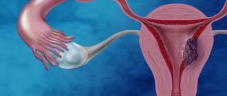
Small hemothorax is dangerous due to the erasure of symptoms, blood infection and chronic organ ischemia.
Causes
The human pleural cavities do not communicate. Hemothorax can only occur on one side, without including the second lung in the process.
Reasons why hemothorax develops on the right or left:
- Malignant tumor in the pleural cavity.
- Pulmonary infarction. Often due to pulmonary embolism.
- Complication after catheterization of the right or left subclavian vein or iatrogenic hemothorax. Often, when the left subclavian vein is damaged, the thoracic lymphatic duct is affected, which leads to chylothorax or the release of lymph into the pleural cavity.
- Traumatic hemothorax due to a direct blow to the chest. Often accompanied by rib fractures, closed or open chest trauma.
Traumatic hemothorax is the reason for contacting and writing a statement to the police, since the injury is classified as grievous bodily harm.
Forecast
With intrapleural bleeding starting from moderate severity, the prognosis can be complex and depends on:
- the severity of the chest lesion that caused the hemothorax;
- rate and duration of blood loss;
- timeliness of diagnostic and therapeutic measures.
The prognosis for bilateral hemothorax is always more difficult. Even if the bleeding is slight, it can become much more intense at any moment. Since both halves of the chest are affected, respiratory decompensation will occur. Also, the severity of the prognosis is aggravated by the coagulated form of hemothorax. The most pessimistic prognosis is for bilateral traumatic coagulated hemothorax with ongoing bleeding. It more often than other types of hemothorax leads to:
- death;
- and if the patient survives - to protracted complications, the relief of which requires more time and more resources both from the patient’s body and from doctors.
The prognosis for life is favorable if the diagnosis and treatment of hemothorax were carried out in the first hours from the moment of its occurrence. After hemothorax, the health prognosis will be favorable if the patient is properly rehabilitated. To avoid late complications (formation of adhesions in the pleural cavity that impair breathing), patients should begin as soon as possible:
- regular swimming lessons;
- race walking;
- performing special breathing exercises.
After suffering a hemothorax, you should expect that the recovery will be long - sometimes it takes at least a year to finally get rid of the consequences of hemothorax.
Kovtonyuk Oksana Vladimirovna, medical observer, surgeon, consultant doctor
20, total, today
( 186 votes, average: 4.60 out of 5)
Review of effective cough remedies for adults
What is an “acute abdomen”: symptoms and first aid
Related Posts
Types of hemothorax
Hemothorax is classified according to the amount of blood loss, so the severity of the injury and the approximate prognosis of the patient can be determined. The volume of blood loss during hemothorax determines whether it is necessary to transfuse blood, albumin, or red blood cells.
Check out the table for the classification of hemothorax according to Kupriyanov.
| View | Volume of blood loss | Localization of blood in the pleural cavity |
| Small | Up to 500 ml | Blood is found exclusively in the pleural sinuses |
| Average | 500-1000 ml | Blood reaches the angle of the scapula |
| Total | From 1000 ml | Blood occupies almost the entire pleural cavity |
ATTENTION! In case histories of hemothorax, it is added whether bleeding into the pleural cavity continues or not.
Based on the location of blood in the pleural cavity, the following types are distinguished:
- apical,
- interlobar,
- paracostal (on the side of the ribs),
- supradiaphragmatic (in the sinuses),
- paramediastinal (near the sternum).
Types of hemothorax are taken into account in the medical history, as well as when choosing a puncture site.
Causes, symptoms and treatment of hemothorax
Hemothorax is an accumulation of blood in a slit-like space, which is located between the visceral and pariental layers of the pleura of the lung.
Complications
Hemothorax can cause complications. The most common of them include the following.
- Lung problems. Blood pressure in the chest can cause the lung to collapse. If this condition progresses, it can lead to respiratory failure.
- Infections. If hemothorax is not treated, it can cause infections of the lungs, pleura, or pleural fluid in the chest cavity.
- Scarring. Pleural membranes and lung tissues with hemothorax are more prone to scarring. Over time, this can lead to fibrosis and immobilization of the ribs.
Clinic
With small hemothorax, the clinical picture is not pronounced. There are no changes in the cardiovascular and respiratory systems. If this happened due to a chest injury, then surgical intervention in the initial stages will not be required. If blood enters the cavity in small portions more than once, the risk of developing posthemorrhagic chronic anemia and iron deficiency increases.
Average hemothorax is characterized by symptoms:
- weakness,
- shortness of breath with difficulty inhaling,
- squeezing chest pain,
- cyanosis or blueness of the skin,
- cold sweat.
The patient has a rapid and weak pulse, rapid and shallow breathing, pale skin and mucous membranes.
A dry cough often occurs after movements, during which the pain intensifies.
Breathing is not audible on auscultation. Similar symptoms develop with closed pneumothorax. It is impossible to clinically distinguish the presence of air or blood in the pleural cavity. This can only be done using x-rays.
During the first 3-6 days, due to the absorption of blood by the pleural layers, the body temperature rises to 38-39 degrees Celsius. Then it gradually decreases. If the temperature persists for more than 10 days, this may indicate infection of the pleura and the onset of systemic inflammatory response syndrome (SIRS).
The clinic can be complicated by pneumothorax - air entering the pleural cavity or cardiac tamponade - blood entering the pericardium. The patient's condition is deteriorating, only surgical treatment and rapid evacuation of blood increase the patient's chances of survival.
Hemothorax: symptoms, diagnosis and treatment
Hemothorax is a condition characterized by the concentration of blood in the space between the visceral (facing the chest) and parietal (in contact with the lung parenchyma) layers of the pleura. Normally, the slit-like area between the inner and outer layers of the pleura contains a small amount of fluid, allowing the sheets to slide freely relative to each other. The tightness of the pleural cavity and the negative pressure created in it keep the lungs in an expanded state. This ensures free transfer of movement from the chest to the lungs. This is how the breathing process occurs. The effusion of blood into the parietal area due to injuries, pathological conditions or surgical interventions disrupts hemodynamics and the respiratory process. The development of hemothorax to an extensive stage can be fatal. Therefore, a patient with signs of a pathological condition requires timely and accurate diagnosis, and as a result, adequate treatment.
This is how the breathing process occurs.
Diagnostics
An observation x-ray is taken. The following signs of hemothorax are detected on x-ray within 5 minutes:
- collapse of the lung (the X-ray shows a decrease in its area due to compression by blood),
- horizontal level of fluid, which is visible as a band of darkness (can be at the level of the sinuses or higher, up to the collarbones),
- shift or flotation of the mediastinum (trachea, heart) to the healthy side.
The darkening on the x-ray is shown as light or white due to the fact that the image itself is negative.
X-ray signs of hemothorax are clearly visible, the study itself is carried out even in paramedic stations and remote villages, and is completed within 5 minutes. This research is cheap.
If the lung is cleared above the horizontal darkening strip, it means that hemothorax is accompanied by pneumothorax, and there is both air and blood in the cavity. The condition is an emergency and is classified as causing grievous harm in the event of injury.
Auxiliary research methods, in addition to x-rays for hemothorax: a general blood test with severe anemia, a decrease in the number of red blood cells, hemoglobin, hematocrit.
During puncture of the pleural cavity: if the resulting blood has clotted, this means that bleeding continues. That is, the Rivilois-Gregoire test is positive. If it does not clot, then the process of fibrinolysis has begun in the pleural cavity, and new blood does not flow. The incoming blood must be pumped out in full.
Signs
With minor hemothorax, the patient is quite active, may feel satisfactory or complain of slight shortness of breath, a feeling of respiratory discomfort, and cough.
With average hemothorax, the clinical picture is more pronounced: a condition of moderate severity, intense shortness of breath, aggravated by physical activity, congestion in the chest, intense cough.
Subtotal and total hemothorax have similar manifestations, varying in severity:
- severe, sometimes extremely serious condition, which is determined by a combination of respiratory failure and hemodynamic disturbances due not only to compression of large vessels of the mediastinum, but also massive blood loss;
- cyanotic staining of the skin and visible mucous membranes;
- severe shortness of breath with slight physical exertion, change in body position, at rest;
- frequent thready pulse;
- severe hypotension;
- chest pain;
- annoying painful cough;
- forced position with an elevated headboard, since suffocation develops in a lying position.
Treatment
Traumatic or hemothorax of another origin requires immediate treatment. Therapy is not always limited to drainage of the pleural cavity. The treatment strategy for hemothorax is to identify and treat the cause, for example, antitumor therapy.
If the hemothorax is small and there is less than 500 ml of blood in the pleural cavity, then no special treatment is required. The liquid will be absorbed by the pleural layers on its own within 7-10 days. The patient will need to have a repeat x-ray to make sure there is no blood and not clotting in the cavity.
If the hemothorax is medium or large, surgical treatment in a hospital is necessary. With this pathology, the patient is treated by a thoracic surgeon. Only he can perform manipulations to drain the pleural cavity; this is not the competence of a paramedic or nurse.
After pumping out the blood using a syringe, it is necessary to rinse the pleural cavity with saline until it becomes slightly pink or colorless. Then the puncture site is re-treated with an antiseptic and a bandage is applied. The dressing is changed once a day. If infection of the contents in the cavity occurs, antibiotics from the cephalosporin group can be administered with saline solution: Ceftriaxone, Cefotaxime or macrolides.
If simple drainages are ineffective, active or passive drainage according to Bulau from the 7th or 8th intercostal space is used.
First aid
For moderate or total hemothorax, pleural puncture is performed. Emergency assistance in the form of drainage of the pleural cavity is provided by a doctor.
If bleeding continues after puncture, as well as when more than 1000 ml of blood is removed through drainage at a time, it is necessary to perform an emergency thoracotomy, dissection of the chest with a search for the bleeding vessel and its ligation, and suturing the wound. Sometimes it is necessary to excise the damaged lung.
As maintenance therapy, venous catheterization and restoration of blood volume are performed. Hemostatic drugs are prescribed: tranexamic or aminocaproic acid, calcium gluconate.
THIS IS INTERESTING! If the patient's blood, which spilled into the pleural cavity less than 1 hour ago, is not infected with microbes, reinfusion can be performed. To determine the infection or purity of blood, Petrov and Efendiyev tests are performed. Anticoagulants are added to uninfected blood and infused intravenously into the patient.
Puncture
Recommendations for surgical tactics during puncture for hydrothorax, including hemothorax or chylothorax:
- puncture site – 6-7 intercostal space,
- puncture line – between the posterior and middle axillary lines,
- injection site, the upper edge of the previous rib to prevent puncture of the intercostal vessels.
- patient's position - lying or sitting.
Treatment of the disease
Modern treatment methods make it possible to quickly eliminate hemothorax. The choice of treatment method depends on the severity of the symptoms, the type of hemorrhage, as well as the reasons that provoked the pathology. Small hemothorax can be eliminated using conservative treatment methods:
- symptomatic treatment is carried out;
- immunocorrection;
- sometimes antibacterial drugs are prescribed;
- disaggregant therapy.
It is important to evacuate the accumulated blood. If the hemorrhage was small, then the human body can cope with it on its own (maximum period - 2 weeks) and there will be no need to use other treatment methods. But throughout this time, the patient should stay in the hospital to eliminate the risk of rebleeding.
If a lot of blood has accumulated, then thoracentesis or drainage of the cavity is performed. Proteolytic enzymes, antibiotics and antiseptics are introduced into the cavity. Full surgical intervention is performed in the case of a clotted hemothorax or if it is not possible to straighten the lung by other means. Urgent surgery is also indicated for damage to large vessels.
What to do?
If you think you have hemothorax
and the symptoms characteristic of this disease, then doctors can help you: a pulmonologist, a surgeon, a therapist.
Source
Did you like the article? Share with friends on social networks:
Complications
Hemothorax involves the release of blood into the pleural cavity. Because of this, pressure on the lungs in the pleural cavity may increase, causing pain in the pleural points.
Then respiratory failure due to the fact that it is difficult for the lungs to expand when inhaling. Shortness of breath occurs during physical activity or at rest.
The blood that is in the cavity can clot, and adhesions begin to form between the layers of the pleura. They arise due to the action of fibrin threads, a blood plasma protein. The condition is called clotted hemothorax or "h. concretus" in Latin, this often occurs when there is little blood output.
The adhesive process develops into limited mobility of the pleura and respiratory failure. Over time, it will be difficult and painful for the patient to inhale and exhale. Less oxygen will enter the tissues, and a state of chronic ischemia .
Regardless of the severity of hemothorax, harm to health is caused by blood infection . If bacteria enter the blood or they begin to multiply, the patient’s temperature rises, pain intensifies and abscesses form . Abscesses worsen respiratory failure, they spread throughout the pleural cavity and cause blood poisoning or sepsis. The final condition is pleural empyema or pleural fibrosis.
CAREFULLY! Less than 1 week may pass from the moment of infection to the moment of generalized sepsis.
Symptoms of hemothorax
Depending on the severity of the bleeding, the amount of blood shed, the displacement of media-building organs and compression of the lung, the symptoms will be more or less pronounced:
- If the hemothorax is small and the blood has not reached the level of the scapula, the victim may complain of pain in the chest area, which intensifies during coughing, as well as mild shortness of breath.
- With hemothorax of large or medium size, the symptoms manifest themselves quite clearly. A person complains, even with calm breathing and a slight cough, of sharp, severe pain in the chest area radiating to the shoulder and back, general weakness occurs, blood pressure drops, and shallow breathing increases. Without treatment, respiratory and cardiovascular disorders continue to worsen; even with little physical activity, the victim experiences severe pain, cannot be in a horizontal position and is forced to take a sitting or semi-sitting position.
- Severe hemothorax is characterized by symptoms such as: tachycardia, dizziness, fainting, cold sweat, severe pain in the chest area, anemia, pale skin.
- If hemothorax is accompanied by a rib fracture, then subcutaneous emphysema, soft tissue hematomas often occur, and hemoptysis appears when a lung ruptures.
- If the spilled blood has coagulated, the person feels severe shortness of breath and unbearable pain in the chest, the lung tissue is subject to sclerotic processes, and respiratory function is impaired.
- With hemothorax, the infected person develops fever with severe chills, lethargy, and general intoxication of the body.
No matter the severity of the hemothorax, there will always be difficulty breathing and pain in the chest area. To diagnose the severity and clarify the diagnosis, an x-ray or computed tomography is performed, which is a more reliable way to detect accumulated blood. In addition, it is possible to additionally identify the cause that caused the hemothorax, for example, to detect a tumor. If necessary, doctors take a puncture of accumulated blood, this allows them to detect bacteria or fungi, as well as the type of cells.
Results
- Hemothorax is an injury that leads to complications: blood infection and pleural empyema, respiratory failure and lung collapse, adhesions, chest resistance.
- According to topographic anatomy, bleeding occurs from the bronchial or pulmonary arteries due to injury, cancer, or against the background of lung collapse.
- There are small medium and large hemothorax according to Kupriyanov, the types of pathological condition are based on the amount of blood loss.
- The clinical picture or symptoms are characterized by weakness, shortness of breath, cold sweat, cough. The patient has difficulty inhaling or exhaling.
- The x-ray shows a horizontal fluid level, mediastinal displacement, and lung collapse.
- The main surgical tactics and emergency care consists of draining the pleural cavity after puncture with a thick needle in the 6-7 intercostal space.
Symptoms
In mild cases, the disease may be asymptomatic
. Percussion reveals a shortening of the resulting sound along the posterior axillary line. Auscultation - decreased excursion of the lungs over the lower parts of the lungs.
With severe hemothorax, symptoms characteristic of internal bleeding appear: tachycardia increases, cyanosis of the skin is visible, cold sweat, pale skin, low blood pressure. The patient notes pain in the side and shortness of breath.
, respiratory failure occurs
. On percussion, a dull sound is heard predominantly over the lower parts of the lungs. Auscultation - weakening of pulmonary sound.