Types of acute leukemia
The international FAB classification differentiates acute leukemia according to the type of tumor cells into two large groups - lymphoblastic and non-lymphoblastic. In turn, they can be divided into several subspecies:
1. Acute lymphoblastic leukemia:
• pre-B-shape
• B-shape
• pre-T-shape
• T-shape
• Other or neither T nor B form
2. Acute non-lymphoblastic or, as they are also called, myeloid leukemia:
• acute myeloblastic, which is characterized by the appearance of a large number of granulocyte precursors
• acute monoblastic and acute myelomonoblastic, which are based on the active reproduction of monoblasts
• acute megakaryoblastic - develops as a result of active proliferation of platelet precursors, the so-called megakaryocytes
• acute erythroblastic, characterized by an increase in the level of erythroblasts
3. Acute undifferentiated leukemia is a separate group
Lymphoblastic leukemia: treatment
Medical management depends on the predominance of early or late lymphoblastic cells. Combination therapy is used, consisting of the following techniques:
- Chemotherapy;
- Use of cytostatics;
- Radiation therapy;
- Radiation exposure;
- Bone marrow transplantation.
The transplant is used after chemo-radiation therapy. For transplantation, both donor and own, purified bone marrow are used.
The treatment regimen consists of two stages: radiation therapy and post-radiation therapy. The purpose of irradiation is to achieve the following results:
- Destruction of leukemia cells;
- Normalization of analyzes;
- Relief of symptoms of the disease.
The efficiency is 40%. Eight out of ten people achieve stable remission. Most cases tend to relapse, requiring repeated irradiation over two to three years. The goal of repeated courses of treatment is to get rid of all lymphoblasts.
Symptoms of acute leukemia
Acute myeloblastic leukemia
Acute myeloblastic leukemia is characterized by a slight enlargement of the spleen, high body temperature and damage to internal organs.
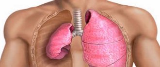
For example, with leukemic pneumonitis, the focus of infiltration and inflammation is in the lungs, causing characteristic symptoms - cough and fever.
Every fourth patient with myeloblastic leukemia experiences leukemic meningitis with severe headaches, fever, chills and neurological symptoms.
At a severe stage of the process, leukemic infiltration of the kidneys may develop, leading to serious renal failure, up to complete urinary retention.
Specific leukemids appear on the skin - pink or light brown formations, the liver enlarges and becomes dense.
When the intestines are damaged, abdominal bloating, stool liquefaction, and severe, unbearable pain appear. In severe cases, ulcers form, and cases of perforation are possible.
Acute lymphoblastic leukemia
This variant of the disease is characterized by an enlargement of the spleen and lymph nodes. As a rule, the pathological process is fixed in the supraclavicular region, first on one side, and then on both. The lymph nodes are dense, painless, and when adjacent organs are compressed, characteristic symptoms may occur.
For example, with enlarged lymph nodes in the mediastinum, a cough may appear, as well as breathing difficulties in the form of shortness of breath. Damage to the mesenteric nodes in the abdominal cavity leads to abdominal pain. Men may experience thickening and pain in the ovaries, most often on one side.
Acute erythromyeloid leukemia
In acute erythromyeloid leukemia, anemic syndrome comes first - a pronounced decrease in the level of red blood cells and hemoglobin, manifested by weakness, pallor and increased fatigue.
Relapses
Recurrence of AL is considered to be the appearance of more than 5% of blast cells in the bone marrow aspirate, regardless of changes in the peripheral blood.
Relapse is also caused by extramedullary lesions. Relapse of acute leukemia is fundamentally different from the debut of the disease, and is considered as the emergence of a new, therapy-resistant clone of leukemic cells. Thus, as a result of tumor progression, secondary resistance develops. Extramedullary relapses are detected in approximately 10% of cases of ALL, much less frequently in acute nonlymphoblastic leukemia (ANLL)
, and most often, blast cells are soon found in the bone marrow [Kovaleva L.G., 1990, Vorobyov A.I., 2002; Appelbaum F. et al, 1998].
There are clinical features of individual variants of OL. The most common symptoms of acute lymphoblastic leukemia are weakness, drowsiness, fever not associated with infection, ossalgia and arthralgia. Infectious complications are uncommon. Sometimes the only complaint, especially in children, is pain in the bones and spine.
Weight loss is rarely significant and is observed only with a long history. In 1% of patients, headache, nausea, vomiting are detected, most often with involvement of the central nervous system [Savchenko V.G. et al., 1998; Volkova M.A., 2001; Bishop JF, 1997].
A physical examination may reveal pallor of the skin, petechial rashes, bruises, bleeding gums, increased body temperature to subfebrile and febrile levels, lymphadenopathy, splenomegaly, hepatomegaly, enlarged kidneys, and painful bones when pounded. In patients with pre-B immunovariant ALL, skin lesions may be detected [Savchenko V.G. et al., 1998; Volkova M.A., 2001].
In 60% of patients with ALL, hyperleukocytosis is detected above 10.0x10⁹/l, of which in 10% of cases it is above 100.0x10⁹/l. In 60% of patients at the time of diagnosis, thrombocytopenia is detected less than 50.0x10⁹/l. In most cases, significant lymphadenopathy and hepatosplenomegaly are detected [Volkova M.A., 2001; Vorobyov A.I., 2002; Burnett AK et al, 1997].
Clinical manifestations of ONLL are very nonspecific. Weakness and malaise may precede diagnosis many months before it is made. Pallor and dizziness can be a manifestation of anemic syndrome. In 15-20% of patients, increased body temperature and sweating are detected.
Often the onset of acute non-lymphoblastic leukemia is associated with sore throat and pneumonia. Various manifestations of hemorrhagic syndrome may be observed. Petechial rashes and ecchymoses are found in 50% of patients. Sometimes the only manifestation of the disease is bleeding. 50% of patients complain of slight weight loss. Ossalgia is detected in 20% of patients.
In 50% of patients, an increase in the size of the liver, spleen, and lymph nodes is diagnosed. Leukemides are detected in 10% of patients. In patients with myelomono- and monoblastic variants of ONLL, gum infiltration is detected. Initial damage to the central nervous system in ONLL is rare [Volkova M.A., 2001; Vorobyov A.I., 2002; Hoeltzer D. et al, 1995].
In 85-90% of cases, blast cells are detected in peripheral blood smears, from 2-3 to 90-95%. Neutropenia, anemia, and thrombocytopenia of varying severity are also diagnosed.
In more than 50% of patients, hyperleukocytosis is noted in peripheral blood smears, however, only in 20% of patients the number of leukocytes exceeds 100.0x10⁹/l [Volkova M.A., 2001]. Hyperleukocytosis in patients with acute non-lymphoblastic leukemia has clinical manifestations - leukostasis in the vessels of the brain leads to the appearance of neurological symptoms, in the vessels of the lungs - to respiratory failure, etc. [Vorobiev A.I., 2002].
Hyperuricemia occurs in 50% of patients with ONLL. In patients with monoblastic and myelomonoblastic AL, severe hypokalemia may be detected due to the high content of lysozyme in the blood serum, aggravating damage to the renal tubules [Chebotarev D.F., Mankovsky N.B., 1982; Vorobyov A.I., 2002].
Stages of acute leukemia
Oncologists distinguish five stages of the disease, which occur with characteristic symptoms:
Initial stage of acute leukemia
This period often occurs latently, without pronounced clinical manifestations. Lasts from several months to several years - at this time the pathological process is just beginning, the level of leukocytes changes slightly (and their number can either increase or decrease), immature forms appear and anemia develops.
A blood test at this stage is not as informative as a bone marrow test, which can reveal a large number of blast cells.
Advanced stage of acute leukemia
At this stage, true symptoms of the disease appear, caused by inhibition of hematopoietic processes and the appearance of a large number of immature cells in the peripheral blood.
In the advanced stage of acute leukemia, two variants of the course of the disease can be distinguished:
• The patient feels well, there are no complaints, but the blood test reveals characteristic signs of leukemia
• The patient experiences a significant deterioration in health, but without significant changes in the peripheral blood
Remission
Remission, that is, the period of subsidence of exacerbation, can be complete or incomplete.
Complete remission is characterized by the absence of symptoms of the disease, as well as blast cells in the blood. In the bone marrow, the level of immature cells should not exceed 5%.
In incomplete remission, the patient may feel relieved and feel better, but the level of blast cells in the bone marrow remains elevated.
Relapse
Relapses of acute leukemia can occur directly in the bone marrow, as well as outside it.
Terminal stage
This stage is characterized by a large number of immature leukocytes in the peripheral blood and is accompanied by inhibition of the function of all vital organs. Most often ends in death.
Morphology
Various forms of acute leukemia have stereotypical morphological manifestations: leukemic infiltration of the bone marrow in the form of focal and diffuse infiltrates of cells with large light nuclei containing several nucleoli. The size and outline of the nuclei, as well as the width of the plasma rim, can vary. Blasts make up 10-20% of brain cells. The cytogenetic affiliation of blasts, as a rule, can only be revealed using special research methods - cytochemical and immunohistochemical. Reactions to peroxidase, staining for lipids with Sudan black, PIC reaction, histoenzyme-chemical actions to detect nonspecific esterase, chloroacetate esterase, and acid phosphatase are used. Immunohistochemically it is possible to determine markers of B-, T-lymphocytes, cells of the myeloid and monocyte series.
In the peripheral blood and bone marrow, the phenomenon of “leukemic failure” (“hiatus leucemicus”) is described, developing due to the presence of only blast and differentiated cells and the absence of intermediate forms.
In the bone marrow tissue, normal hematopoietic cells are displaced by tumor cells, reticular fibers are thinned and reabsorbed, and myelofibrosis often develops. With cytostatic therapy, the bone marrow is devastated with the death of blast forms, the number of fat cells increases and connective tissue grows.
Leukemic infiltrates in the form of diffuse or focal accumulations are found in the lymph nodes, spleen and liver. This leads to an increase in the size of these organs. The liver is characterized by the development of fatty degeneration. Leukemic infiltration of the mucous membranes of the oral cavity and tonsil tissue is possible.
Diagnosis of acute leukemia
A doctor may suspect a diagnosis of “acute leukemia” based on changes in a blood test, but the key criterion is an increase in blast cells in the bone marrow.
Changes in peripheral blood tests
In most cases with acute leukemia, patients develop anemia with a sharp decrease in the level of red blood cells and hemoglobin. There is also a drop in platelet levels (restores during remission and falls again during exacerbation of the pathological process).
As for leukocytes, a twofold picture can be observed - both leukopenia, that is, a decrease in the level of leukocytes, and leukocytosis, that is, an increase in the level of these cells. As a rule, pathological blast cells are also present in the blood, but in some cases they may be absent, which does not exclude leukemia.
Leukemia, in which a large number of blast cells are recorded in the blood, is called “leukemic,” and leukemia with the absence of blast cells is called “aleukemic.”
Changes in red bone marrow
Examination of red bone marrow is the most important criterion for diagnosing acute leukemia.
The disease is characterized by a specific picture - an increase in the level of blast cells and inhibition of the formation of red blood cells.
Another important diagnostic technique is bone biopsy. Bone sections are sent for biopsy, which in turn makes it possible to identify blastic hyperplasia of the red bone marrow and confirm the disease.
Criteria for diagnosing acute leukemia:
• 30% or more of all cellular elements of the red bone marrow are blasts
• The level of erythrokaryocytes is more than 50%, blasts - at least 30%
• In acute promyelocytic leukemia, the appearance of specific hypergranular atypical promyelocytes in the bone marrow is noted
Clinical manifestations
Clinical manifestations are the same for all variants of acute leukemia and can be quite polymorphic. The onset of the disease can be sudden or gradual. There is no characteristic onset or any specific clinical signs for them. Only a thorough analysis of the clinical picture makes it possible to recognize a more serious disease hidden under the guise of a “banal” disease.
A combination of bone marrow failure syndromes and signs of a specific lesion is characteristic.
Due to leukemic infiltration of the mucous membranes of the oral cavity and tonsil tissue, necrotizing gingivitis and tonsillitis (necrotizing tonsillitis) appear. Sometimes a secondary infection occurs and sepsis develops, leading to death.
The severity of the patient's condition may be due to severe intoxication, hemorrhagic syndrome, respiratory failure (due to compression of the respiratory tract by enlarged intrathoracic lymph nodes).
The use of active cytostatic therapy influenced the course of acute leukemia, that is, it led to drug-induced pathomorphosis. In this regard, the following clinical stages of the disease are currently distinguished:
- first attack
- remission (complete or incomplete),
- relapse (first, repeated).
Bone marrow failure
It manifests itself in the form of infectious complications, disseminated intravascular coagulation syndrome, hemorrhagic and anemic syndromes.
Development of infectious complications
occurs due to immunodeficiency caused by dysfunction of leukocytes. Most often, infectious complications are of bacterial origin; fungal and viral infections are less common. A sore throat, gingivitis, stomatitis, osteomyelitis of the maxillofacial area, pneumonia, bronchitis, abscesses, phlegmon, and sepsis may develop.
Hemorrhagic syndrome
in acute leukemia it is caused by thrombocytopenia, damage to the liver and vascular walls. It manifests itself as hemorrhagic diathesis of the petechial-spot type. “Bruises” and small petechiae appear on the skin and mucous membranes. The appearance of hemorrhages is easily provoked by the most insignificant influences - friction of clothing, slight bruises. Nosebleeds, bleeding from the gums, metrorrhagia, and bleeding from the urinary tract may occur. Hemorrhagic syndrome can lead to very dangerous complications - brain hemorrhages and gastrointestinal bleeding.
Anemic syndrome
manifests itself in the form of pallor, shortness of breath, rapid heartbeat, and drowsiness.
DIC syndrome
most often occurs in promyelocytic leukemia.
Specific lesion
Signs of intoxication are noted: weight loss, fever, weakness, sweating, loss of appetite.
Infiltration of the gums by leukemic cells may be observed, while the gums are hyperplastic, hang over the teeth, and are hyperemic.
Proliferative syndrome
may manifest itself as an increase in the size of the lymph nodes (lymphadenopathy), spleen, and liver. In some cases, leukemids appear on the skin - formations of soft or dense consistency that rise above the surface of the skin. Their color can match the color of the skin or be light brown, yellow, or pink.
Damage to the central nervous system ( neuroleukemia
) occurs especially often in ALL and significantly worsens the prognosis. Metastasis of leukemia cells occurs into the membranes of the brain and spinal cord or into the substance of the brain. Clinically, manifestations of varying severity are possible - from headaches to severe focal lesions.
The manifestation of acute leukemia can be sudden or subtle.
Treatment of acute leukemia
The treatment regimen for acute leukemia depends on the age and condition of the patient, the type and stage of development of the disease, and is always calculated individually in each specific case.
There are two main types of therapy for acute leukemia - chemotherapy and surgical treatment - bone marrow transplantation.
Chemotherapy consists of two successive stages:
• The goal of the first stage is to induce remission. With the help of chemotherapy, oncologists achieve a decrease in the level of blast cells
• Consolidation stage necessary to destroy remaining cancer cells
• Reinduction, as a rule, completely repeats the scheme (drugs, dosages, frequency of administration) of the first stage
• In addition to chemotherapy drugs, the general treatment regimen contains cytostatics.
According to statistics, the total duration of chemotherapy treatment for acute leukemia is about 2 years.
Chemotherapy in combination with cytostatics is an aggressive method of exposure, causing a number of side effects (nausea, vomiting, deterioration of health, hair loss, etc.). In order to alleviate the patient's condition, concomitant therapy is prescribed. In addition, depending on the condition, antibiotics, detoxification agents, platelet and red blood cell mass, and blood transfusions may be recommended.
Bone marrow transplantation
Bone marrow transplantation provides the patient with healthy stem cells, which later become the ancestors of normal blood cells.
The most important condition for transplantation is complete remission of the disease. It is important that the bone marrow, cleared of blast cells, is refilled with healthy cells.
In order to prepare the patient for surgery, special immunosuppressive therapy is carried out. This is necessary to destroy leukemia cells and suppress the body's defenses to reduce the risk of transplant rejection.
Contraindications for bone marrow transplantation:
• Impaired functioning of internal organs
• Acute infectious diseases
• Relapse of acute leukemia that does not go into remission
• Elderly age
Lymphoblastic leukemia: causes
The exact etiology is unknown. It is believed that the disease appears as a result of a combination of many predisposing factors: heredity, polluted environment.
Malignant degeneration of leukemia cells begins in the prenatal period. In childhood beta leukemia in young children, the mutation of cancer cells is completed before birth. The development of other types of leukemia requires additional predisposing factors.
They are as follows:
- Ionizing radiation;
- The result of an abnormal immune response to infection. Some viruses cause spontaneous disturbances in the formation of blood cells;
- Hereditary congenital diseases may carry a predisposition to leukemia.
- It develops more often in people with white skin color;
The cell type is determined using immunophenotyping. Depending on the markers, several types of lymphoblasts are divided. They correspond to the stages of leukemia development. The strategy for choosing treatment also depends on the predominance of pro- or prelymphoblasts (early and late cells). The lower the blast cell concentration, the better the prognosis.
How does it manifest?
Acute leukemia in adults and children does not differ in symptoms. The first signs begin to appear at the initial stage of development, but they show a vague character, so it is difficult to recognize the disease. A person experiences anemia due to a decrease in the level of red blood cells. Specific symptoms in adults and children manifest themselves in the same way; the disease in the early phases is characterized by the following conditions:
- sudden weight loss;
- severe fatigue and lethargy;
- aching joints;
- enlarged lymph nodes;
- constant drowsiness;
- heaviness in the stomach;
- pressing pain in the left hypochondrium;
- increased sweating;
- slight increase in temperature.
Symptoms increase with each subsequent stage and depend on the level of immature cancer cells. They significantly reduce the level of the body's immune defense. Thus, acute leukemia provokes frequent infectious diseases, which significantly worsens the patient’s general condition. With the development of pathology, nonspecific symptoms also appear:
- pale skin;
- nosebleeds;
- frequent vomiting;
- convulsions;
- dyspnea;
- swelling of the hands.
Return to contents
Myeloid leukemia: symptoms
The predominant clinical picture is damage to one or another organ system. The main symptoms of myeloblastic leukemia in the acute period are as follows:
- Paleness and sweating;
- hyperthermia up to 40 degrees;
- Severe weakness;
- Body pain;
- Enlarged liver;
- Swelling of the skin;
- Abdominal pain, enlarged liver;
- Lymph nodes are swollen;
- Unreasonable bleeding appears;
- Neurological disorders;
- Frequent infections and fungal infections.
The first to appear are dysfunctions of organs that have a good blood supply. This is due to the entry of a large number of cancer cells with the blood.
Leukemia in children: classification
Forms of leukemia:
- Acute (up to two years);
- Chronic (more than two years).
The rapid growth of blast cells in geometric progression leads to the need for complete destruction of atypical cells. The clonal (reproduction of one cell) theory of the pathogenesis (development) of leukemia is based on this principle. Mutating cells quickly replace normal hematopoietic germs. Therefore, in order to cure leukemia it is necessary to destroy all pathological foci of hematopoiesis.


