Content
- 1 Etiology
- 2 Classification 2.1 By etiology
- 2.2 By the nature of the inflammatory process
- 2.3 According to clinical signs
- 3.1 Acute nonspecific tenosynovitis
Diagnostics
The diagnosis of Achilles tendon tenosynovitis is made on the basis of patient complaints, anamnestic data, and the results of a physical examination. Instrumental and laboratory methods can be used as auxiliary methods, but, as a rule, in the diagnosis of the described pathology they are dispensed with.
During physical examination the following is carried out:
- upon examination - assessment of the motor activity of the ankle joint, the contours of the Achilles tendon, the condition of the skin over it;
- upon palpation (palpation) - identification of pain and swelling.
Of the instrumental methods, puncture may be informative if the development of a purulent process is suspected. In this case, the aspirated (sucked out) contents are sent to the laboratory for testing.
Laboratory research methods that are used in the diagnosis of the described pathology are as follows:
- general blood test - an increased number of leukocytes and ESR indicate the development of an inflammatory process; a further increase in these indicators may signal suppuration;
- bacterioscopic examination - if there is a punctate, it is examined under a microscope, the pathogen that led to the occurrence of the described pathology is identified;
- bacteriological examination - the punctate is sown on nutrient media, the species of microorganisms that appear is determined from the colonies that appear, and sensitivity to antibiotics is also determined.
Etiology
Independent aseptic tendovaginitis (so-called professional tendovaginitis) occurs as a result of prolonged microtraumatization and overstrain of the tendon sheaths in workers of certain professions (loaders, typists, pianists, heavy industry workers and others), who for a long time perform the same type of movements involving a limited group of muscles; Similar tendovaginitis can develop in representatives of some sports (skiers, speed skaters and others) during overtraining. The cause of nonspecific tendovaginitis is the spread of the inflammatory process to the synovial sheath of the tendon from a purulent focus, which can be osteomyelitis, purulent arthritis, felon. Specific tendovaginitis develops in some infectious diseases (gonorrhea, brucellosis, tuberculosis), and hematogenous spread of pathogens is more often observed. Rheumatic diseases (rheumatoid arthritis, rheumatism, systemic scleroderma, ankylosing spondylitis, Reiter's syndrome) can also cause the development of tendovaginitis due to reactive inflammation of a toxic nature [1].
Treatment of tendovaginitis
Therapeutic measures begin with ensuring complete rest of the limb. This can be achieved through strict adherence to bed rest or immobilization with rigid fixation orthopedic products.
Acute forms of tenosynovitis require cooling of the inflamed surface; this can be done using frozen foods, a heating pad with cold water, or a hypothermic “Snowball” pack, which can be purchased at a pharmacy. The chronic course is treated with warming procedures in the form of medicinal compresses or ointments.
Drug therapy for tendovaginitis, which your doctor will prescribe, is selected taking into account the clinic of the disease by the attending doctor:
- Non-steroidal anti-inflammatory drugs (Ketaprfen, Diclofenac, Ibuprofen), prescribed in high dosages for a long period.
- Colchicine or Indomethacin is used if the pathology is caused by gout.
- For severe pain that is not relieved by NSAIDs, glucocorticosteroids (Betamethasone, Triamcinolone) are administered into the cavity of the inflamed tendon. This procedure is carried out according to strict indications, since the procedure can lead to tendon rupture.
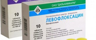
- Antibiotics (Ampicillin, Omoxicillin) are used for infectious forms of inflammation to combat pathogenic microbes.
- Specific treatment may be required for lung damage with Koch's bacillus or sexually transmitted infections.
Surgical treatment of tendovaginitis may be required for persistent pain and limited movement, most often in the shoulder joint. During the operation, scar tissue is excised and the tendon is then sutured. During the rehabilitation period, physical therapy sessions are indicated to restore the functioning of the tendon.
Conservative treatment of tendovaginitis is complemented by a course of massage, UHF, and ultrasound treatment. Particular importance is given to swimming and performing a special set of exercises in water, which is compiled by a medical specialist taking into account the stage of the disease and the functional state of the patient.
Therapeutic exercise is carried out taking into account the therapeutic load on the diseased limb. The set of exercises is constantly changing to increase the load on the tendon. Correct dosing of the intensity of movements determines the speed of recovery of affected tissues. Excessive efforts may negate all previous treatment.
Clinical picture
The clinical picture of tenosynovitis depends on its form.
Acute nonspecific tenosynovitis
Characterized by an acute onset and rapid appearance of painful swelling at the location of the affected tendon sheaths. Most often, acute tenosynovitis is observed in the tendon sheaths on the dorsum of the hands and feet, less often in the synovial sheaths of the fingers and in the sheaths of the flexor tendons of the fingers. Edema and painful swelling spread from the foot to the shin and from the hand to the forearm, which is also accompanied by limitation of movements; in some cases, flexion contracture of the fingers may develop. In cases where the inflammation is purulent in nature, there is a rapid rise in body temperature, the appearance of chills and the development of regional lymphadenitis and lymphangitis. Purulent tenosynovitis is most often localized in the area of the sheath of the flexor tendons of the hand [1] [2].
Acute aseptic (crepitating) tenosynovitis
Characteristic lesions include synovial sheaths located on the dorsum of the hand, less commonly the foot, and in some cases the intertubercular synovial sheath of the biceps brachii muscle. The disease begins acutely: a swelling quickly appears in the area of the affected tendon, upon palpation of which crepitus is felt. There is limited movement of the finger, as well as pain when moving. Acute aseptic tenosynovitis can develop into a chronic form of the disease[1][2].
Chronic tenosynovitis
Most often observed in the sheaths of the flexor and extensor tendons of the fingers in the area of their retinaculum. The most common clinical picture is chronic tendovaginitis of the common synovial sheath of the flexor muscles of the fingers, which is located in the carpal tunnel - the so-called carpal tunnel syndrome. In this case, a tumor-like painful formation of an elongated shape is determined in the area of the carpal canal, which has an elastic consistency, often takes the shape of an hourglass and moves slightly with movement. Sometimes you can palpate dense formations (“rice bodies”) or determine fluctuations. Characteristic limitation of tendon movements[1][2].
A unique form of chronic tendovaginitis is the so-called stenosing tendovaginitis, or de Quervain’s tenosynovitis [1].
Tuberculous tenosynovitis
Clinically it is characterized by the formation of “rice bodies” along the extensions of the tendon sheaths, which can be determined by palpation [1] [2].
Types and classification
When diagnosing elbow pain, it is important to understand what causes the inflammation. For this purpose, special tests and examinations are carried out, as a result of which the type of disease can be determined. There are several types of tendovaginitis, the difference between which is their cause (type of pathogen), symptoms and recommended treatment:
- due to development - aseptic (non-purulent) and purulent;
- by the nature of the inflammatory process - serous (with liquid non-purulent contents), serous-fibrinous (with a thick effusion without impurities of pus) and purulent;
- by type of course - acute and chronic.
Each of the main types of tenosynovitis should be considered separately. Despite the fact that the pathological process affects the same tissues, their manifestations will differ.
We recommend you read: Inflammation of the elbow tendon
Acute aseptic tenosynovitis develops with a sharp increase in the load on the joint. It can be caused by training, competitions and improper exercise. The painful sensations are acute, intense, affect the performance of the joints, and are accompanied by severe swelling. If you limit the stress on your elbow, the symptoms will subside within a few days. If you do not follow the recommendations of doctors and continue to load the joint, the process can become chronic.
Acute post-traumatic tenosynovitis is an inflammation that occurs as a result of a blow or fall on the elbow. Symptoms occur immediately after injury, and in some cases a hematoma appears. For treatment, you will need to fix the limb with a tight bandage - careless movements can aggravate the situation.
Chronic aseptic tendovaginitis develops as a complication of acute inflammatory processes. There is degeneration of the tendon sheaths, which is manifested by frequent pain in the elbow area. A characteristic symptom is crepitus (crunching) when palpating the damaged area. The treatment is long-term, physical activity must be strictly dosed.
Reactive tendovaginitis is a consequence of systemic diseases that manifest themselves as inflammation of the joints. These include rheumatoid arthritis, ankylosing spondylitis, rheumatism, scleroderma and others. The treatment is complex and consists of measures both against the underlying disease and against pain in the elbow joint.
Acute nonspecific infectious tenosynovitis is a purulent inflammation that develops as a result of bacterial microflora entering either from the outside (during open damage to the joint) or through the bloodstream from any part of the body. An abscess forms in the area, which causes acute throbbing pain. The treatment is complex, its main stage is treatment with antibiotics.
Specific infectious inflammation of the tendon sheaths is one of the stages in the development of dangerous systemic diseases. These include tuberculosis, brucellosis, gonorrhea and other diseases. Tenosynovitis is accompanied by other characteristic symptoms, and treatment is aimed at eliminating the main pathogen.
There are both basic and mixed types of tenosynovitis of the elbow joint. In addition, in the absence of timely treatment, safe aseptic forms can develop into complicated purulent inflammation.
Notes
- ↑ 1 2 3 4 5 6 7 8 9 Skripnichenko D. F.
Tenosynovitis // Great Medical Encyclopedia: In 30 volumes / Chief editor B. V. Petrovsky. — 3rd edition. - M.: Soviet Encyclopedia, 1985. - T. 24. Vascular suture - Teniosis. - pp. 539-540. — 544 p. — 150,800 copies. - ↑ 1 2 3 4 Kletkin M. E.
Tenosynovitis.
Medical j
(2013). Retrieved October 8, 2013.
Forecast
The prognosis for tendovaginitis of the Achilles tendon is generally favorable - the described treatment of the disease is effective.
The prognosis may worsen in advanced conditions, as well as in complicated cases, against the background of which scarring of the tendon often occurs, followed by its functional failure.
Kovtonyuk Oksana Vladimirovna, medical observer, surgeon, consultant doctor
7, total, today
( 37 votes, average: 4.59 out of 5)
Fracture of the nasal bones: causes, symptoms, treatment
Pain in the neck and lower back due to osteochondrosis
Related Posts
Reasons for the development of inflammation
Sometimes the cause of the development of tenosynovitis is purulent diseases or wounds of surrounding tissues, through which microbes penetrate into the vagina.
But in some cases, the infection has nothing to do with the development of the disease, and the cause may be the load on the tendons, for example, in musicians, typists, athletes and others who are forced to constantly use their hands. Tenosynovitis can occur as a result of severe trauma. There are primary and secondary (infectious) diseases. Secondary tenosynovitis is a consequence of an inflammatory disease of infectious etiology, which is extremely rare. The most common is stenosing tenosynovitis or professional one. In addition, another cause of the disease is called varicose veins, which manifests itself in connection with degenerative changes in the synovial membrane of the tendon sheath.
Acute post-traumatic tenosynovitis
Post-traumatic tendovaginitis occurs with sprains and bruises of the wrist joint. The history includes a characteristic injury: a fall on an arm that is sharply bent or straightened at the wrist joint, less commonly a bruise in the wrist area. There is pain and swelling in the affected area.
Immobilization is prescribed using a tight bandage, plaster or plastic splint. On the first day after the injury, cold is applied to the affected area, then thermal procedures are performed and UHF therapy is prescribed. In very rare cases (with significant hemorrhage in the tendon sheath), a puncture is performed to remove accumulated blood.
Symptoms of post-traumatic tendovaginitis completely disappear within a few weeks.
What is tenosynovitis and how to deal with it
Tenosynovitis is an inflammation of the tendon tissue and the sheath that covers it (tendon sheath). Unlike tendonitis (simple inflammation of the tendon), tendovaginitis develops only in certain anatomical places, where the tendons are covered with a sheath: the area of the forearm, wrist joint, hand, ankle joint and foot.
This is a very common disease, especially among women and workers who, due to the nature of their work, are forced to perform the same type of hand movements every day. The consequences of tendon tendonitis can be very serious. If the acute form responds well and quickly to treatment, then the chronic form can lead to dysfunction of the hand, which is why you even have to change your place of work.
According to ICD-10 (International Classification of Diseases, 10th revision), tenosynovitis has the code M65.9.
What is the essence of the disease
A tendon is a dense, non-elastic connective tissue formation that connects muscles and bones or two bone structures. During muscle contractions, these structures move relative to the tissues surrounding them. According to official medical data, the tendon in the area of the wrist joint makes 10 thousand movements or more per day.
With tenosynovitis, the tendon and its synovial membrane become inflamed
This is a colossal load that would lead to damage to the bone of this soft tissue formation and its rupture. But in the area where the tendons come into contact with the surrounding tissues, there are special protective sheaths, sheaths. They consist of 2 synovial membranes.
The inner one fits tightly to the tendon, and the outer one encloses it all in a kind of capsule. Between the two membranes there is a small amount of synovial fluid, which reduces the force of friction and impact. Thus, the tendon is perfectly protected from damage due to friction.
With tenosynovitis, both the tendon itself and its sheath become inflamed. The process can be due to many reasons, including infectious and aseptic. Due to swelling and accumulation of inflammatory fluid inside the tendon sheaths, all movements become difficult, become painful, and the function of the limb suffers.
If the disease is not treated, it can become chronic with the development of specific complications, and in the case of purulent inflammation, the infection can spread to neighboring tissues with the development of phlegmon and sepsis.
Causes of tenosynovitis and its types
Depending on the causes of occurrence, all tendovaginitis can be divided into 2 large groups:
- aseptic,
- infectious.
Aseptic option
Occurs in most cases. It develops mainly in people whose profession or hobby involves multiple similar movements of the hands. At risk are pianists, computer typing operators, tailors, people who work with levers, cooks, etc. The same muscles are involved, respectively, and their tendons.
Such intensive work leads to depletion of synovial fluid reserves, increased friction, microtraumatization of connective tissue and the development of aseptic inflammation. Serous and sometimes hemorrhagic exudate begins to accumulate inside the vagina. The latter changes cause characteristic symptoms: swelling, smoothness of the contours of the anatomical area and pain.
Work that involves constant similar movements most often leads to the development of tenosynovitis.
Infectious variant
It is also called septic or purulent tendovaginitis. Caused by pathological microorganisms entering the tendon coupling. They can penetrate directly from the external environment during injuries and open wounds or be carried through the blood and lymphatic fluid from other foci of infection in the body.
Important! This is a very dangerous condition, since pus from the tendon sheaths can quickly spread to adjacent tissues of not only the hand, but also the forearm, causing the development of phlegmon. Treatment in this case is only surgical; amputation of the limb is not excluded.
All infectious tendovaginitis can be divided into two categories:
- nonspecific, which are caused by nonspecific microbes (staphylococci, streptococci, E. coli);
- specific, which include tuberculosis, syphilitic, gonorrheal and brucellosis variants of pathology.
If in the first case standard antibacterial therapy is used for treatment, then in a specific process, targeted treatment is needed for the infection that is complicated by the development of tendon inflammation.
Acute tenosynovitis
The acute aseptic form develops after overload of a certain area of the body (hand or foot). The most commonly affected muscles are the forearm flexor tendons.
Swelling or slight smoothing of the contours appears in the diseased area, so not all patients pay attention to this. Skin color does not change. Pain appears with active and passive movements of the hand. Its location depends on which tendon is affected.
Most often this is the area of the thumb and wrist joint (damage to the flexor tendons of the 1st and 2nd fingers).
Purulent acute tenosynovitis
Another symptom that may indicate this problem is the appearance of a specific crunching or clicking sound in this area during movements (crepitating tenosynovitis).
In the acute purulent form, pronounced signs of inflammation appear. The sore finger turns red, the skin over it is hot, tense, shiny, and may have a bluish tint. Pain is present not only during movements, but also at rest. Takes on a pulsating or twitching character.
At the same time, signs of general malaise appear:
- reactive inflammation of regional lymph nodes;
- fever;
- general weakness;
- headache;
- lack of appetite.
With the development of purulent complications, the patient’s general condition worsens significantly; signs of inflammation from one finger spread to the entire hand and/or forearm. Septic shock may occur.
Chronic tenosynovitis
It develops only with aseptic lesions. It may have a primary chronic course or be a complication of the acute form of the disease if it is not treated.
As a rule, such patients complain only of pain that occurs when performing certain movements. There is also pain on palpation along the inflamed area, and sometimes crepitus can be detected.
A special clinical form of the chronic version of this disease is stenosing tenosynovitis, or de Quervain's tenosynovitis. With it, the inflamed tendon is compressed in the osteofibrous canal, which leads to constant and quite severe pain. Nerves that pass nearby can also become damaged, leading to complications such as carpal tunnel syndrome.
De Quervain's tenosynovitis often affects women
Treatment of tendovaginitis
Treatment measures primarily depend on the cause of tendovaginitis and can be conservative or surgical.
Conservative therapy
The first step is to limit the load on the affected limb. For this purpose, it is recommended to give up your main activities for 10-14 days.
To limit painful movements, the patient is recommended to wear a special orthosis that fixes the first finger of the hand and the area of the wrist joint.
In the future, it can be worn to prevent relapses when performing the necessary work.
Orthosis for fixation of the wrist joint
A good analgesic effect can also be achieved by applying cold compresses to the inflamed area.
The basis of treatment is the use of medications:
- analgesics and non-steroidal anti-inflammatory drugs to eliminate pain and inflammation (tablets, injections, ointments);
- in case of infectious tenosynovitis, antibiotics are used;
- if the pain does not go away, the doctor can perform a blockade with a local anesthetic and a long-acting glucocorticosteroid in the area of the inflamed tendon;
- Enzyme preparations can also be prescribed to resolve the source of inflammation and prevent the formation of adhesions.
Conservative treatment must be supplemented with physiotherapeutic procedures (shock wave therapy, laser therapy, electrophoresis, phonophoresis, etc.). After eliminating acute pain, therapeutic exercises are prescribed to strengthen muscles and prevent re-inflammation. Also, the course of treatment can be supplemented with proven folk remedies.
Surgery
Surgery for tendovaginitis is prescribed only for complications:
- purulent inflammation that does not respond to antibiotic therapy, or the presence of spread of infection (cellulitis, abscesses);
- stenotic process, when there is constant pain, a person cannot perform his duties because of it;
- development of neurological complications (carpal tunnel syndrome);
- development of finger contractures due to adhesions.
The type of surgical intervention is chosen by the surgeon based on the specific situation. Typically, the tendon sheath is incised and removed, and the tendon is released. If necessary, its plastic surgery is performed. The operation can be either open access or performed using endoscopic microsurgical techniques, which do not require extensive skin incisions.
The prognosis for tendovaginitis is favorable provided that treatment is started on time and its volume is adequate. If the process becomes chronic, dysfunction of the hand or foot may occur, which can only be corrected by surgery.
Source: https://surgicalclinic.ru/tendovaginit
Classification of tenosynovitis
Taking into account the etiological factor, the following are distinguished:
- Aseptic tendovaginitis, which, in turn, can be professional, reactive and post-traumatic.
- Infectious tendovaginitis, which is divided into specific and nonspecific.
Taking into account the nature of the inflammatory process, they distinguish:
- Serous tendovaginitis.
- Serous-fibrinous tendovaginitis.
- Purulent tendovaginitis.
Taking into account the course, acute and chronic tendovaginitis are distinguished.
Chronic aseptic tendovaginitis
It can be primarily chronic or develop after acute aseptic or post-traumatic tendovaginitis. The cause is chronic microtraumatization followed by degeneration of the tendon sheaths. The course is recurrent.
A patient with tenosynovitis complains of pain that worsens with movement. There is usually no swelling. Palpation reveals pain along the tendon and crunching or crepitus during movements.
A special form of chronic aseptic tenosynovitis is stenosing tenosynovitis, in which the tendon is partially blocked in the osteofibrous canal. There are several syndromes caused by stenotic tendovaginitis.
Carpal tunnel syndrome develops when this tunnel, which is located on the palmar surface of the wrist joint, narrows. This compresses the finger flexor tendons and the median nerve. Upon examination, pain is revealed along the tendons and sensory disturbances in the area of the I-III and inner surface of the IV fingers, loss of the ability to make precise and subtle movements and decreased hand strength.
De Quervain's disease is a stenosing tendonitis of the extensor brevis and abductor longus tendons of the first finger, which are compressed in the osteofibrous canal located at the level of the styloid process. There is impaired movement, swelling and pain in the area of the “anatomical snuffbox”.
With stenosing ligamentitis, the first, third and fourth fingers of the hand are most often affected. The disease develops as a result of sclerotic changes in the area of the annular ligaments and is accompanied by some difficulty in straightening the finger - as if at a certain moment it is necessary to overcome some obstacle for further movement.
During the period of exacerbation of tendovaginitis, the limb is immobilized, physiotherapy is prescribed (phonophoresis with hydrocortisone, electrophoresis with potassium iodide and novocaine), and therapy with anti-inflammatory drugs is administered. In case of severe pain, blockades with glucocorticosteroids are performed.
During the recovery period, patients with tendovaginitis are prescribed ozokerite in combination with dosed therapeutic exercises.
If there is no effect of conservative therapy, dissection or excision of the affected tendon sheath is performed.
Prevention of tenosynovitis
Tenosynovitis, the symptoms and treatments for which are described above, can be prevented. In order to reduce the likelihood of developing the disease to a minimum, the following recommendations must be followed:
- distribute workloads evenly; if there are occupational hazards, think about changing jobs;
- Healthy food;
- lead an active lifestyle, walk more, exercise;
- observe the work and rest schedule;
- be regularly examined by a doctor in order to detect possible diseases early;
- observe the rules of hygiene, treat possible skin damage in a timely manner;
- For open or severe wounds, apply an antiseptic bandage to the injury site.
Health to you!
Tenosynovitis: symptoms
The disease is characterized by symptoms of any other inflammation. The main signs of acute tenosynovitis are:
1. Pain. It appears clearly at the site of localization of the inflammatory process and decreases with distance from the lesion. The pain is acute, constant, and when suppuration appears, pulsation occurs. When you try to move the affected tendon, the pain intensifies.
2. Dysfunction.
3. Edema. It develops as a result of the dilation of blood vessels and the flow of blood from them into the lesions. With tendovaginitis, the swelling is usually tense and severe. Sometimes there is even separation of the skin in places of small cracks.
The spread of edema occurs quickly; for example, when the tendons of the upper extremities are damaged, the edema spreads from the fingertips to the entire hand in two hours. Increased swelling in the affected area causes additional tissue compression, which leads to increased pain.
4. Hyperthermia. This is a local increase in temperature, which is caused by a sharp dilation of blood vessels and the influx of warm blood into the lesion.
5. Redness. Appears as a result of vasodilation. At first, redness appears clearly along the contour of the affected tendon capsule, but then quickly begins to expand, occupying more and more new areas of the skin. When you feel the area of redness, you can hear a characteristic light crunch.
6. Residual effects of tendovaginitis. These include contractures that develop after the inflammatory process. The appearance of contractures is facilitated by keeping the injured limb in a gentle position for a long time. In some places, the formation of transverse adhesions is possible.
Treatment of the disease
General and local therapy is used to treat tendovaginitis. The general includes measures to combat infection using antibacterial and immune-strengthening agents. If there is no infection, anti-inflammatory drugs, such as indomethacin, are prescribed. Local treatment includes resting the affected limb through immobilization and compresses.
After acute symptoms have been relieved, physiotherapeutic procedures will be useful - UHF, ultrasound, paraffin, ozokerite, etc., therapeutic exercises, massage. Purulent complications must be opened and the tendon sheath drained. Next, antibiotics are prescribed. Treatment with folk remedies is allowed in mild forms and in cases of intolerance to conservative treatment. Infusions, decoctions, compresses and ointments are used.
To prevent the disease, you should follow some recommendations:
- do not overwork your limbs;
- avoid injuries and strains of tendons;
- treat microtraumas in a timely manner, this also applies to the skin;
- avoid panaritiums;
- monitor hand and foot hygiene;
- personal hygiene is important;
- during work, take breaks for rest and finger exercises;
- the first symptoms are a reason to visit a doctor.
Tendon tendonitis: symptoms, types of crepitus and purulent, treatment
Tenosynovitis is an inflammatory disease.
It affects the tendon tissues and their sheaths. In another way, the sheath covering the tendon is usually called the sheath. It consists of connective tissue and is a kind of soft tunnel.
This is precisely the main difference from tendinitis and tendinosis, in which the pathological process affects only the tendon tissue.
Mechanism of occurrence
The disease does not develop in all tendons, but only in those that have a vagina. Tenosynovitis of the foot, ankle, knee, and hand joints is most often diagnosed.
Tenosynovitis - external manifestations of pathology
Chronic tendovaginitis is often found in people whose activities involve monotonous work. Until tendon tenosynovitis becomes chronic, treatment is easy, but as the pathology progresses, this process becomes more complicated.
Tenosynovitis of the knee joint or other joint develops in the inner layer of the membrane, which produces a special fluid needed to lubricate the tendons. The outer leaf is not involved in the process. Inside, due to inflammation, not normal lubricating fluid is produced, but prostaglandins, which cause pain, swelling and redness of the skin in this area.
Causes of the disease
The etiology of tenosynovitis of the hip joint or other joints is varied. The main factors that can provoke a violation are:
- Past tendon injuries;
- Nonspecific infections that have been present in the body for a long time, but have not caused the disease;
- A specific infectious process accompanied by the presence of an abscess in nearby bones;
- Long-term exposure to tendon microtraumas;
- Systemic pathologies of the body.
On a note!
Any injury reduces the protective forces of joints and tendons, so you should consult a doctor.
Infection can also enter the synovium and tendon structure through the bloodstream. This route of infection is called hematogenous. Any past pathologies, after which the infectious agent remains in the body, can cause the development of tenosynovitis.
The disease can be triggered by other inflammatory diseases. Sometimes the cause is rheumatism or rheumatoid arthritis.
Reasons for the development of tenosynovitis
Classification of the disease
The ICD-10 code for tenosynovitis is as follows: M65.2, M75.2-3, M76.0-76.7. The doctor enters the exact diagnosis into the medical record after the examination.
Classification involves dividing the disease into types, depending on the causes, the nature of the inflammatory process and the duration of the course. Depending on the cause, the following categories of disease are distinguished:
- Infectious type;
- Aseptic.
Crepitant tendovaginitis refers to the aseptic form. Accompanied by serous-hemorrhagic inflammation, pus accumulates. Based on the nature of the flow, the following forms are distinguished:
- Purulent;
- Serous;
- Serous-fibrous.
The purulent form of tenosynovitis is the most dangerous. A severe infection process begins inside the tendon. An accumulation of pus begins.
In the serous form of inflammation of the tendon sheath, the pathological process covers the inner sheet of the synovial membrane of the tissue. Serous fluid is released.
In serous-fibrous forms, fibrin plaque forms on the sheets of the membrane. Because of this, tendon friction increases, which increases inflammation and unpleasant symptoms.
Tenosynovitis can be acute, subacute and chronic. The acute form lasts for one month; in subacute cases, symptoms persist for up to six months. Chronic tendovaginitis is a form of the disease that lasts more than 6 months.
Symptoms of tenosynovitis
The sooner a person pays attention to signs of damage to the tendon sheath, the faster the condition can be improved. Symptoms depend on which tendon is involved in the inflammatory process and in what form the disease occurs.
Features of the acute form of pathology
Acute tendovaginitis of the Achilles tendon develops against the background of an injury. The color of the skin over the inflamed tissues does not change; slight swelling appears. Pain occurs only when performing active movements. At rest, there are often no symptoms.
If the pathology begins to progress, the symptoms become brighter. The symptoms are especially noticeable in the purulent form of the disease. The following manifestations occur:
- The skin turns red;
- Local temperature rises;
- The skin becomes stretched from swelling and shines;
- The pain is disturbing even at rest.
Less commonly, patients note a general deterioration in health, loss of appetite and weakness. Sometimes the lymph nodes become enlarged.
Acute course of tenosynovitis
Features of the chronic form
A chronic course is possible with aseptic tendovaginitis. The pain is not severe, it occurs locally at the site of inflammation. If you palpate this area, you can detect the beginning of crepitus. There are no obvious signs of the disease. The nature of the symptoms depends on which tendon is involved.
With stenosing tenosynovitis of the finger flexors, pain occurs in the wrist joint. More often a field of injury is formed. If the popliteus muscle is affected, discomfort appears when walking up the stairs or after jogging. Inflammation of the synovium of the femoral tendon occurs more often in women than in men.
Diagnostics
The doctor will tell you how and how to treat tendovaginitis only after a thorough diagnosis. As you can see in the photo of the disease, the symptoms are clearly visible. An experienced rheumatologist, orthopedist or traumatologist can immediately make a diagnosis. You may see severe redness and swelling.
Symptoms of tenosynovitis
To make an accurate diagnosis, the doctor will prescribe the following diagnostic procedures:
- MRI;
- General blood test and biochemistry;
- CT.
Bacteriological seeding of exudate, which accumulates in areas of inflammation, can be carried out. Such an analysis will allow us to identify the nature of the pathogen and select appropriate therapy.
The list of necessary diagnostic procedures is determined by the doctor. Not all methods described are always necessary.
An x-ray is needed to rule out arthritis and osteomyelitis. Some external signs are similar, but after a diagnostic procedure the diagnosis will become obvious.
Treatment of pathology
Treatment of tendovaginitis is carried out after an accurate diagnosis has been made. Patients undergo therapy in a hospital setting. Only joint efforts will prevent the development of complications, including tendobursitis, and the progression of the disease. It is worth considering the most commonly used techniques and their effectiveness.
Drug therapy
Treatment of tenosynovitis always includes the use of medications. However, this method is not enough to get rid of the purulent form of pathology.
The following medications are used:
- Anti-inflammatory and painkillers (Diclofenac, Paracetamol, Nimesil);
- Hormonal painkillers (Dexamethasone);
- Antibacterial agents (Cefazolin, Ceftriaxone).
Puncture
Puncture is an intermediate technique between surgical treatment and drug therapy. Although it is possible to reduce symptoms with this method, complete cure cannot be achieved.
A puncture is performed on the inflamed area. Before this, an anesthetic is injected into the injection site. This allows you to stop the progression of pathology and protect surrounding healthy tissue.
Operation
The operation is performed if the following indications exist:
- Purulent inflammatory process;
- Persistent tendon deformation that cannot be corrected with medications.
The operation is carried out urgently. Preparation includes monitoring blood sugar levels, blood pressure and blood tests. Anesthesia can be local or general. Layer-by-layer incisions are made for tendovaginitis, rinsing with antiseptics is done, and the tissue is sutured.
Physiotherapy
Shock wave therapy is an important step in recovery after surgery. Ultrasound, electrophoresis, and UHF are carried out. Usually 7 to 14 procedures are required.
Folk remedies
Treatment of tendovaginitis with folk remedies is permissible only at the recovery stage. The acute form should be treated medically.
After surgery, you can use an alcohol compress. Do not get on the wound surface. The alcohol concentration should not exceed 20%. Blood microcirculation improves and the lymphatic drainage system activates.
Mud applications on the affected tendon and iodine mesh have a beneficial effect. Any folk recipes should be used only after a doctor’s prescription.
During treatment it is important to reduce the load. Compression bandages that support large joints can be used.
Timely therapy allows you to maintain the ability to move. If you begin proper treatment of the acute form of the pathology, within 2 weeks you will be able to defeat the disease, and the person will return to your normal lifestyle.
Source:
Tendon tendonitis: causes, symptoms and treatment methods
Tendon tendonitis is an inflammation of the inner (synovial) membrane of the fibrous sheath of the muscle tendon. The disease develops as a result of infection, excessive physical exertion, and performing similar movements at work. Acute or chronic tendovaginitis often accompanies joint pathologies: arthritis, osteoarthritis, gout.
Clinically manifested by painful sensations that intensify during movement, swelling, and hyperemia. The infectious disease occurs against the background of symptoms of general intoxication of the body. Tenosynovitis responds well to conservative treatment methods. Surgical intervention is indicated for patients with progression of pathology and rapid development of complications.
The essence of the disease
It is important to know! Doctors are shocked: “An effective and affordable remedy for joint pain exists...” Read more...
Tendons connect bone and muscle structures; Thanks to their density and elasticity, the musculoskeletal system can withstand severe loads.
When moving, muscle fibers contract and the tendon shifts relative to adjacent tissues. The part of the tendon that is adjacent to the muscle is covered with a connective tissue sheath.
Its inner surface is lined with a membrane that constantly produces synovial fluid with an oily consistency. It provides:
- easy tendon sliding;
- protects the cord from abrasion and rapid wear.
Source: https://dpo57spb.ru/lechenie/tendovaginit-suhozhiliya-simptomy-vidy-krepitiruyushhij-i-gnojnyj-lechenie.html