Dislocation of the lower jaw - main symptoms:
- Labored breathing
- Pain in the lower jaw
- Inability to fully open mouth
- Facial asymmetry
- Difficulty speaking
- Bloody discharge from the ear
- Protruding chin
- Chin pulled back
- Difficulty swallowing
- Crunching sound when moving jaws
- Inability to close your mouth
- Backward displacement of the tongue root
- Chin bevel
- Parotid pain
- Head tilted forward
- Inability to close lips
- Clicking noise when moving jaws
- Flattening of the cheeks
- Inability to close teeth
- Slurred speech
Causes of jaw dislocation
Most often, the mechanism of dislocation of the lower jaw is associated with sudden movements of the jaw itself or a rough external influence on it.
Spontaneous dislocation of the lower jaw can be caused by excessive opening of the mouth during yawning, screaming, biting off a large piece of food, vomiting, singing, laughing, etc. In some cases, dislocation of the lower jaw occurs during various types of medical procedures - tooth extraction, taking impressions of teeth , gastric probing, bronchoscopy, gastroscopy, tracheal intubation, etc. The cause of dislocation of the lower jaw can be various bad habits: for example, the habit of opening bottles with your teeth, gnawing nuts or opening various packages. In addition, acute traumatic dislocation can occur as a result of forced violent movement in the joint: a direct blow to the lower jaw, a fall on the chin, etc.
Pathological and habitual dislocations of the lower jaw usually occur in patients with concomitant diseases (gout, rheumatism, rheumatoid polyarthritis, epilepsy, chronic arthritis and deforming arthrosis of the TMJ, tumors and osteomyelitis of the jaws, etc.); with jaw deformities, malocclusion, overstretching of the articular capsule, traumatic reduction of an acute dislocation or insufficient time of immobilization of the lower jaw. They require little external influence to occur; Sometimes such dislocations occur for no apparent reason as a result of gradual displacement of the articular surfaces. Congenital dislocation of the lower jaw is caused by anomalies in the development of the TMJ.
Factors predisposing to the occurrence of dislocation of the lower jaw are weakening of the ligamentous apparatus of the TMJ, flattening of the articular head and a decrease in the height of the articular tubercle, joint hypermobility, complete edentia, and elderly patients.
Dislocation of the lower jaw is accompanied by depletion of the ligamentous apparatus and deformation of the articular surfaces. The articular disc also suffers, changing its anatomical shape and structure. The cause may be a mechanical injury to the jaw or a very open mouth during chewing, yawning, vomiting, or screaming.
Main reasons:
- injury;
- unqualified dental procedures;
- arthrosis and arthritis of the TMJ.
The disease most often affects the female half due to the shallower depth of the articular fossa and the severity of the bone tubercle. Posterior and anterior dislocations are noted. Complete, incomplete, unilateral, and bilateral injuries are also considered. If there is repeated, constantly recurring popping of the head, then this is a habitual dislocation that needs urgent restoration. Jaw dislocations in children are more common due to trauma or unsuccessful impact.
Most often, the principle of dislocation of the lower jaw is associated with rapid movements of the jaw or rough influence on it. In some cases, the problem occurs during tooth extraction, taking dental impressions, during examination of the stomach, gastroscopy, bronchoscopy, and tracheal intubation. The root cause of dislocation of the jaw joint can also be bad habits such as opening corks with teeth, chewing nuts, opening various packages.
Injury in an acute form can be observed due to forced movement in the joint: a direct blow to the bone, falling face down. Pathological and habitual jaw dislocations are observed in people with concomitant pathologies:
- rheumatism;
- epilepsy;
- chronic arthritis;
- rheumatoid polyarthritis;
- tumors;
- osteomyelitis of the jaws and other pathologies.
Factors predisposing to the manifestation of the problem are depletion of the ligamentous apparatus of the TMJ, joint hypermobility, toothlessness, advanced age and others.
Causes
In order for a person to dislocate his jaw, it is necessary to apply a force greater than that which the bursa can withstand. Each has its own strength of endurance. In some cases, even a powerful blow does not damage the ligamentous apparatus. And sometimes a strong slap in the face contributes to the formation of a dislocated jaw. The following factors can cause damage:
- weakening of the ligament muscles after a previous dislocation,
- frequent cramps,
- diseases: arthritis, polio, rheumatism, joint inflammation,
- biting hard objects - such as nuts, crackers.
- excessive opening of the mouth during yawning.
A disorder of the jaw joint can be a congenital pathology. Age also causes muscles to weaken, causing minor impacts to dislocate the jaw.
General information
Dislocation of the lower jaw is a persistent violation of the anatomical relationships of the elements of the temporomandibular joint, accompanied by the development of a characteristic pathological symptom complex. Dislocations of the lower jaw make up 1.5-5.5% of the total number of dislocations encountered in traumatology. Middle-aged and elderly women are more susceptible to dislocation of the lower jaw, which is explained by the peculiarities of the anatomy of the TMJ (small depth of the mandibular fossa of the temporal bone, smaller size of the articular tubercle, relative weakness of the ligamentous apparatus supporting the joint). Orthopedic dentistry and maxillofacial surgery deal with the issues of conservative and surgical treatment of dislocations of the lower jaw.
Dislocation of the lower jaw
Diagnostics
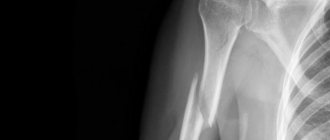
Diagnosing a dislocation is not difficult: a misalignment of the jaw is visually visible, palpation reveals a depression or tubercle and displacement, that is, loss of bone from the socket of the temporal bone. The second method is radiography in three projections of the maxillofacial area. The photographs show the dislocation, its location and shape.
In more complex cases, MRI and computed tomography are performed to exclude tumor formation, fracture and ligament rupture. Computed tomography is the final argument for performing surgery to restore a damaged joint or joints. A comprehensive diagnosis will help to make a correct diagnosis, excluding many complications from chronic pathologies, and will also help to exclude a fracture, which is identical in symptoms to a dislocation.
Classification of dislocations
First of all, it is necessary to distinguish between complete and incomplete dislocation (subluxation) of the lower jaw: in the first case, the contact of the articular surfaces is completely disrupted and the articular head is located outside the mandibular fossa of the temporal bone; in the second (with subluxation) – the contact of the articular surfaces is partially preserved. When a dislocation of the lower jaw is combined with a fracture of the condylar process, they speak of a fracture-dislocation.
Depending on the time and causes of occurrence, congenital and acquired dislocations of the lower jaw are distinguished; the latter can be traumatic, pathological and habitual in origin. Taking into account the direction of displacement of the head of the lower jaw, dislocations are divided into anterior and posterior. Based on the symmetry of the lesion, unilateral and bilateral dislocations of the lower jaw are distinguished.
Dislocation of the lower jaw, from the moment of occurrence of which no more than 5-10 days have passed, is considered acute; from 1.5 weeks and longer – chronic (old). If a dislocation of the lower jaw is not accompanied by damage to the skin, it is regarded as simple; in case of rupture of blood vessels, tendons, soft tissues, or skin, the dislocation is considered complicated. The most common cases in clinical practice are anterior bilateral subluxations and dislocations of the mandible.
It is important not to confuse complete and partial dislocation of the jaw. In the first case, there is an absolute disruption of the connection of the articular surfaces. The articular capsule is located outside the mandibular fossa of the temporal bone. In the second case, the contact of the two joints is partially intact.
We invite you to familiarize yourself with: Coxarthrosis of the hip joint, grade 4
Depending on the root causes and oldness of the problem, congenital and acquired injuries of the lower jaw are distinguished. Acquired ones can be habitual, pathological and traumatic in nature. Taking into account the direction of the shift, injuries can be anterior or posterior. Based on the symmetry of the damage, dislocations are divided into unilateral and bilateral.
If 5-10 days have passed since the fall or blow, the injury is acute. An injury is considered chronic if more than 1.5 weeks have passed since the injury. If the injury is not accompanied by damage to the epidermis, then it is simple. If ruptures of capillaries, soft tissues, and tendons occur in parallel, then the injury is complicated.
Symptoms and signs
Due to its mobility, the head of the joint can move in different directions. Subluxation of the temporomandibular joint is when the head is partially displaced from the articular cavity, and dislocation is the complete release of the head of the lower jaw from the joint cavity.
These pathological conditions are divided according to the time of formation into acute and chronic - up to a week from the injury and later.
Based on the amount of involvement of other tissues, the injury is divided into simple and complex - with a complicated injury, the joint capsule, skin, muscles, blood vessels and nerves are additionally damaged.
Dislocation and subluxation can be post-traumatic and acute. The first is formed as a result of external influence on the joint, and acute or chronic is its relapse when there is a sprain, or the muscles of the upper or lower jaw are injured.
Depending on the nature, subluxations and dislocations can be divided into unilateral, bilateral, posterior, lateral or anterior dislocations, depending on the direction of the exit of the head.
Subluxation is very similar in symptoms to dislocation, it’s just that the symptoms of injury are not so pronounced:
- Jaw problems. Sometimes it is closed so the patient cannot open it. It is difficult to close the teeth completely, it is difficult or impossible to speak and chew. The jaw may be shifted to the right or left. The habitual bite changes, the joint on the left or right may seem to be “jammed”;
- The patient experiences pain behind the ears and temples, tissue swelling occurs;
- Speech is very difficult and slurred;
- There is increased salivation, the person has difficulty swallowing;
- Externally, the face is asymmetrical, a deviation to the side is visually noticeable. There is a clicking sound in the joint when chewing or talking;
- The feeling of the joint is painful, there is a feeling of numbness or “pins and needles”.
Symptoms of dislocation of the lower jaw
With an anterior bilateral dislocation of the lower jaw, the patient’s mouth is open, the lips and teeth do not close, speech is difficult and slurred, so the patient tries to explain himself with gestures. There is hypersalivation, severe pain in the parotid region, and a change in the configuration of the face due to the displacement of the chin anteriorly. Examination reveals tension in the masticatory muscles, flattening of the cheeks; The displacement of the heads of the condylar processes is determined by palpation. Attempts to forcefully close the mouth by pressing upward on the chin are ineffective and are accompanied only by low-amplitude springy movements of the lower jaw and increased pain.
The clinical picture of unilateral dislocation of the lower jaw is similar. The patient's mouth is slightly open, the chin is shifted to the healthy side from the midline, and the lower part of the face is skewed. Habitual dislocation is accompanied by deviation of the lower jaw, crunching, clicking and pain in the joint.
Unlike an anterior dislocation, when the articular head of the lower jaw is displaced backward, the patient cannot open his mouth, which also makes swallowing, breathing and speech difficult. The main complaints are associated with sharp pain in the parotid areas. The patient's position is forced, with the head tilted forward. There is a posterior displacement of the chin and root of the tongue; in this case, the lower incisors rest against the anterior part of the hard palate, and the lower molars do not close with the antagonist teeth. Bleeding from the external auditory canal is possible due to damage to its bone wall.
With complicated dislocations of the lower jaw, pain and swelling of the periarticular tissues, subcutaneous hematomas, fractures of the lower jaw and temporal bone may be detected. On palpation, the head of the lower jaw is identified in the area of the mastoid process.
If a person has a bilateral dislocation of the lower jaw, then the symptoms are as follows:
- the person’s mouth is slightly open, lips and teeth cannot close;
- difficulty speaking;
- there is severe salivation;
- excessive pain in the parotid area;
- change in the contour of the face due to a shift in the chin.
During the examination of the patient, tension in the masticatory muscles, alignment of the cheeks, and shift of the heads of the condylar processes are observed. Attempts to force the mouth to close are unsuccessful. They are accompanied by severe pain and springy sliding of the lower jaw.
With unilateral jaw dislocation, the symptoms are identical. The mouth cannot close completely, the chin is not symmetrical, and the lower part of the face faces the other way. Habitual dislocation is accompanied by clicking and cracking sounds.
Unlike anterior dislocation, when the jaw joint is displaced from the lower side, the patient cannot close his mouth. This makes breathing difficult, swallowing and speech are reduced to nothing. The main symptom of jaw joint dislocation is severe pain in the parotid areas. There may be blood discharge from the ear. In case of complicated injuries, pain, swelling of the periarticular tissues, subcutaneous bruises, and fractures of the lower jaw may occur.
Treatment
Treatment includes first aid and realignment of the jaw joint (in the case of fresh subluxation, when no more than an hour has passed since the injury). Then, to consolidate the result, the patient is prescribed mouthguards and splints for 10–15 days in order to strengthen the ligamentous apparatus and restore the joint capsule. The lower jaw should be immobilized during this period. Pathological causes of subluxations after reduction of the joint are eliminated with medications (depending on the type of disease).
In case of chronic displacement of the TMJ, surgical intervention is necessary. The capsule of the jaw joint stretches, becomes covered with growths of connective tissue and scars. To restore the integrity of the TMJ, excision of the altered tissue will be required during surgery.
Reduction methods
Acute injury can be easily reduced. The first time, the help of a traumatologist is necessary; in the future, the patient can master the reduction technique independently. But it is not recommended to carry it out on yourself without medical supervision.
For anterior subluxation , when the head of the articular bone protrudes forward, the Hippocratic or Blechman-Gershuny technique is effective: with two thumbs you need to press on the molars, while simultaneously lifting the chin. Then the head of the bone moves backward. At the end of the manipulation, the doctor will hear a slight click. After the manipulation, the lower jaw is fixed with a circular bandage for 3 weeks.
In case of posterior subluxation , which happens much less frequently, the following manipulation is performed: the thumbs are placed on the chewing surface of the wisdom teeth (or outer teeth), the remaining fingers cover the chin. During adjustment, the thumbs move downwards, and the rest pull the jaw forward.
Diagnosis of dislocations of the lower jaw
Most often, visual observation and palpation of the affected area is sufficient to diagnose a dislocation of the anterior jaw. The doctor will determine the diagnosis based on the symptoms of jaw subluxation. But to make a distinctive and competent diagnosis of dislocation of the lower jaw, it is necessary to:
- radiography of the temporomandibular joint;
- CT scan of the TMJ.
On the lateral image, in the case of anterior dislocation of the lower jaw, an empty glenoid cavity and a shift of the head of the jaw are noted. In the case of a posterior dislocation, the x-ray shows a backward shift of the articular head. It is located under the lower wall of the bony auditory canal. Thanks to the results of examinations obtained during the diagnosis of dislocation of the lower jaw, it is possible to distinguish dislocation of the lower jaw from cracks. After diagnosing a dislocated jaw, treatment is prescribed only by a doctor.
To recognize a dislocation of the lower jaw, as a rule, an external examination and palpation examination are sufficient. At the same time, clarifying and differential diagnosis is impossible without radiography of the TMJ, and in complex cases, without CBCT or CT of the temporomandibular joint. In case of anterior dislocation of the lower jaw, lateral radiographs reveal a free articular cavity, a displacement of the head of the jaw anterior to the articular tubercle; with posterior dislocation - the articular head, having shifted posteriorly, occupies a position under the lower wall of the bony auditory canal, between the mandibular fossa and the mastoid process.
The obtained clinical and radiological data make it possible to differentiate a dislocation of the lower jaw from a fracture of the condylar process.
Rehabilitation and recovery
After reduction, you should proceed to rehabilitation measures to restore the joint and damaged tissues. These include:
- Immobilization of the joint by wearing a bandage or special devices;
- Medicines (vitamins, chondroprotectors), hydrocortisone ointments and anti-inflammatory gels;
- Physiotherapy (electrophoresis, laser and magnetic therapy);
- Diet and massages.
Child with an apple
First aid for sprain
When considering how to straighten your jaw yourself, it is worth remembering the main rule - it is strictly forbidden to try to straighten the joints spontaneously. This can only lead to complications and worsening of the victim’s condition. It is important to be able to provide first aid to the patient before the ambulance arrives. Actions include:
- Cover your mouth with a bandage or handkerchief to prevent dust and other foreign bodies from entering the upper respiratory tract. This will make breathing difficult and cause pain.
- Be sure to support the jaw with a bandage and apply cold compresses.
- If the patient complains of severe pain, then give an analgesic drug. This could be Analgin, Ketanov.
- Refer the patient to a doctor immediately.
It is strictly forbidden to apply warm compresses so as not to increase bleeding and provoke a hematoma. For any type of facial joint dislocation, it is very important to fix the lower jaw, so try to apply a bandage that will fix the joint in one position. In this position, the patient does not need to be forced to talk, drink or eat.
Symptoms
Symptoms depend on the location of the dislocation, the type of injury and the general condition of the patient + his reaction to pain. But all types of dislocation are associated with pain, misalignment of the jaw, stopping of joint movement and swelling in the dislocation area.
List of symptoms:
- displacement of the jaw with a specific distortion;
- the appearance of a pit or tubercle in the area of jaw displacement;
- acute pain radiating to the skull, teeth and ear;
- restriction of movement;
- excessive salivation;
- disturbances in speech, chewing and swallowing;
- the oral cavity does not close completely;
- swelling in the ears and temple area.
Some side symptoms such as headache, choking, fever, dizziness, and nausea are added to the list of symptoms. Patients with this diagnosis have increased irritability or tearfulness. After reduction, the symptoms immediately disappear, except for minor pain, swelling and cyanosis after bruising, which will gradually subside after treatment or massaging with NSAID-based gels.
Treatment of lower jaw dislocation
Treatment of victims with acute dislocation consists of reducing the dislocation of the lower jaw and immobilizing it for up to two weeks by applying a bandage. When considering how to set a jaw when it is dislocated, you need to know that there are several methods.
We suggest you read: Gouty arthritis treatment 5 methods to cure gouty arthritis
The main methods of jaw realignment are:
- Hippocratic method. Traumatologists, as well as orthodontists and orthopedists can straighten the jaw using this method. After using the analgesic, the specialist wraps a bandage around his hands, finds the victim’s molars by touch, clasps the lower jaw, puts pressure on the bone tissue, pulling the chin down. As a result, relaxation of the masticatory muscles is observed, after which the specialist moves the jaw. If everything went correctly, the result is a click.
- Blekhman-Gershuny method. In the patient's mouth, coronoid processes are observed. During injury they change their location. The specialist presses on them, pulls them down and moves them away.
- Popescu method. This technique is not common. Usually the method is effective for old dislocations. Before starting the manipulations, the doctor administers anesthesia, after which the person assumes a horizontal position. Fabric rollers are placed in the patient's mouth. Afterwards, the specialist presses on the chin, moving it back and up. As a result, the joint returns to its place.
- Operative method. If, as a result of conservative therapy, the reduction of the dislocation of the lower jaw does not produce the desired effect, then they stop at surgery. It occurs only after the administration of a high-quality analgesic. An incision of up to 2.5 cm is made in the zygomatic arch. The jaw is cut through it. A hook is inserted into the hole, which is used to catch the edge of the tenderloin and pull it down. The doctor presses his hand on his chin. As a result, the head takes the desired position.
- Prosthetics. If a dislocated jaw remains in a chronic form for many days, then prosthetics are indicated. In this case, the injury may occur even when yawning. To carry out the manipulation, fixed or temporary orthodontic devices are used. They are placed on the teeth. Such devices prevent the oral cavity from opening too wide, which relieves the joint from stress.
The most safe effective method is determined only by a doctor.
With the help of special exercises, you can normalize jaw mobility, strengthen muscle tissue and overcome tension. This will improve blood circulation and oxygen flow to the joints. With the help of the tasks performed, you can eliminate the symptoms of jaw displacement.
Jaw strengthening
Consider exercises that will strengthen your jaw:
- When opening the oral cavity, perform resistance tasks. Such exercises will help cope with unpleasant symptoms. Place two fingers under your chin and press slightly against it, opening your mouth. Throughout the day, perform 6 sets of 6 repetitions of the exercise. But you should not start procedures if they cause severe pain. If pain occurs, you should consult your doctor.
- While closing your mouth, perform a resistance exercise. Open your mouth and place two fingers on your chin directly under your lower lip. When closing your mouth, press down lightly with your fingers. This exercise strengthens muscle tissue. During the day, also perform 6 sets of 6 repetitions.
- Chin retraction. Stand straight, straighten your shoulders and pull your chin to your chest, as if you need to make it double. Stay in this position for three seconds. This exercise will help strengthen muscle tissue. Perform 10 times a day.
If discomfort occurs during exercises, do not force them.
In addition to strengthening, it is important to learn how to relax your jaw. Exercises have been developed for this:
- Try to keep your teeth from closing all the time. This will significantly reduce the load on the lower jaw. Keep the tip of your tongue between your teeth throughout the day. This will help keep your jaw position under control and prevent you from grinding your teeth. Before going to bed, relax your jaws as much as possible and do not close them together. Your personal dentist may recommend a custom mouth guard.
- Open and close your mouth. Try to keep your tongue on the roof of your mouth, slowly opening and closing your mouth. Such procedures will help relieve tension and relax the jaw. They are an integral part of any set of daily jaw strengthening exercises. Lower the jaw so that the muscles do not strain. There is no need to hold your mouth open. During the day, perform 6 sets of 6 repetitions.
- Try the “Goldfish” exercise. This activity helps to cope with stress. Place two fingers in the area where the jaws connect. Place the finger of the other hand under the chin. Open your mouth slightly, pressing lightly on the joint. Perform the exercise the same number of times a day. When the mouth opens, there is no need to resist the chin. The procedure is intended only to relax the jaw.
- Chin retraction. You can try to retract your chin to relax your jaw. You will need to move your shoulder joints back, push your chest forward, and try to retract your chin, making it double. Hold in this position for 3 seconds. Repeat the exercise 10 times.
- Breathing exercises. Stressful situations very often make a person clench his jaw. To cope with tension, you need to slowly inhale through your nasal passages for 5 seconds. Then exhale also for 5 seconds. Perform the exercise as often as the body allows.
We invite you to familiarize yourself with: Osteochondrosis of the lumbosacral region code according to ICD 10
Such exercises will help not only cope with problems with the jaw, but also make speech more literate.
There are a number of exercises that can help improve mobility and relieve pain. Let's look at the main ones:
- Place an additional object between the teeth to practice moving the lower jaw back and forth. Place the part so that it is at a right angle to the face. Move the lower jaw forward so that the protruding object is directed towards the ceiling. Having mastered classes with one object, you can begin to use thicker objects. Try to choose those items that are intended to be placed in the oral cavity.
- Correction of posture. Many people stretch their heads slightly forward when moving. For this reason, natural posture is disrupted. You will need to stand against the wall, bring your shoulder blades together and gradually push out the lower part. The lower jaw should touch the chest. This will help bring the spine into a natural position.
Each exercise must be given special attention and performed correctly so as not to aggravate the problem.
Jaw subluxation, the symptoms and treatment of which are determined by a doctor, is a dangerous injury that requires special attention and medical care. With timely treatment, most complications can be avoided. But if no measures are taken, this risks secondary dislocation and other pathologies.
First aid consists of reversing the dislocation of the lower jaw under infiltration or conduction anesthesia. To reduce the anterior dislocation of the lower jaw, the methods of Hippocrates, Khodorovich, Blechman, Gershuni, Popescu (for old dislocations) are used. The classic way to reduce bilateral dislocation of the lower jaw is the Hippocratic method: the patient sits on a low chair, so that the back of the head is supported, and the lower jaw is located at the level of the elbow joints of the dentist or traumatologist/surgeon. Standing opposite the patient's face, the doctor places his thumbs, wrapped in a towel or a thick layer of gauze, on the lower molars, and with the rest covers the lower jaw from the outside. Gently pressing down with his thumbs, the doctor moves the jaw back with a small push, quickly removing his fingers from the teeth to avoid biting. The movement of the articular heads of the lower jaw into place is accompanied by a characteristic click and intense closure of the jaws.
When reducing a posterior dislocation after the lower jaw has been displaced downward, it is moved anteriorly. In order to avoid recurrent dislocation of the lower jaw and limit movement in the TMJ after the reduction procedure, it is necessary to immobilize the jaw using a chin sling for 7-10 days (for anterior dislocation) and for 2-3 weeks (for posterior dislocation). Until recovery, the patient is advised to stop eating solid food and follow a gentle diet. If it is impossible to reduce the dislocation of the lower jaw using conservative methods, they resort to the surgical method. For chronic dislocations of the lower jaw, resection of the articular heads of the lower jaw may be required, followed by mechanotherapy.
Patients often learn to adjust habitual dislocations of the lower jaw on their own. Further treatment should include therapy for the underlying disease, wearing it for 2-3 months. medical orthopedic devices and splints that limit mobility in the joint (Petrosov apparatus, Burgonskaya-Khodorovich apparatus, Yadrova splint). According to indications, it is necessary to carry out selective grinding of teeth, prosthetics of missing teeth, blockade of masticatory muscles, massage, therapeutic exercises, physiotherapy (electrophoresis of medicinal substances, galvanization).
Surgical treatment of habitual dislocation of the lower jaw can be aimed at strengthening the ligaments, deepening the articular cavity, increasing the height of the articular tubercle, repositioning and fixing the intra-articular disc.
Treatment and reduction methods
Treatment of a dislocation always begins with its reduction, which should only be carried out by a qualified specialist. There are several reduction methods, but each of them is intended mainly for the reduction of a certain type of dislocation.
Hippocratic method
This technique is used more often than others when reversing a dislocated jaw. The technique is that the doctor wraps the fingers (thumbs) with a sterile bandage (you can use a towel or other clean cloth) and stands in front of the victim, seated on a chair.
The doctor places the prepared thumbs in the patient’s mouth on top of his chewing teeth, while the remaining fingers clasp the dislocated jaw from below.
After this, the doctor begins to press down on the jaw with his thumbs, and with the remaining fingers creates pressure on the chin area in an upward direction. Reduction of the dislocation can be determined by the characteristic click of the joint.
After reduction, the patient’s jaws close involuntarily, which is why it is necessary to wrap the fingers with some kind of cloth before the procedure. This will avoid possible injury from the victim’s teeth.
After reduction, the doctor applies a special bandage to fix the jaw , while the patient is prohibited from opening his mouth sharply and widely, as well as eating solid foods. During the first 15 days, food should be soft, preferably pureed and not require chewing.
Blechman-Gershun technique
Here you can make adjustments in two ways:
- The doctor feels the ends of the damaged joint from the inside, inserting his fingers into the victim’s mouth, and then performs a reduction by pushing the jaw back and, at the same time, down. When the joint is in place, you can hear a characteristic click.
- The doctor also feels the ends of the damaged joint, but from the outside, after which, grasping the injured jaw and chin, he performs the reduction with the same movements as in the first option. The external reduction technique, as a rule, causes much less discomfort to the patient and is more convenient for the doctor himself.
Popescu technique
This method is used only for the reduction of old (chronic) jaw dislocations, if it is displaced forward. Reduction is performed under local anesthesia, with the patient placed on his back. After preparing the patient, the doctor places rolls of sterile cotton wool, the diameter of which is about 2 cm, in his mouth between the teeth and cheek and then carries out the reduction by lifting the jaw up and moving it back.
It should be remembered that this technique does not always help. If it was not possible to correct an old dislocation using this method, then an operation will be required and further wearing of special devices to fix the jaw in the desired position.
How to straighten a dislocation at home
When wondering how to straighten your jaw at home, it is worth remembering that it is strongly recommended not to engage in spontaneous realignment. Only a doctor will diagnose a dislocation of the lower jaw and determine the severity. Only if a person is endowed with first aid skills and knows the methods of reduction, can one try to reduce the joint at home.
If it does not fall into place the first time, then it is important to immediately take the patient to a medical facility. At home, you can alleviate a person’s condition by providing first aid.
Types and characteristics of dislocations
When examining a patient with a dislocation of the lower jaw, the specialist must determine in which direction the head of the joint has shifted:
- forward;
- back;
- to the side.
If the dislocation occurs simultaneously on both sides, it is called bilateral, and if only on one side, it is called left-sided or right-sided.
Additional Information. Most often, doctors have to treat anterior bilateral dislocation of the jaw.
There is also a classification of the disease according to its causes:
- Traumatic - a single case involving injury.
- Habitual - appears constantly due to careless movements under the influence of certain diseases.
And finally, according to the severity of the condition, medical science divides dislocation of the lower jaw into two types:
- Light —displacement occurs without damaging adjacent tissues.
- Complex - in which it is necessary to additionally treat neighboring tissues.
It happens that the head of the mandibular bone does not completely come out of the joint socket. Doctors call this condition subluxation. Then the treatment is much simpler than the reduction of a full-fledged dislocation.
Forecast and prevention of dislocation of the lower jaw
With timely reduction of acute dislocations of the lower jaw and compliance with the terms of immobilization, the outcome is favorable; relapses are unlikely. With concomitant diseases and early load on the jaw, the development of habitual dislocations and stiffness in the joint is possible.
Prevention of TMJ dislocation involves controlling the amplitude of mouth opening while eating, singing, brushing teeth, and performing medical interventions; eliminating predisposing factors, preventing injuries to the lower jaw. After reduction of a dislocation or surgery on the TMJ, it is necessary to follow the recommended regimen and complete rehabilitation.
The prognosis for therapy is positive, but only if all the instructions of the attending physician are followed. But no one is safe from re-dislocation. For preventive purposes, it is recommended to do the following:
- use special dentures that limit the opening of the oral cavity;
- correction of teeth, which can provoke displacement of the lower jaw;
- timely therapy and prosthetics of teeth responsible for the normal position of the jaw;
- myogymnastics to strengthen the masticatory muscles.
To avoid dislocations and other injuries, it is important to monitor the amplitude of mouth opening and carry out preventive measures.
Treatment based on prostheses
Recurrent jaw luxation or subluxation can be managed with treatment that limits mouth movement. For this purpose, special orthopedic structures (splints) are installed on the teeth. Over time, they secure the joint in place. However, the doctor cannot give guarantees that after treatment the dislocation will not happen again.
These tires come in several varieties and can be removable or non-removable. What will be best in a particular case is determined by the dentist or traumatologist performing the treatment.
First aid
If your jaw is dislocated, you should not try to put it back into place yourself unless you are a medical professional with experience in this field. Many complications can arise during reduction. First of all, it is necessary to immobilize the bone with a supporting bandage or bandage, as well as with a scarf, handkerchief or cloth. Next, before the ambulance arrives, apply a heating pad with ice; if you don’t have this item, you can even apply a bag of frozen food.
When applying a bandage, try to fix the jaw and not damage the nerves and blood vessels, moderately tightening the bandage.
Rehabilitation
For any dislocation of the jaw joint, long-term chondoprotectors are prescribed. During the recovery period, nutrition should be light, mostly liquid.
In case of limited joint mobility, development begins after removal of the prostheses. Therapeutic gymnastics is agreed with the doctor; exercises should be done without fanaticism, otherwise another dislocation will follow. With successful treatment, re-injury is minimal, but it is necessary to visit a doctor once a year so that he can assess the condition of the jaw apparatus.
Important points in the postoperative period
Fracture + dislocation is treated through surgery. The indication for surgery is displacement of fragments, crushing of muscles, ligaments, blood vessels and nerves. After the operation, you must follow some rules: do not eat solid foods, try to eat only crushed or grated food. Do not open your mouth wide, even when visiting the dentist. When sneezing, coughing, loud laughter or screaming, you should hold your jaw with your hands to prevent dislocation. It is recommended to avoid hypothermia and overheating of the temporomandibular joint, as well as impacts and falls. The joint needs to be constantly massaged, compresses applied, and physiotherapy performed. If all the conditions are met, you can forget about repeated dislocations forever.