Destruction of the vitreous body of the eye, or DVT, is a pathological process in which the functioning of the ocular apparatus is disrupted. The disease is widespread, as it occurs in approximately 50% of people over 70 years of age. In old age, this condition is normal. Doctors believe that it is almost impossible for older people to avoid it. However, it can also appear in young people. For this reason, today we would like to tell you the main features of this disorder and how it affects the patient’s condition.
What it is
Destruction of the vitreous body is a change in the structure of the vitreous body, accompanied by its clouding and dysfunction. Symptoms appear at an early stage of the disease, and with special treatment its progression can be stopped.
Brief description of the disease
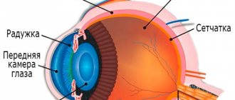
The vitreous body is a gel-like substance that completely fills the space between the lens and the retina. It consists of several main components:
- water;
- hyaluronic acid;
- proteoglycans;
- collagen.
It is collagen and hyaluronic acid that make the vitreous body thick and gel-like in structure.
The vitreous body in an absolutely healthy person is transparent and colorless. What makes it so is the special structure of the molecules of the substances from which it consists.
Due to some diseases, the properties of molecules change and they break down into smaller particles.
In this case, not only the structure of the vitreous body changes, but its size also decreases. As a result, the vitreous body is filled with cloudy, opaque formations, which are products of the breakdown of cholesterol and proteins.
Their shadow is projected onto the retina, causing the human eye to see them as dots or flickering lightning.
Prevalence and significance
Destruction of the vitreous body is a common pathology that occurs in almost 70% of middle-aged people seeking ophthalmological care.
People aged 40-65 years are most often affected by the disease. Women get sick more often than men, but the reasons for this correlation of incidence with gender have not been established.
In recent years, there has been a tendency towards a decrease in the average age of manifestation of CTD: more and more children and young people are suffering from this disease.
Risk factors
For some groups of people, the risk of developing vitreous destruction is higher than the population average. Here are the main categories of people susceptible to DST:
- people over 40 years old;
- female persons;
- patients with high myopia;
- people whose work involves high eye strain or exposure to harmful substances;
- pregnant and lactating women;
- people suffering from all kinds of chronic diseases.
Anatomy of the vitreous
The vitreous body looks like a transparent mass that has no color.
Its consistency resembles jelly. This anatomical connection is located behind the lens and ligament of cinnamon, and approximately two-thirds makes up the majority of the contents inside the eyeball. The vitreous body is in contact with the outer part of the flat process of the ciliary body, and the rest with the retina. Posteriorly, this anatomical formation comes into contact with the optic nerve head. If the total size of the eyeball weighs 7 grams, then the vitreous body occupies 4 grams of this weight. This means that the vitreous body itself occupies the largest part in the eyeball. The shape of this part of the visual organ is spherical, but slightly flattened on the sides. The anterior portion of the vitreous has a small notch that corresponds to where the posterior surface attaches to the lens. Along the edges of the recess there is a low shaft segment that connects to the fibrous ligament. This is followed by connection with the capsular formation of the lens along the length of its equator. Next, a capillary fissure passes between the lens and the vitreous body, which is called the retrolentral space. The connection between the vitreous body and the ocular membrane is the strongest among all other anatomical components in the eye. This strong connection is called the base of the vitreous. If the detachment process begins, then the ciliary epithelium extends immediately behind it.
On the other hand, the vitreous body has a strong connection with the macula. In general, in this part of the visual organ the following three zones can be noted:
- Behind the lens
- Posterior
- Ciliary department.
Inside there are also interfibrillar spaces that are filled with viscous, liquid and amorphous contents. Peripheral seals are composed of delicate, long and thin submicroscopic transparent fibrils. These microscopic connective tissues in the area of the base of the vitreous body are located more densely, and this corresponds to the posterior boundary layer, which becomes thinner the further you go. Anteriorly, the vitreous body has a more compacted shape, which indicates the presence of an anterior boundary layer. Next, the limiting membrane is formed. In the center of the lens it becomes very thin, and near the posterior pole of the lens this membrane practically disappears. The older a person is, the denser his boundary layer. The cloquet canal runs nasally from the central axis of the vitreous body. It appears as an enlargement near the optic disc. The cloquet canal ends at the base of the nose.
Causes
There are a huge number of reasons known that cause destruction of the vitreous body. In most cases, DST develops under the influence of several causes. Causes are usually divided into physiological and pathological. Physiological ones include:
- age-related degenerative changes;
- changes in hormonal levels associated with natural conditions (pregnancy, puberty, lactation);
- high physical or psychological stress;
- malnutrition, which caused a deficiency of vitamins and microelements.
The following reasons are considered pathological:
- insulin-dependent and metabolic diabetes;
- infectious diseases of the organs of vision;
- advanced myopia;
- cardiovascular diseases associated with metabolic disorders (hypertension, atherosclerosis);
- cervical osteochondrosis, accompanied by compression of the arteries;
- parasitic diseases (toxoplasmosis, toxocariasis, opisthorchiasis, echinococcosis);
- suffered injuries to the organs of vision, skull and brain;
- surgical interventions on the eyes and brain;
- the effects of radiation on the body;
- autoimmune processes (systemic lupus erythematosus, vasculitis, scleroderma);
- genetic abnormalities associated with damage to the nervous system;
- neuroinfections;
- kidney and liver diseases.
Factors leading to destruction
The reasons may be different:
- Changes in the eye associated with a person's age;
- The patient has diabetes;
- Chronic inflammatory diseases;
- Severe myopia;
- Diseases of the circulatory system.
- Infection with helminths;
- Hormonal imbalance when carrying a child, menopause, taking hormones.
- Stressful situations, depression;
- Injuries and operations on the eyes, nose, head;
- Avitaminosis;
- Radioactive exposure.
Diseases in the internal organs also provoke the development of this process. For example, kidney or liver disorders. In this case, the structure of the vitreous body is disrupted. “Flying floaters” indicate that retinal detachment has begun. This may result in loss of vision.
Kinds
There are several types of vitreous destruction:
- Filamentous destruction. In this form of the disease, a hardware examination reveals elongated fibers in the vitreous body, located a short distance from each other. This form usually occurs in people suffering from vascular and metabolic disorders, myopia, as well as in elderly people. The severity of the disease can vary from mild to extremely severe.
- Granular destruction. In this case, upon examination with a slit lamp, round particles resembling a fine suspension are detected. The suspension consists of pigment cells, protein bases and leukocytes. Patients with diabetes mellitus and chronic infectious eye diseases are most susceptible to this form of DST.
- Crystal destruction. This form is the rarest. Microscopic examination reveals particles shaped like crystals. Calcium and magnesium salts and cholesterol breakdown products accumulate in the vitreous body.
Symptoms and diagnostic methods
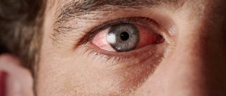
Destruction of the vitreous body is easy to suspect based on the patient’s complaints. As mentioned above, the main symptom that worries patients is the appearance of various visual effects reminiscent of the flickering of “flies”.
This symptom is characteristic of dozens of other diseases, but some signs make one suspect destruction of the vitreous body :
- visual effects are permanently saved;
- “flies” do not change their shape and size;
- “floaters” are noticeable only when the patient is in bright natural light or looks at white objects.
The severity of the disease can be judged by the severity of symptoms. The more noticeable the manifestations, the larger the particles flashing in the eyes, the more severe the damage to the vitreous body.
It is very important not to ignore these symptoms, since without proper treatment, DST can lead to vitreous detachment and irreversible loss of vision.
In addition to collecting anamnesis, doctors use hardware techniques to make an accurate diagnosis. Most often used:
- ophthalmoscopy;
- slit lamp examination;
- automated visual acuity testing.
To get a complete picture of the disease, the patient is asked to undergo an examination by a neurologist, therapist, endocrinologist and rheumatologist.
Forecast
The prognosis for development is favorable; usually the volume and type of opacities stabilize after a period of their occurrence and development lasting several months. In this case, remission during the disease, as a rule, does not occur; floating opacities remain in the eye cavity in a terminal state. Traditionally in ophthalmology it is believed that the disease does not affect the ability to work and does not lead to the development of complications dangerous to the patient’s health. In practice, the development of fairly dense, opaque vitreous opacities significantly worsens the patient’s quality of life. Floaters are constantly in motion and in various ways interfere with viewing objects and performing daily activities and work duties. However, in most cases, patients stop noticing opacities after 6-12 months.
| Check information. It is necessary to check the accuracy of the facts and reliability of the information presented in this article. There should be an explanation on the talk page. |
Treatment
Unfortunately, in most clinical situations, none of the currently existing treatment methods has sufficient effect. In some cases, the situation resolves on its own without any intervention, but most often, disturbances in the functioning of the vitreous remain for life.
The question of the need for treatment should be decided individually for each patient, taking into account the severity of manifestations and the form of DST. There are no radical and specific ways to restore the structure of the vitreous body. All treatment methods are aimed at eliminating the causes of DST and relieving unpleasant symptoms.
Drug treatment
For destruction of the vitreous body, the following drugs are prescribed:
| Type of medication | Examples |
| Medicines that have a resolving effect | Instillation of potassium iodide into the eye, Traumeel orally |
| Antioxidants that help restore blood circulation and saturate tissues with oxygen | Emoxipin |
| Vascular drugs that restore blood circulation in the brain | Cavinton |
Surgery
Surgical treatments should be used with great caution and only when the expected benefit outweighs the risk. This is due to the high probability of developing severe postoperative complications in patients with DST. Surgeries are contraindicated in patients over 65 years of age due to progressive age-related changes.
The following types of operations are carried out:
- vitreolysis (laser surgery that involves the destruction of cloudy particles in the vitreous);
- vitrectomy (removal of the natural vitreous and its complete replacement with an implant).
Traditional methods of treatment
Alternative medicine offers to treat the destruction of the vitreous body by instilling homemade drops into the eyes. To make such drops use:
- honey and water;
- honey and aloe juice;
- propolis.
Traditional methods of treatment should be used with caution, as sometimes they can only worsen the situation.