general description
General description of ectodermal dysplasia syndrome
Ectodermal dysplasia syndrome is a hereditary disease characterized by underdevelopment of ectoderm derivatives - hair, teeth, nails, etc. The syndrome manifests itself already in early childhood, however, the degree of its severity may vary.
In general, there are several types of ectodermal dysplasia syndrome, which have different manifestations and require different approaches to therapy. Among them are Christ-Siemens syndrome (anhidrotic or hypohidrotic ectodermal dysplasia), Clauston syndrome (hydrotic ectodermal dysplasia), one of the subtypes of which is Witkop syndrome (nail dysplasia).
Manifestations and symptoms
How to understand and recognize the disease
- Symptoms of ectodermal dysplasia syndrome
With anhidrotic or hypohidrotic ectodermal dysplasia, the facial structure characteristic of ectodermal dysplasia syndrome is noted. In sick children, a high forehead is noted, often protruding beyond the general line of the facial part of the skull. The nose with this disease is usually wide with sagging in the middle part, the lips are enlarged in size and often turned outward, the ears are often also turned outward and are located slightly lower than their localization in healthy people.
Since Christ-Stevens syndrome is an anhydrotic or hypohidrotic form of ectodermal dysplasia, and due to aplasia of the sweat glands, thermoregulation is impaired, patients are extremely sensitive to increases in external temperature, and since the ability to regulate body temperature is limited, fever is observed. The skin of patients with Christ-Stevens syndrome appears dehydrated (dehydrated), often cracked and wrinkled – including in childhood.
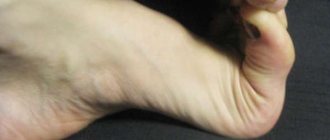
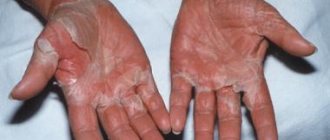
Also a characteristic symptom is a violation of hair growth and its normal structure; areas of baldness are often observed, both on the scalp and in the eyebrow area; hair on other parts of the body is present in very small quantities or is completely absent. Teeth are also affected, the number of which is reduced and the shape is changed; in some cases they may be completely absent. Nails, as an element of ectodermal origin, are another marker of damage in ectodermal dysplasia syndrome - they are significantly flattened, often flake and crumble, and are also characterized by small sizes - both on the fingers and toes.
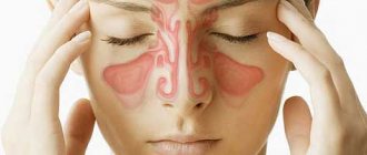
Patients with Christ-Stevens syndrome have a tendency to allergic diseases and the development of secondary infections of the upper respiratory tract due to impaired mucus secretion due to underdevelopment or complete absence of secreting glands. Symptoms of allergic damage to the respiratory tract are coughing, nasal discharge and sneezing due to high sensitivity to various allergens at different times of the year (in spring and summer there is an allergy to pollen from flowering plants, in autumn and winter - to house dust mites, mold, etc.) . Secondary bacterial infections are characterized by similar symptoms - rhinitis, often purulent, wet or dry cough, fever. Additional signs of this syndrome are retardation in mental and physical development.
With Clauston's syndrome - hydrotic ectodermal dysplasia - the most common lesions of the nails and hair are similar to those with the hypohidrotic syndrome described above (thin exfoliating nails, short, dry, sparse and brittle hair), as well as hyperpigmentation and hyperkeratosis of the nails, periorbital area, skin in the area of the knee and elbow joints. Mental retardation is also noted, which, however, is often expressed implicitly and can be corrected using generally accepted methods, in contrast to mental retardation with some other symptoms, including those associated with immunodeficiency states. With Witkop syndrome, a clear sign of the disease is the absence of teeth or their underdevelopment - large distances between adjacent teeth, irregular shape, etc. Other symptoms mentioned above may also be present.
Diagnostics
What to check and what tests to do
Diagnosis of ectodermal dysplasia syndrome
Diagnosis of ectodermal dysplasia syndrome is based on the presence of characteristic clinical signs and the results of examination of the patient. Laboratory tests (blood tests, immunograms, bacterial cultures, etc.) may also be recommended.
d.) – to confirm the diagnosis and identify concomitant diseases or complications of ectodermal dysplasia syndrome. Instrumental studies such as x-rays, ultrasound, ECG and others can also be useful.
Prenatal diagnosis can also be carried out (DNA analysis of chorionic villus or other methods).
Treatment and prevention
Methods of treatment and prevention
Treatment of ectodermal dysplasia syndrome
Treatment for this syndrome is largely symptomatic, because It is not possible to correct genetic disorders in order to influence the cause of the disease. Thus, in the absence of teeth or their improper development, dentures are performed; various ointments and multivitamin complexes are prescribed to improve the condition of nails and skin.
With anhydrotic ectodermal dysplasia, sudden changes in ambient temperature and exposure to climates with elevated average temperatures should be avoided, which is necessary to prevent the development of fever due to the inability to adequately regulate body temperature.
In the presence of concomitant diseases of allergic or infectious origin, appropriate therapy, etiotropic and symptomatic, should be carried out.
Prevention of ectodermal dysplasia syndrome
Prevention of this syndrome is not carried out, since it is hereditary.
Photos and illustrations
Enlarged lips and underdeveloped teeth in ectodermal dysplasia
Ectodermal dysplasia
MedClub.ru
Causes of development of dysplasia of the integumentary epithelium
Ectodermal dysplasias are caused by abnormal development of embryonic ectodermal structures. Genetic defects are responsible for approximately 30 of all identified ectodermal dysplasias.
X-linked recessive anhydrotic ectodermal dysplasia is caused by mutations in the EDA gene, which encodes the protein ectodysplasin. This protein activates NF-kappaB and JNK/c-fos/c-jun signaling pathways. Ectodysplasin plays an important role in cell survival, growth and differentiation.
Autosomal dominant and autosomal recessive anhydrotic ectodermal dysplasias are caused by mutations in the DL gene, which encodes the EDA receptor. Autosomal recessive anhydrotic ectodermal dysplasia can also result from mutations in the EDARADD gene, which encodes a protein that interacts with the EDA receptor.
Heterozygous mutations in the TRAF6 gene have been described in patients with hypohidrotic ectodermal dysplasia.
Claston syndrome, which is an autosomal dominant form of ectodermal dysplasia, is caused by mutations in the GJB6 gene, which encodes connexin-30, a component of intercellular gap junctions.
The etiology of anhidrotic ectodermal dysplasia is the presence of mutations of certain genes. The most common form of the disease is caused by damage to the EDA gene, located on the X chromosome.
It encodes a protein called ectodysplasin-a, disturbances in the structure of which lead to the pathological development of ectoderm derivatives. At the moment, both the functions of this protein and the pathogenesis of disorders due to mutation of the EDA gene are unknown.
Causes of anhidrotic ectodermal dysplasia
The etiology of anhidrotic ectodermal dysplasia is the presence of mutations of certain genes. The most common form of the disease is caused by damage to the EDA gene, located on the X chromosome.
It encodes a protein called ectodysplasin-a, disturbances in the structure of which lead to the pathological development of ectoderm derivatives. At the moment, both the functions of this protein and the pathogenesis of disorders due to mutation of the EDA gene are unknown.
Types of ectodermal dysplasia
There are two types of ectodermal dysplasia, recognized by experts as classical:
- Hidrotic, Clouston's syndrome. A hereditary disease, the characteristic feature of which is dysplasia of ectodermal derivatives: nails (convex shape), hair (baldness), hyperpigmentation of the skin. In some cases, patients experience strabismus and dementia. Unlike the second type, patients do not have disturbances in the functioning and structure of the sebaceous and sweat glands and abnormalities in the development of teeth.
- Hypohydric, Christ–Siemens–Touraine syndrome. A less common syndrome of this type is Rapp-Hodgkin syndrome with labial cleft, alveolar process, hard and soft palate.
| class="fa fa-info-circle"> | In modern medicine, more than several dozen different forms of ectodermal dysplasia have been described, the classic ones being the most common. |
Ectodermal dysplasia. Symptoms and manifestations
Let's talk about the symptoms of hydrotic ectodermal dysplasia. The disease is characterized by the main signs of nail dystrophy and the presence of hypotrichosis, palmar-plantar keratosis (increased keratinization of the skin) is less pronounced, and there are signs of pigmentation disorders. Let's take a closer look:
- Impaired nail development is the main symptom. The plates are fragile, thickened, deformed, brown, atrophied in places, and grow slowly.
- The hair is dry, brittle, thinning, sometimes to the point of total alopecia.
- Disturbances in the development of teeth are absent or slightly manifested.
- Foci of de- and hypopigmentation may develop on the skin.
Let's look at the symptoms of hypohydric (anhidrotic) ectodermal dysplasia. The main features of the disease are expressed visually. Large frontal bone, asymmetrically set ears, flattened nose, overly pronounced supraorbital ridges, the middle part of the face is depressed, the lower third of the face is reduced due to insufficient development of the alveolar bone, protruding lips.
| class="fa fa-info-circle"> | In the oral cavity, oligodontia, the absence of teeth, is clearly expressed. The existing teeth are conical and pointed. Hypoplasia of the labial frenulum is observed, and the median diastema is wide. |
Some types of ectodermal dysplasias are mild, while others are devastating. Abnormalities of teeth, hair, and nails usually become apparent during infancy or childhood. Other signs and symptoms that may occur variably in people with ectodermal dysplasia include:
- Hyperthermia with fever and seizures
- Xerophthalmia and conjunctivitis
- Hearing or vision impairment
- Xerostomia and caries
- Developmental delay or mental retardation
- Dysphagia
- Signs of narrowing of the airways
- Frequent pharyngitis, otitis and rhinitis, nasal congestion, hearing loss and hoarseness
Physical examination
In general, the clinical picture will depend on the specific type of ectodermal dysplasia:
- Dry, hypopigmented skin. Chronic eczematous dermatitis may also be present.
- Anhidrosis.
- Thin, brittle hair with alopecia is a feature of most types of ectodermal dysplasia.
- Nail dystrophy.
- Hypodontia or anodontia, deformed, vestigial teeth and/or enamel defects, frequent caries.
- Reduced lacrimation and salivation.
- Craniofacial anomalies.
Anhidrotic ectodermal dysplasia
The greatest disturbances in the dentofacial region are observed with anhidrotic ectodermal dysplasia (AED). Its diagnosis is based on identifying a complex of clinical symptoms, none of which individually is pathognomonic for this disease. Pathognomonic symptom complex of AED: anhidrosis, hypotrichosis, multiple congenital adentia, facial and cranial dysplasia, dysmorphogenesis of soft tissues of the oral cavity. Additional diagnostic signs are: hypoplasia of the glands of the mucous membranes, hypoplastic and dystrophic changes in the skin, hypoplasia of the nipples, and areolas of the mammary glands. The listed signs, although characteristic of AED, are not always clearly expressed. When substantiating the diagnosis, each of the listed signs must be analyzed and documented with a detailed description with references to data from special tests and examinations. Anhidrosis. 1. History: a) attacks of “unexplained fever” from the first weeks of life; b) an increase in body temperature in an infant during sucking; c) rapid fatigue and lethargy during games, especially in the heat or in a warm room, poor transfer
the prevalence of febrile diseases, deterioration of well-being in children, adolescents and adults, feeling of heat in a warm room, in hot weather, in the sun, during physical work, during intense movements, eating hot food and drinking (motor restlessness, palpitations, shortness of breath, nausea, dizziness, headache, fainting), the need in summer to often use a cold shower, moisten clothes, sleep outside, often rest during work; d) a sharp decrease or absence of sweating during heat and physical work throughout life. 2. Examination of the skin: a) lack of sweating, dry skin; b) absence or sharp decrease in the number of sweat pores. 3. Counting sweat pores: if sweat pores are detected during visual inspection, then they are counted using a magnifying glass. 4. Sweating tests (absence or sharp decrease in sweating). 5. Tests for thermoregulation: violation of thermoregulation. 6. Histological examination of the skin: hypoplasia or absence of sweat glands.
Hypotrichosis. 1. History: a) lack of hair since birth; after a few months, sparse, thin hair appears; b) rarely gets his hair cut; c) wears a wig. 2. Inspection: a) the hair on the head is sparse, short, thin, fluff-like, dry, brittle, usually straight and poorly pigmented; b) eyebrows are absent or sparse; c) eyelashes are sparse, short, sometimes absent; d) hair on the limbs and body is absent or sporadic; e) hair in the armpits and pubic area is absent or sparse. 3. Microscopic examination of the hair: the hair on the head is vellus-like, there are no other structural changes in the hair shaft. 4. Histological examination of the skin of the scalp: hair follicles are scanty and hypoplastic. Congenital multiple edentia.
1. History:
a) delayed teething (over 1-3 years of age); b) the sequence of teeth eruption is often disrupted; teeth erupt first on the upper jaw, then on the lower jaw; c) multiple or, less commonly, complete edentia; d) teeth are located at large intervals; e) conical teeth.
2. Inspection: a) insufficient number of teeth: at the age of 2.5 to 6 years (period of temporary occlusion), with multiple edentia, there are from 2 to 10 teeth on both jaws instead of 20 normally; at the age of 6 to 13 years (period of mixed dentition), in some patients 1-2 molars, sometimes fangs, often the upper ones, are cut; at the age of 13 years and older (period of permanent dentition), temporary canines (usually lower) become loose and fall out, the number teeth are reduced to 2-8 instead of 32 normally: on the upper jaw there are often central incisors, canines, 1-2 temporary molars, on the lower jaw - temporary incisors, canines, 1-2 molars; b) the size of the teeth is less than the average norm, the shape is disturbed; the incisors are narrow, short, with a smoothed shape of their angles, a weakly defined dental cusp, often fang-shaped with a sharp end of the cutting cusp; the fangs are narrow, but with a well-defined crown equator and a sharp cutting cusp; molars of atypical shape with a reduced number of weakly defined cusps and small intercuspal grooves; c) the enamel of the teeth is smooth, shiny, sometimes with transverse striations closer to the cusps of the teeth; d) the color of temporary teeth is white, permanent teeth are yellow; e) teeth are located at large intervals; the longitudinal axes of the upper central incisors are often deviated in the vestibular, lingual or lateral direction, the upper canines - in the mesial direction, the lower ones - in the lateral direction; f) contacts between the dentition are broken, there are two or three pairs of teeth interlocking in the usual occlusion; g) bite height is reduced; h) teeth are often mobile due to their functional overload. X-rays reveal short tooth roots. The periodontal gap is widened, especially in the area of the teeth in contact with the teeth of the opposite jaw. The alveolar processes of the jaws are hyzoplastic, low, and rise only in the area of existing teeth and their rudiments. Orthopantomography reveals that in the edentulous areas of the upper jaw the structure of the bone tissue is disrupted (especially in the area of the tuberosities), the alveolar process is underdeveloped or absent. The vertical dimensions of the body of the lower jaw are sharply reduced due to underdevelopment and atrophy of the alveolar process. The internal and external oblique lines are layered and projected at the level of the upper contour of the body of the lower jaw. The articular heads of the lower jaw are thickened. With age, the described disorders intensify. A telex-ray examination of the head in a lateral projection reveals a concave profile of the facial skeleton, a concave bottom of the nasal cavity, increased transparency of the paranasal sinuses (frontal, sphenoid, maxillary), insufficient development of the nasal bones and anterior nasal protrusion, underdevelopment or absence of the alveolar processes of the jaws, a violation of the proportionality of the size ratio the body and branches of the lower jaw, reduced vertical dimensions of the body of the upper and lower jaws.
Ectodermal dysplasia. Diagnostics
The disease is diagnosed based on the clinical picture.
In the early stages of development, dysplasia of the integumentary epithelium is diagnosed using DNA analysis of chorionic villi; the study is carried out at the 9th week of embryo development. At week 20, an ultrastructural examination of the fetal skin is performed.
Histological studies reveal atrophy of the epidermis, hypoplasia of hair follicles and sebaceous glands, and aplasia of sweat glands.
Laboratory research
In general, laboratory tests are not helpful in diagnosing ectodermal dysplasias.
Visualization
X-rays of the arms and legs can demonstrate specific skeletal deformities. Renal ultrasound, cystography, and intravenous pyelography may be useful in the evaluation of children with ectodermal dysplasia associated with cleft lip and/or palate, who often have genitourinary tract abnormalities.
Other tests
A skin biopsy may demonstrate anhidrosis and a reduction in the number of eccrine glands.
Diagnosis of anhidrotic ectodermal dysplasia
Despite the great rarity of the disease, an experienced specialist will certainly be able to make a fairly accurate diagnosis. Since the symptoms of this disease manifest themselves mainly externally, all the signs can be seen with the naked eye, combining them into a single clinical picture. To make a diagnosis you will need the following:
- a complete examination of the patient to identify characteristic signs and complete a complete clinical picture
- take a blood test
- do a chest x-ray, ultrasound and ECG
- conduct genetic research to identify mutations in the gene
- take a special sweat test
- conduct a skin biopsy to examine the condition of the sweat glands
- examine the structure of a patient's hair under a microscope
- conduct an X-ray of the jaw to determine whether there are rudiments of teeth or whether they are absent altogether
Based on external signs and laboratory tests, the doctor will be able to correctly determine the disease and its form and suggest further actions to combat the disease.