Varicose veins of the esophagus (phlebectasia) manifest themselves against the background of impaired blood outflow from the common venous system of the liver. It is often associated with a complication of portal hypertension and is considered dangerous. About half of the cases are fatal. In the remaining patients, the disease reappears within three years after treatment. According to experts, esophageal varicose veins (EVV) are most often diagnosed in male patients over 50 years of age who suffer from alcohol addiction and cirrhosis of the liver.
Classification
In medicine, there are several classifications of the disease depending on the nature of the course, diameter and location of the organ’s vessels.
By degree
Today, the classification created back in 1997 is used. Up to this point, it has changed a large number of times. There are three degrees:
- The first
is characterized by an increase in vessels in diameter up to 5 mm. They are elongated and located at the bottom. - With the second
, there is an increase in the lumen of the veins to 10 mm. Located in the middle part of the esophagus. - The third
degree is considered the most severe and manifests itself in the form of thinning of the walls of blood vessels and an increase in their diameter to more than 10 mm. They are located quite close to each other, and “red markers” are formed on their surface.
The degree is determined based on the results of a diagnostic examination.
Varicose veins of the stomach
When VRVP affects the stomach, another classification is used, which also includes three degrees:
- The veins are difficult to distinguish on the gastric mucosa; their diameter is up to 5 mm.
- The vessels acquire a solitary-polypoid character and increase in size to 10 mm.
- There is the appearance of nodes from the veins, which are polypoid in nature. The diameter is more than 10 mm.
At the first stage of development, it is possible to slow down the processes of change with the help of complex therapy.
According to Vitenas and Tamulevichiute
Experts also use the classification of phlebectasis of the esophagus according to Vitenas and Tamulevichute. They distinguish four degrees, but not related to the gastric veins:
- The vessels acquire a bluish tint, have a linear orientation, and their diameter ranges from 2 to 3 mm.
- The veins begin to form nodules, the size of which is more than 3 mm. Their great tortuosity and unevenness are observed.
- The nodes are clearly visible during examination, reach the gastric vault and rise above the surface of the mucosa.
- Vascular nodes acquire a polypoid shape or become like a bunch, reducing the lumen of the esophagus.
At stage 4 of the disease, the mucous membrane that covers the formed nodes begins to become covered with a venous network. Thus, varicose veins form in an area already affected by the disease.
Scientific Center for Chemistry of the Russian Academy of Medical Sciences
A separate classification, implying three degrees of development of the disease, is proposed by the Scientific Center for Chemistry of the Russian Academy of Medical Sciences:
- The veins are enlarged to 2-3 mm in diameter.
- There is an expansion of the lumen of blood vessels up to 5 mm.
- The diameter of the veins is more than 5 mm.
The characteristics of the degrees are quite scarce, and the classification is used in rare cases.
Zdenek Marzatka
Like most classifications, there are three degrees of development of phlebectasia:
- The vessels rise slightly above the mucosa.
- The veins twist, the lumen increases.
- The veins half block the lumen of the esophagus. They have a pseudotumor appearance.
Regardless of the classification used and the degree of development of the pathological process, esophageal varicose veins are considered an incurable pathology. The symptoms also cannot be ignored. Thanks to modern drugs, it is possible to slow down the process and alleviate the patient’s condition, eliminating the development of complications. Lack of therapy causes death.
Main classification
International ICD 10 distinguishes only 2 types of varicose veins of the esophagus: with and without bleeding. In clinical practice, a classification is used that differentiates varicose veins of the esophagus into 3 degrees, according to the pathological changes that occur.
This classification of varicose veins of the esophagus was introduced in 1997, and as the main diagnostic criterion implies the degree of change in the condition of the veins, the expansion of the natural diameter:
- To diagnose grade 1 varicose veins of the esophagus, it is enough that the veins located in the lower section reach 5 mm and are elongated compared to the physiological state.
- The diagnosis of grade 2 varicose veins of the esophagus assumes a wider diameter of the vein, increased under the influence of pathological reasons to 10 mm, the veins begin to twist and are located in the middle part of the organ.
- To determine the third, not only the diameter of the veins is important (it must exceed the centimeter mark). Grade 3 varicose veins of the esophagus are already thinned venous walls, formed twists, fully formed varicose nodes and red marks on the outside, which are considered markers for determining the severity of the condition.
Causes
There are several causes of the disease. Among them, experts identify a number of main factors that significantly increase the risk of developing varicose veins:
- Cardiovascular failure.
- Tension of the vein that occurs during the formation and growth of tumors.
- Liver pathologies that cause circulatory disorders.
- Blood clots in blood vessels.
- Gastropathy.
All diseases require timely treatment, since ignoring their symptoms leads to the development of serious complications, including grade 1 varicose veins of the esophagus. Over time, the pathology only progresses.
How to treat
If a patient has varicose veins of the esophagus, it is necessary to treat first of all the pathology that provoked its appearance. Additionally, medications are prescribed that reduce the risk of massive bleeding, vitamin complexes and drugs necessary to suppress the production of acid in the stomach.
Important information: How to treat to get rid of cellulite on the legs with varicose veins and which anti-cellulite cream to choose
In some cases, the introduction of colloidal solutions is justified. Drugs that are often included in the treatment regimen also include drugs that help narrow the lumen of blood vessels and strengthen their walls. Medicines that stimulate hematopoiesis may also be recommended. In severe cases, when conservative methods do not achieve a positive result, surgical treatment may be prescribed.
Endoscopic methods
For a disease such as varicose veins of the esophagus, treatment is often performed using low-traumatic endoscopic methods. The most widespread are intranasal and paravasal methods of administering sclerosant. In the first case, a special substance is introduced into the area of the node. In the second case, the sclerosant is introduced into the submycotic layer.
At the site of injection of the substance, blood flow is blocked and damaged tissue is replaced with connective tissue. This approach to treatment allows you to eliminate dilated veins with minimal risk of complications, but there is a high probability of relapse of the disease.
Balloon tamponade
This treatment method is used as part of surgical care when the patient develops massive bleeding from a dilated vessel. The balloon tamponade procedure for a disorder such as esophageal varicose veins involves the insertion of a special probe equipped with balloons into the esophagus. Air is pumped into these cavities of the apparatus.
Thus, the vessel is pressed, which leads to stopping bleeding. Nowadays this treatment method is used extremely rarely, because it is associated with a high risk of complications. There is a high probability of bleeding. This way you can block one bleeding area, but open up several more. Open bleeding can be treated using less traumatic methods.
Important information: Is it possible to do Nordic walking with varicose veins and are there any contraindications?
Surgical methods
When a patient's esophageal varices progress, treatment may require surgery. Often this therapy is resorted to when the vessels begin to bleed. These methods include:
- devascularization;
- venous bypass;
- transjugular intrahepatic portosystemic shunt.
The method of surgical intervention is selected individually, taking into account the degree of neglect of the process. When using this approach to treatment, the patient requires rehabilitation. The risk of re-expansion of the veins is extremely low.
Clinical picture
Phlebectasia develops individually, and the course directly depends on the presence of concomitant disorders and the cause. In certain cases, the pathology can be characterized by rapid development and pronounced symptoms. The main manifestations of the disease include:
- heartburn;
- belching;
- difficulty swallowing food;
- discomfort in the chest area;
- increased heart rate;
- bleeding, which manifests itself as the presence of blood in the stool.
At stage 1 of the development of esophageal varicose veins, signs may not appear, and the disease may proceed secretly. Moreover, the earlier the symptoms were detected and treatment started, the greater the patient’s chances of improving the condition.
Diagnostics
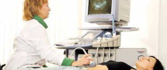
Varicose veins are often detected during an X-ray examination during the diagnosis of another disease. When studying the images, the specialist pays special attention to changes in the contours of the esophagus and the presence of uneven areas. X-rays provide sufficient information, but esophagoscopy is performed to confirm the diagnosis and determine the degree of development of the pathology.
This diagnostic method is performed with great caution, since there is a possibility of damage to the walls of the organ, which may result in bleeding.
What do esophageal varices look like?
Introduction
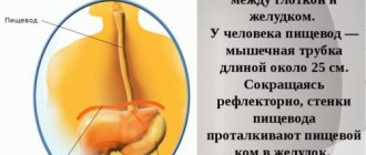
Esophageal varices (abbreviated as esophageal varices) are a pathological increase in the diameter of the venous vessels located in the lower part of the esophagus. Most often, this pathology is caused by portal hypertension (abbreviated as PH) - an increase in pressure in the portal vein (v. portae), which flows into the liver and collects blood from almost the entire intestine.
Compared to other types of varicose veins, varicose veins have completely different causes, symptoms and treatment. What unites these different pathological conditions is only the presence of enlarged veins.
The presence of varicose veins is only one of the symptoms of severe diseases leading to portal hypertension. Its appearance is most often caused by cirrhosis of the liver - a dangerous and almost irreversible disease. Typically, the treatment provided only slightly alleviates the patient's condition, but cannot completely cure it.
The problem of varicose veins is dealt with by hepatologists, gastroenterologists, and surgeons.
Treatment
The disease must be treated immediately after its detection. To achieve positive results, complex therapy is carried out, including medication and therapeutic treatment. When stage 3 or 4 of the development of varicose veins is established, surgical intervention is prescribed. But even after it is carried out, there is a high risk of relapse of the pathology within three years.
Therapeutic treatment
The therapy method is carried out in combination with medication or surgery. The essence of treatment is that the patient follows a specially designed diet, limiting physical activity, giving up bad habits and changing lifestyle and diet.
Therapeutic treatment will not help get rid of symptoms, but it will significantly speed up the body’s recovery process and increase the effectiveness of other methods.
Surgical intervention
One of the most common operations when phlebectasis of the esophagus is established is sclerosis. The method involves introducing a special solution into the injured vessel. The procedure is repeated after five days, then after four weeks and three months. To achieve a sustainable effect, it is recommended to carry out the procedure about 4 times a year.
When varicose veins are diagnosed, the following may also be prescribed:
- Vein lining.
- Portosystemic stent shunting. The procedure refers to endoscopic methods and is carried out by introducing a stent into the liver, which connects the hepatic and portal vessels.
- Anastomosis.
- Devascularization. During the procedure, injured vessels are completely removed and replaced with a prosthesis.
The method of surgical intervention depends on the degree of development of the disease, the presence of contraindications and the wishes of the patient.
Drug treatment
The use of medications helps reduce the negative impact on vascular walls. The patient is prescribed:
- Preparations to reduce acidity levels.
- Astringents.
Taking vitamin complexes to improve immunity is indicated. Painkillers may also be used after surgery. Daily dressings and a course of antibacterial drugs are prescribed.
Diet
Patients with phlebectasis should follow a special diet. Experts recommend eating 4-6 times a day. It is important that the last meal is taken no later than 3-4 hours before bedtime. The temperature of all dishes should not be too low or too high.
In order to prevent acidic fluid from the stomach from entering the esophagus, the head of the bed should be raised by at least 10 centimeters.
You can also help your body by eating right.
. While eating, there is no need to be distracted by talking or watching TV.
Experts recommend drinking about two liters of liquid per day. Patients need to give up strong black tea, confectionery, sweets, alcohol, coffee and baked goods.
Dietary nutrition must be combined with physical therapy. Daily exercise will help restore blood circulation and prevent blood clots.
Degrees of the disease
Classification of varicose veins of the esophagus according to clinical and morphological characteristics divides the disease into four degrees:
- The first degree of the disease is detected accidentally during endoscopy. An increase in the lumen in one of the parts of the vessel by no more than 3 ml is recorded. The patient is not worried about anything, only complaints about primary diseases are possible.
- The second degree is characterized by tortuosity of the veins, their unevenness, and no narrowing of the lumen. During the examination, it is possible to identify protrusions or stretched sections of veins. There are no specific symptoms of the disease; some patients at this stage of the disease complain of discomfort when swallowing.
- In the third degree, the lumen of the esophagus narrows due to the bulging of individual sections of the veins. The mucous layer is changed and more closely resembles the folds of the stomach. Patients complain of gastroesophageal reflux - heartburn, belching, pressure in the upper abdominal cavity.
- The fourth degree of the disease is diagnosed when numerous vein nodules are detected in the esophagus, not collapsing and with a thinned surface. Numerous erosions are found on the mucous layer. Patients register, in addition to signs of esophagitis, a salty taste in the mouth. The fourth degree most often leads to spontaneous bleeding.
Prevention measures
To prevent the development of esophageal varices, you should monitor the condition of your liver and undergo regular medical examinations.
The main recommendations of experts include:
- Avoiding heavy physical activity.
- Proper nutrition.
- Healthy lifestyle.
- Timely examination and treatment if unpleasant symptoms occur.
There are no special preventive measures to prevent the development of varicose veins. Doctors advise following general rules, avoiding overeating, consuming large amounts of fatty foods, and exercising daily.
Causes of pathology
The main factor in the development of pathology is the complexity of the functional structure of the blood supply to the abdominal organs. The veins of the esophagus connect with similar vessels of the spleen, the portal vein and the veins of the abdominal organs.
What normally works, when there are obstacles to blood flow, turns into a pathological formation. It causes a variety of negative symptoms that indicate problems.
Having diagnosed the first signs of the disease, you must immediately consult a doctor.
Conventionally, the factors causing pathology are divided into three large groups: intra- and extrahepatic, and mixed.
Reasons for formation in a particular case:
- Intrahepatic blockage occurs in the portal vein of the liver. It is provoked by cirrhosis, thrombosis, splenomegaly, cancer or purulent disease.
- Extrahepatic blockade is associated with obstruction of the bile ducts and vascular changes. It can often be caused by formations in the esophagus or thyroid gland.
- The mixed form appears if other organs are involved (for example, the heart with cardiovascular failure). It appears when there is already an existing violation of systemic blood flow.
- Modern medicine cannot yet explain the idiopathic form (congenital varicose veins of the esophagus and some others).
The causes of varicose veins of the esophagus most often lie in diseases of the digestive or hepato-biliary systems.
Possible complications
The danger of the disease is that it cannot be treated. Thanks to modern methods of therapy, it is only possible to relieve symptoms and slow down the pathological process.
The most dangerous consequence of esophageal varicose veins is bleeding, which occurs even with minor exertion or overeating. This condition is a threat to life and health and leads to chronic anemia.
Also among the complications are the development of an inflammatory process on the esophageal mucosa and rupture of small vessels, which leads to increased bleeding.
Forecast
The prognosis for VRVP is disappointing. Phlebectasia has a fairly high mortality rate. The death of the patient often occurs against the background of the development of concomitant diseases and progression of liver cirrhosis.
Life expectancy on average is about 5-7 years. But with varicose veins there is a high risk of re-development of the pathology. Relapse is observed in 80% of cases. If the treatment was effective, the patient receives the appropriate degree of disability.
Timely diagnosis of pathology significantly reduces the risk of death. When establishing varicose veins of the esophagus, you must follow all the doctor’s recommendations and eat right. Lack of therapy leads to the development of serious complications and consequences. That is why you should not ignore the symptoms, and if they appear, you should immediately contact a specialist.