Antibodies to hepatitis A - class M or G - are an objective indicator of the immune response to the causative agent of this infectious disease. Determination of antibodies of various classes confirms the acute phase of the disease or the result of a previous illness, as well as the effectiveness of vaccination.
For detection, the enzyme-linked immunosorbent assay (ELISA) method is used, which is available in almost any modern diagnostic laboratory. An appointment to determine antibodies to the hepatitis A virus is given by a family doctor, pediatrician or infectious disease specialist.
Indications for analysis
The need for a blood test for antibodies to the hepatitis A virus (IgG or IgM) is determined by the attending physician. It is necessary to take into account not only the results of a serological study, but also the entire complex (general clinical, biochemical, instrumental), since the mere fact of detecting antibodies does not mean a final diagnosis. Among the possible indications for this study, the most significant are:
- assessment of the results of vaccination;
- confirmation of the diagnosis of “acute viral hepatitis A”;
- exclusion of hepatitis A when examining an unclear patient with jaundice syndrome;
- retrospective analysis of the incidence of hepatitis A in any social group or organized team;
- in preparation for vaccination against hepatitis.
The study can be performed on a patient of any age, but requires some simple preparation.
Decoding the test for detection of the Epstein-Barr virus
The information content of the studies conducted on the presence of this pathogen is quite high. But if there is any other viral pathology, it is necessary to first carry out a number of additional diagnostic measures for the presence of the most likely pathogens in the body, and when taking an analysis during pregnancy, for toxoplasmosis, in order to understand the reasons for possible distortion of the results.
Reference values for research indicators for Epstein-Barr virus
| Name of the study | Index | Meaning | Note |
| Epstein Barr Virus, DNA real-time PCR | negative | Informative for the entire time the virus is in the body | |
| Epstein Barr Virus capsid protein (VCA) | signal/cutoff ratio | 0 — 0,9 | With recent infection, the excess of the norm is maximum |
| Epstein Barr Virus early antigens (EA), IgG | positivity rate | 0 — 0,99 | Most effective at the onset of the disease |
| Epstein Barr Virus nuclear antigen (EBNA) | positivity rate | 0 — 0,79 | The increased rate may persist for a long time after recovery. |
Preparing for analysis
No special preparation is required for the analysis - the rules are similar to those for most biochemical and serological studies. The patient should schedule a visit to the laboratory in the first half of the day - before 12.00. The meal should be in the evening before the day of blood donation, that is, the period of “fasting” should be at least 8-10 hours.
2-3 days before the planned visit to the laboratory, it is necessary to exclude excessively fatty foods, since venous blood may be technically unsuitable for analysis. Taking medications (even long-term) does not have a significant effect on the results of testing antibodies to the hepatitis A virus.
Types of tests for Epstein-Barr virus
These biochemical and molecular studies can detect antibodies to Epstein-Barr virus DNA (EBV, Epstein-Barr virus DNA) in the patient’s blood:
- Real-time PCR reaction (Epstein Barr Virus, DNA real-time PCR) - the result can be positive or negative. It is carried out throughout the entire period of treatment of diseases caused by the pathogen.
- VCA reaction to IgM capsid protein
(Epstein Barr Virus capsid protein (VCA), IgM) is carried out in the early stages of infectious mononucleosis. - EA - reaction to early antigens IgG
(Epstein Barr Virus early antigens (EA), IgG is carried out for the differential diagnosis of infections similar to infectious mononucleosis. - Quantitative reaction to nuclear antigen
(Epstein Barr Virus nuclear antigen (EBNA), IgG positivity rate).
If the test results go beyond the permissible reference (acceptable, normal) values, this means that the study needs to be repeated after 14 days. If pathogen DNA is detected, a repeat analysis is carried out after 30 days to determine the dynamics of treatment.
Since Paul-Bunnel antibodies are formed in the body during infectious mononucleosis, one of the types of laboratory diagnosis of the virus is based on their detection. In addition, the MFA method determines antibodies to the viral envelope antigen:
- A high titer of IgM antibodies indicates a recent infection, since this indicator is maximum two weeks after the onset of the disease, and then it decreases.
- An increase in IgG titer indicates the beginning of the period of convalescence. The rate may remain quite high for some time after recovery.
- Anti-EBNA IgG appears almost after recovery, and can be in the body even several years after the infection. Their appearance in the blood of a person who is still sick indicates the subsidence of the acute phase and the beginning of the recovery period.
Decoding the results
The assessment of the results obtained is quite clear - antibodies in the blood are present or absent, but what is important is which class of antibodies IgM or IgG. Modern diagnostic test systems are quite sensitive, so false positive or false negative responses are rare. Reference values are indicated on the packaging of the test system.
In accordance with the response, a further examination and treatment plan for the patient is planned.
Anti HAV IgG negative
This response indicates the absence of antibodies to the hepatitis A virus. The patient has not encountered this pathogen, meaning there is still a possibility of infection. If the test is prescribed to evaluate the effectiveness of vaccination, this demonstrates the absence of an immune reaction to vaccine injections. The patient is not protected from infection with the hepatitis A virus.
Anti HAV IgG positive
This result indicates that antibodies to the hepatitis A virus were detected in the patient’s blood. If the patient has been vaccinated against hepatitis A, then the vaccination result is positive - the patient is protected from penetration of the pathogen. In addition to this qualitative study, it is advisable to also make a quantitative determination - to establish the titer (concentration) of protective antibodies.
If the patient has not been vaccinated, but class G antibodies are detected in the blood, this indicates that the person has previously suffered from viral hepatitis A. The patient is reliably protected from the virus; repeated cases of viral hepatitis A have not been described. There is no need for vaccination, as well as emergency specific prophylaxis with human immunoglobulin after contact with a patient with hepatitis A.
Anti HAV IgM positive
The detection of class M immunoglobulins in the patient’s blood confirms the acute phase of the disease, that is, at the current time the person is sick with viral hepatitis A. This result is the basis for making a diagnosis of “acute viral hepatitis A”, since it is impossible that HAV IgM antibodies are present , but there is no virus.
Biochemical studies are necessary to assess the functional state of the liver.
Anti HAV IgM negative
This result indicates the absence of acute viral hepatitis A. If the patient has signs of hepatitis, then another virus that has caused liver damage should be looked for. It is advisable to screen for viral hepatitis with a parenteral transmission mechanism - hepatitis B and C, as well as examination for herpes viruses (herpes simplex viruses type 1 and 2, cytomegalovirus, Epstein-Barr virus).
A negative test result for antibodies to the hepatitis A virus is used in difficult diagnostic cases when carrying out differential diagnosis of diseases with jaundice syndrome.
Questionable results
A variant of the answer can be called doubtful in which both class M immunoglobulins and class G immunoglobulins were found in the patient’s blood. Perhaps this is a rare situation when the patient is already beginning to recover, that is, a gradual seroconversion occurs - replacement of IgM with IgG.
The second possible option is a technical error during the study, or the low quality of the test system used. To eliminate doubts and possible errors, the test for antibodies to the hepatitis A virus should be repeated in another diagnostic laboratory.
Presence of the virus in young children
Barra virus can be detected in the body of a newborn baby only using a PCR test. The fact is that a young body is unable to resist a pathogenic infection. This diagnosis is carried out only if the mother was previously exposed to an attack by viral microflora.
Adult children are susceptible to domestic infection with this disease. It is quickly transmitted by airborne droplets. When this pathology is detected in the patient’s blood, the main task of doctors is to reduce the level of progression of the disease to the lateral phase. Continuous therapy allows you to control the activity of the Barr virus.
Infectious mononucleosis
Reactivation of infection
Reactivation of Epstein-Bar infection is possible, which can lead to various clinical forms:
- chronic recurrent infection;
- chronic active infection such as chronic infectious mononucleosis;
- a generalized form of chronic active Epstein-Barr infection with damage to the central nervous system, myocardium, kidneys, etc.;
- Epstein-Barr virus-associated hemophagocytic syndrome;
- erased or atypical form of infection in the form of prolonged low-grade fever and clinical manifestations of secondary immunodeficiency;
- oncological lymphoproliferative processes;
- autoimmune diseases (systemic lupus erythematosus, rheumatoid arthritis, Sjogren's syndrome, etc.);
- chronic fatigue syndrome.
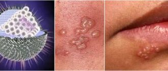
What is herpes virus type 4?
Herpesvirus type 4, as one of the types of herpesvirus infection, was described 53 years ago by the English virologist Michael Epstein. The professor was assisted in his work on the project by his graduate student Yvonne Barr. The virus owes its name to these people. However, 15 years after becoming familiar with the virus, its scientific name was changed to human herpesvirus 4, and a year ago the virus began to be called human gamma virus type 4.
But what is the Epstein-Barr virus? Like any other virus, the virion (viral particle) of herpesvirus type 4 consists of genetic material (in this case, double-stranded DNA) and a surrounding protein shell (capsid). Additionally, the virus is surrounded by a membrane, which helps it easily penetrate into the host cells.
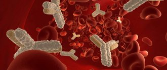
Any virus is a non-cellular form that is an infectious factor and cannot develop and multiply outside the cells of a living organism. The favorite habitat of herpesvirus type 4 is the epithelial cells of the nasopharynx. They do not disdain leukocytes, preferring one type called B-lymphocytes. It is B cells that actively participate in providing the body's immune defense. Upon contact with an antigen, which in our case is the herpes virus type 4 (more precisely, its antigens), B lymphocytes produce antibodies (immunoglobulin proteins). They are the ones that can be detected in the patient’s blood by testing for the Epstein-Barr virus (EBV).
The fourth type of herpes virus has 4 antigens that appear strictly in a certain sequence:
- EA is an early antigen that appears at the initial stage of the disease, when viral particles are in the stage of synthesis (primary acute infection or reactivation of a latent virus with decreased immunity),
- VCA is a capsid antigen, which is contained in the protein shell and also belongs to the early ones, because clinically the disease in this period may not even manifest itself yet,
- MA – membrane antigen, appears when the virion is already formed,
- EBNA - nuclear (polypeptide or nuclear) antigen is one of the late antigens, antibodies to which can be detected even several months after the disease and remain in the blood throughout life.
Herpesvirus type 4 is very insidious. Since the virus is inactive outside a living organism, you can only become infected with it from a person who is the source of the infection. And it is not at all necessary that he exhibit all the symptoms of the disease; the infection may have an erased form, masquerading as ordinary fatigue. For example, chronic fatigue syndrome in most cases is associated with the Epstein-Barr virus.
Individual virions can be found in blood, saliva, semen, vaginal secretions, and tissues of various organs. Virus particles, along with saliva and blood, can enter objects outside of our environment, where they will remain in an inactive state until they somehow enter the human body. In the vast majority of cases, infection occurs through airborne droplets or contact (through kissing). But intrauterine transmission of the virus from mother to fetus, infection during a blood transfusion procedure (if the donor blood contained virus virions), or through sexual contact is also possible.
Consequences of acute infection
- recovery;
- asymptomatic virus carriers;
- latent infection;
- Epstein-Barr virus reactivation;
- chronic recurrent infection;
- oncological lymphoproliferative process: lymphomas, nasopharyngeal carcinoma, leukoplakia of the tongue and oral mucosa, stomach cancer and colorectal cancer;
- autoimmune diseases: systemic lupus erythematosus, vasculitis, rheumatoid arthritis, Sjogren's syndrome;
- chronic fatigue syndrome;
- splenic rupture.
There are several options for “exiting” an acute infectious process. After the primary Epstein-Barr infection, persistence of the virus is observed in the body, which may not be clinically manifested in any way (asymptomatic virus carriage or latent form of infection).