Xeroderma pigmentosum is one of the hereditary dermatological diseases. It is extremely rare: 2 cases per 1 million births. Symptoms of the disease appear in early childhood and steadily progress without comprehensive treatment. The danger of xeroderma pigmentosum lies in its high oncogenic potential. Skin manifestations of the disease are an obligate precancer, that is, they are prone to malignant degeneration and in almost all cases turn into cancer.
At the same time, the first dermatological symptoms of xeroderma pigmentosum in a child are most often regarded by parents as a phenomenon of some kind of allergic dermatitis and do not cause unnecessary concern. Lack of alertness leads to late seeking medical help and delayed treatment.
- Causes and mechanism of development
- Kinds
- Clinical picture
- Complications
- Diagnostics
- Treatment methods
- Forecast
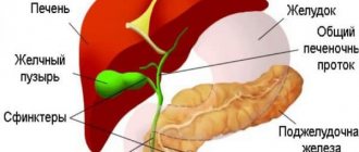
Etiology of the disease
Since xeroderma is a disease of genetic etiology, it is characterized by a pathology inherited by children, which affects the enzymes responsible for protecting the skin from ultraviolet radiation. In the case of xeroderma, such enzymes are either completely absent or do not work properly.
With age, the number of cells that have undergone genetic mutation increases, which leads to the development of cancer. Strong susceptibility to the influence of ultraviolet radiation and radiation are factors that influence the progress of the formation of pathogenic cells and, as a consequence, xeroderma pigmentosum.
BezOkov recommends: genetics and mechanism of disease development
Among the variety of malignant tumors, 10% is allocated to skin cancer. The development of such pathology mainly occurs in people who have entered retirement age.
The most commonly affected areas of the body are uncovered areas, often the face and neck. This is a clear confirmation of the theory that increased ultraviolet radiation is a trigger for the development of a dangerous disease.
In the vast majority of people, normally the DNA of skin cells damaged by sunlight is constantly restored thanks to genetically determined processes. One of the main mechanisms is excision reproduction. Its action is based on recognizing the damaged area and removing it using the enzyme DNA polymerase. At the same time, based on the intact nucleotide strand, the segment is restored and placed in the place of the removed one.
Another mechanism that can restore a damaged chain is photoreactivation. At least 7 enzymes are involved in this process. Its essence is to completely restore the damaged section of DNA strands without removing or replacing it. The damaged area returns to normal.
Over the years and the aging of the body, regeneration processes slow down significantly. There are more and more DNA segments that have been damaged. This leads to the accumulation of mutating cells and the growth of dangerous tumors.
The main reason for the development of pathology is a decrease in the protective function or the inability to restore cells. As a result, the accumulation of mutated segments and damage to the main DNA strands occurs.
Pathology can be transmitted through generations. That is, completely healthy parents may have a baby with damaged DNA. This happens due to a copy of a mutated gene that makes the regeneration process impossible.
What are the influence of provoking factors?
Prolonged exposure to the sun with hypersensitivity to ultraviolet radiation and radiation can cause the following pathologies:
1. Impaired pigmentation.
2. Keratization of the skin.
3. Atrophy of the epidermis.
4. Thinning of connective tissues.
5. Atypia at the cellular level.
6. Precancerous condition.
Like any pathology, xeroderma has a number of concomitant diseases, such as syndactyly (fusion of fingers), tissue degeneration (baldness, growth retardation and deterioration in the condition of teeth).
Diagnostics
The relevance of xeroderma pigmentosum today is very high. After all, an increasing number of people are trying to be in the sun more often, without any fear of the effects of UV rays. And that's wrong. Even if a person is not threatened by this disease, it is best to expose the skin to a minimum degree of active solar radiation. How can this disease be detected?
- Examination of the skin using a monochromator, a special instrument that determines the level of photosensitivity of the skin.
- The next step is a biopsy. In this case, particles of tumors on the patient’s skin are examined.
- Tissue samples taken during the biopsy are examined histologically.
Causes of xeroderma pigmentosum
This disease occurs as a result of the transmission of an autosomal gene from a parent to a child. To do this, the parents must be closely related (in other words, incest). In this case we are talking about a family disease. Another cause of xeroderma is a small amount or complete absence of UV enzyme in fibroblasts (connective tissue cells).
Also, deficiency of RNA polymerase (the enzyme that produces ribonucleic acid) can cause the development of xeroderma. In this case, the disease is provoked by abnormal ultraviolet exposure.
Some scientists believe that xeroderma pigmentosum (the photo below demonstrates how the disease manifests itself on the face) may also appear as a result of an increase in the amount of porphyrins in the human body and the entry of photosensitizers into the blood.
Clinical course and symptoms of the disease
Xeroderma has three stages of development:
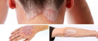
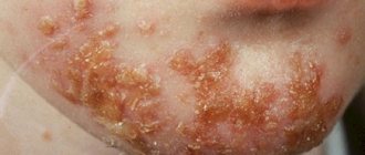
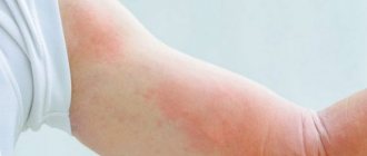
1. The first symptoms appear in children under three years of age. Their manifestation is usually associated with prolonged exposure to the sun, especially during periods of high solar activity (in spring and summer). The skin begins to peel off, after which it becomes covered with uneven hyperpigmentation spots and freckles. The symptoms of xeroderma pigmentosum are not limited to this.
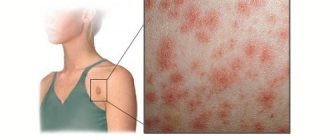
2. After several years, clinical signs of xeroderma appear. From this moment on, doctors diagnose the second stage of the disease. Signs of atrophy, spider veins and pigmentation spots appear on the skin of a sick person. In appearance, the manifestations on the skin resemble signs of radiation dermatitis. Ulcers, cracks, spots and crusts appear on some parts of the skin. In some cases, wart-like growths may appear. In addition to the skin, damage occurs to cartilage and connective tissue. Atrophy of the cartilage of the nose and ears occurs, and deformation of the mouth and nose occurs. The disease is accompanied by the occurrence of blepharitis (inflammation of the ciliary edge of the eyelid), ulcers on the mucous membrane of the eyes, and eversion of the eyelids. Eyelashes may stop growing, resulting in clouding of the cornea of the eye, tearing and photophobia.
3. The third stage of xeroderma begins during puberty. It is characterized by the appearance of neoplasms of various etiologies (benign or malignant). These can be fibromas, melonomas, keratomas, angiosarcomas, and so on. Warts that appear in the second stage can become malignant and metastasize to peripheral organs.
Xeroderma pigmentosum may be accompanied by severe mental retardation of the sick child.
Complications
The main complication of the disease is the appearance of skin and visceral (internal) malignant tumors. Multiple warty neoplasms have the highest risk of oncogenic transformation. The incidence of malignant tumors also correlates with the severity of skin pathological processes. With the early onset of the disease and its aggressive course, oncological tumors appear in adolescence (12-14 years) and quickly metastasize to internal organs.
Among malignant tumors, squamous or basal cell carcinoma most often develops, and melanoma of the skin or eye is the least common. Cases of more than 50 different types of oncology have been described: fibrosarcomas, angiosarcomas, histiocytes and others.
The disease can also lead to severe deformities of the face (mouth, nose, ears). The result is social maladjustment of the child and psychological problems.
Signs of severe form
There are two syndromes: Reed's and De Sanctis-Cacchione's. The first slows down bone marrow growth, accompanied by microcephaly (reduction in the size of the child's skull) and delayed mental and mental development. A more severe form is considered De Sanctis-Cacchione syndrome, or xerodermic idiocy. When it occurs, there is a disruption in the functioning of the central nervous system with the following symptoms: mental retardation, microcephaly, seizures, deafness, atrophy of the pituitary gland and cerebellum, delayed puberty, miscarriages during childbearing age. What is the diagnosis of xeroderma pigmentosum?
Establishing diagnosis
To detect xeroderma pigmentosum, first of all, a monochromator is used, a special optical-mechanical device for examining the skin. This analysis reveals the degree of sensitivity of the skin to ultraviolet radiation.
To confirm the diagnosis, the dermatologist gives the patient a referral for a biopsy. This is a study of tissue in laboratory conditions to identify neoplasms. For analysis, material is taken from recently appeared formations. However, they can be completely removed if their location on the body allows it. The material is collected with a scalpel or using surgical power instruments. Small tumors are taken whole, large tumors are taken partially, including a healthy area of skin.
The puncture is taken with a syringe with a needle-tube. The so-called punctate (liquid from the neoplasm) is examined. Histology is performed on the sample taken, the purpose of which is to clarify the diagnosis, identify the possibility and type of subsequent therapy, determine the stage of the disease and the degree of cell damage. The histological conclusion includes a description of the study and a nosological conclusion.
Treatment of xeroderma pigmentosum
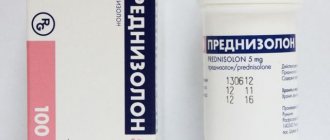
If xeroderma was identified at the first stage, its treatment is carried out on an outpatient basis. The child is registered with a dermatologist with mandatory regular visits to the doctor for consultation. Therapy is carried out by taking antimalarial drugs such as Hingamin, Rezokhin and Delagil. They reduce sensitivity to ultraviolet radiation.
In addition, it is important to strengthen the immunity of a child suffering from xeroderma. Therefore, vitamins A, PP (nicotinic acid), as well as B vitamins are prescribed. Peeling on the skin is treated with various ointments, which include corticosteroids, and warty formations are treated with ointments based on cytostatics. These ointments slow down the growth and reproduction of cells of all types.
Registration with an oncologist
When the cells become malignant, the patient is registered with an oncologist. If a patient exhibits the De Sanctis-Cacchione syndrome described above, treatment is carried out in special clinics under the supervision of a neurologist. In order not to miss the moment of transition of the disease to the malignant stage, constant monitoring is necessary with a number of specialists - a neurologist, dermatologist, oncologist and ophthalmologist.
Papilomatous (warty) formations that are prone to malignancy (transition to malignancy) must be removed surgically. Cryodestruction (freezing with liquid nitrogen), electrocoagulation and laser treatment are widely used in this area. The latter is carried out by exposing a problem area of skin to a beam of a certain length. Electrocoagulation is the banal cauterization of formations using electric current. With this type of therapy, not only the skin, but also the blood vessels are cauterized, which avoids bleeding.
Appearance of the disease
What does xeroderma pigmentosum look like? Human skin has a special pigmentation. This all happens as a result of exposure to ultraviolet radiation on the skin of the patient. But if in healthy people special enzymes work that prevent the occurrence of these spots, then in such patients they are not active. This is all due to a mutation in the proteins that are responsible for tissue restoration after such an influence. Mutated cells gradually accumulate in the body, resulting in skin cancer. In addition to ultraviolet radiation, it should be noted that the patient’s skin is also very sensitive to radiological (ionizing) radiation.
Child protection methods
Methods for protecting children with xeroderma pigmentosum include staying outside the home only in the evening and at night. Windows in the room must be tinted to prevent excess sunlight. When outdoors during the daytime, you need to use dark glasses and hats, and clothing should be made of thick fabric that covers as much exposed skin as possible. Various factors should also be excluded, such as:
1. Inhalation of tobacco smoke.
2. Radiation – X-ray, gamma radiation, ionizing and naturally ultraviolet.
3. Exposure to mold due to the toxins it produces.
4. Chemical irritants such as nitrites, nitrates and so on.
Clinical picture
Genetic skin diseases include the following diseases: xeroderma pigmentosum, reticular progressive melanosis, Pick's melanosis, which, in fact, is the same disease. The clinical picture is divided into three main types:
- Inflammatory. On exposed areas of the skin, spots similar to freckles appear. Gradually, scales similar to lentigo appear.
- Hyperkeratic stage. Islands of accumulations of freckles, scales, and lentigo-type elements alternately form on the skin. Everything resembles the picture of chronic radiation dermatitis. Sometimes warty formations may occur. All these atrophic changes gradually lead to depletion of the cartilage of the nose and ears, and the natural openings may become deformed. Also at this stage, baldness and eyelash loss are possible. The cornea may become cloudy, photophobia and tearing may occur.
- At the last stage, the problem crosses the cancer border. Both benign and malignant neoplasms appear on the skin.