Cerebral aneurysm, intracranial, or cerebral aneurysm, is the name given to a small formation that appears on a blood vessel in the brain. The tumor grows quickly and fills with blood.
An aneurysm is a neoplasm that forms on the blood vessels of the brain.
Aneurysm is quite rare. It is diagnosed in 5% of the world's inhabitants (40 people out of a thousand). The disease most often occurs in women aged 30-60 years. In children, the neoplasm is diagnosed very rarely (2% of all cases), mainly if it is congenital. In 5-10 people out of 10 thousand, an intracranial aneurysm ruptures.
The tumor can appear in any part of the brain. But most often it forms at the point where arteries branch.
Classification of aneurysms
Depending on the external signs, there are 3 types of intracranial aneurysm:
- saccular, or “berry” - looks like a round sac filled with blood, attached to an artery, also resembles a berry hanging on a stem;
- lateral – it can be found in the wall of the vessel; in appearance it resembles a tumor;
- fusiform - originates in the place where the expansion of the blood vessel occurs.
Mostly saccular neoplasms are formed.
Aneurysms are classified according to external signs
Depending on the origin of the aneurysm, there are:
- true - an aneurysm is formed when the artery wall protrudes;
- false - the cavity of the neoplasm, located near the artery, is not part of the blood vessel, blood enters it through a hole that appears in the wall of the vessel;
- exfoliating - a cavity is formed inside the wall of a blood vessel, blood enters it through a hole in the wall.
If an aneurysm is caused by infectious diseases and purulent damage to the wall of a blood vessel, then it is called infected, or mycotic.
Based on the time of appearance, congenital (about 20%) and acquired neoplasms are distinguished.
Depending on the number, aneurysms can be single or multiple.
Based on the number of chambers, neoplasms are divided into single and multi-chamber.
Depending on the size of the aneurysm, there are:
- small (less than 11 millimeters in diameter);
- medium (diameter varies between 11-25 milliliters);
- gigantic (over 25 millimeters in diameter).
Based on location, arterial and arteriovenous neoplasms are distinguished.
Arterial aneurysms occur when the walls of the arteries bulge into a sac or sphere. They are mainly located in the area of the circle of Willis. It is in this place that the maximum branching of the arteries is observed.
Arteriovenous aneurysms form in those places where the veins expand to form a ball and communicate with the arteries.
A type of arteriovenous aneurysm is the vein of Galen aneurysm. They are very rare. But a third of such cases are diagnosed in newborns and young children. Moreover, such neoplasms occur 2 times more often in boys than in girls. There are no signs of illness in half of the children. In some cases, heart failure or hydrocephalus may develop.
In 90% of cases, children diagnosed with vein of Galen aneurysms die. In addition, embolization does not change the situation much; the mortality rate decreases slightly (to 78%).
Aneurysm of the vein of Galen in newborns
An anomaly that develops while the baby is in the womb is associated with congenital diseases. In a large vessel - the vein of Galen - deformations, interlacing, and the formation of glomeruli are possible, which disrupts the flow of blood. In this case, a disorder of brain function and heart failure develops. An aneurysm in a newborn is accompanied by symptoms:
- swelling of individual parts of the body;
- fatigue that occurs during breastfeeding;
- pulmonary edema.
Article on the topic: Pantocalcin - instructions for use for children and adults, indications, release form, analogues and price
Brain aneurysm: causes
Aneurysms occur when the three-layer structure of the walls of arteries and veins is disrupted and the disappearance of the muscle layer or submucosal membrane is observed.
The formation of a cerebral aneurysm can result from:
- genetic predisposition;
- interweaving of veins and arteries;
- tortuosity and abnormal bends of blood vessels;
- damage to collagen fibers and elastic membrane;
- pathological changes in the muscle layer of blood vessels;
- blood circulation disorders;
- coarctation of the aorta;
- vascular calcification;
- thromboembolism;
- diseases developing in connective tissue;
- head injury;
- hypertension;
- infectious diseases of the brain;
- inflammatory diseases;
- atherosclerosis;
- malignant neoplasms and metastases;
- mycotic, bacterial or tumor embolism;
- physical and emotional stress.
Under the influence of the above factors, the vessel wall loses its mechanical strength and elasticity, begins to stretch, fill with blood and protrude like a hernia.
The risk of developing a tumor increases with:
- being overweight;
- taking oral contraceptives;
- frequent stressful conditions;
- smoking;
- alcohol abuse;
- drug use;
- radioactive radiation;
- development of polycystic kidney disease;
- the presence of hypoplasia of the renal arteries.
Brain aneurysm: symptoms
A person with an unruptured aneurysm can live their entire life without knowing it exists. In general, small cerebral aneurysms do not manifest themselves in any way and can only be detected when they increase in size and manifest themselves under pressure exerted on surrounding tissues, or after they rupture.
Only 25% of patients experience symptoms of the disease, characterized by:
- pain that appears in the eye area;
- numbness or paralysis of part of the face;
- squint;
- double vision;
- the appearance of ptosis - drooping of the upper eyelid;
- narrowing or expanding the field of view;
- blurred vision;
- distortion of objects;
- dilated pupils.
Signs of the disease appear when the protruding part of the aneurysm puts pressure on surrounding tissues and nerve endings.
A rupture of an aneurysm, leading to cerebral hemorrhage, is especially dangerous. The larger the tumor, the higher the risk of hemorrhage.
A rupture of an aneurysm is especially dangerous, as it causes hemorrhage in the brain
The signs that accompany a ruptured aneurysm are very similar to those of a stroke.
If a patient has a hemorrhage:
- very severe pain attacks occur;
- dizzy;
- there is a feeling of heat;
- blood pressure drops;
- cognitive disorders are observed: a person loses memory, becomes unable to process information and learn, cannot make decisions on his own, logical thinking is impaired, the patient experiences difficulties in reading, writing and counting;
- psychological disorders develop: sudden mood swings occur, irritability and anxiety increase, insomnia manifests itself;
- behavior is disrupted: the reaction slows down, the patient experiences aggression or fearfulness;
- the processes of urination and defecation become difficult;
- vision is impaired, photophobia appears;
- a person becomes sensitive to sharp sounds;
- a strong whistling noise appears in the ears;
- there is unilateral hearing loss;
- nausea and vomiting occur;
- the tone of the muscles of the back of the head increases, preventing the head from being lowered to the chest;
- general weakness is noticed;
- swallowing becomes difficult, food may enter the respiratory tract;
- the body becomes dehydrated;
- perception is disrupted: the patient becomes unable to take objects, does not understand the world around him;
- Speech disorders occur: understanding and reproducing words and sentences becomes difficult;
- movement disorders develop: the patient has difficulty moving, coordination is impaired, convulsions and paralysis appear;
- the person may lose consciousness;
- in rare cases, the patient falls into a coma.
Sometimes headaches appear several days or weeks before the rupture.
The symptoms of hemorrhage depend on the location of the neoplasm, its size, and the presence of complications. If an aneurysm forms on the carotid artery, it leads to visual impairment. If it is located on the anterior cerebral artery, then the patient’s psyche is disturbed and leg paresis is observed, and if it is on the middle cerebral artery, then speech is impaired and hemiparesis appears. When the aneurysm is located in the vertebrobasilar system, the patient experiences ataxia, nystagmus, dysarthria, dysphagia, damage to the trigeminal nerve and paresis of the facial nerve. A neoplasm localized in the cavernous sinus does not cause hemorrhage into the cranial cavity, since it is located beyond the boundaries of the dura mater.
If an acute headache occurs, combined with any of the above symptoms, you must immediately contact the clinic. Only timely treatment can prevent terrible consequences.
Cerebral aneurysm - symptoms
The insidiousness of the described disease lies in its hidden progression. Noticeable signs of a brain aneurysm appear only when it reaches an impressive size, when it puts pressure on the nerve endings, or ruptures. Until complications appear, a person is not aware of the presence of pathology. Rarely, the following mild symptoms of a cerebral aneurysm may occur:
- short-term numbness of part of the face;
- dizziness;
- photophobia;
- nausea;
- hearing impairment;
- severe headaches;
- weakness;
- blurred vision;
- double vision, “veil”;
- permanently dilated pupils.
Diagnosis of aneurysm
Sometimes an aneurysm is discovered when a person is being examined for other illnesses. If an aneurysm is found in one of the family members, then all close relatives are recommended to undergo examination.
Angiography of cerebral vessels is a promising method that provides accurate diagnosis of aneurysm
Several methods are used to diagnose intracranial aneurysm:
- X-ray of blood vessels, during which contrast agents are used, is called angiography. The procedure is performed using a catheter, which is inserted into the artery and advanced to the affected area. A contrast agent enters the blood and fills the vessels of the head. Then x-rays are taken several times. They will help determine the presence of narrowed or damaged areas of blood vessels, as well as find the exact location of the tumor, determine its size and shape.
- Computed tomography allows you to diagnose the presence of an intracranial aneurysm and its rupture, as well as find out whether there was hemorrhage.
- Computed tomographic angiography differs from computed tomography in that contrast agents are injected before the procedure.
- To conduct magnetic resonance imaging, computer radio waves and a magnetic field characterized by high power are used.
- Magnetic resonance angiography differs from previous diagnostics in the use of contrast agents.
- Sometimes, to determine if an aneurysm has ruptured, the patient is referred for a cerebrospinal fluid test. The patient is given local anesthesia, and then cerebrospinal fluid is removed using a surgical needle, which is examined to detect the presence of bleeding in the brain.
How to treat an aneurysm
If the aneurysm has not yet ruptured, then only the attending physician can determine the advisability of treating the disease. Treatment of the disease in question can only be done through surgery, but this also entails certain risks. Therefore, not only the attending physician, but also specialists from other medical fields should decide to perform an operation for a diagnosed cerebral aneurysm. Before surgery, the location of the aneurysm, its size, the general health of the patient and many other factors are taken into account.
Please note: if the aneurysm is less than 10 mm in size, then doctors do not recommend surgery - the risk of aneurysm rupture is extremely low.
If the decision to perform surgery has already been made, then the surgeon’s work can be carried out in two ways:
- The operation is endovascular. A catheter is inserted into the femoral artery, at the end of which there is a balloon or capsule. Under constant monitoring using computed tomography, the catheter is advanced to a vessel with an existing aneurysm. A balloon or capsule is left at the site of the aneurysm - this helps to stop blood circulation specifically in the damaged area of the vessels. Moreover, such an intervention does not have any effect on the blood supply to the brain - this process occurs in full.
Please note: endovascular operations for cerebral aneurysms are considered preferable - they are less traumatic and more effective.
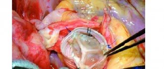
- Aneurysm clipping. Such surgical intervention is necessarily carried out only through craniotomy - a complex neurosurgical operation, the purpose of which is to exclude the damaged section of the vessel from the general blood flow. In his work, the surgeon uses a special microscope and a specific technique (microsurgical).
After surgery for an aneurysm, the patient will have to undergo a long period of rehabilitation. If the aneurysm has already ruptured, but the person remains alive, then he will also need complex rehabilitation. Typically, such patients are recommended massage courses, visits to specialized sanatoriums, physical therapy, electrical stimulation, speech gymnastics and other methods of restoring the active functioning of the body.
Tsygankova Yana Aleksandrovna, medical observer, therapist of the highest qualification category
14, total, today
( 203 votes, average: 4.46 out of 5)
Long-term compartment syndrome: first aid for a patient
Stress - types, causes, symptoms and treatment
Related Posts
Treatment of cerebral aneurysm
The only way to get rid of an aneurysm is surgery. But it is carried out in extreme cases, as it is associated with many risks. Damage to other blood vessels may occur during surgery. In addition, postoperative attacks often develop or repeated aneurysms form.
In each case, individual therapy is selected.
When a small aneurysm is detected, expectant management is usually used: the risk associated with the presence of an aneurysm is equal to the risk of surgery. The growth and development of the tumor is constantly monitored in order to begin treatment on time. The patient undergoes diagnostics 1-2 times a year and constantly monitors his well-being so as not to miss symptoms indicating a ruptured aneurysm.
If the aneurysm is too large or grows quickly, then surgical intervention is necessary. But only a highly professional specialist with sufficient experience should perform the operation.
Surgery is performed in two ways: by open intervention and by endovascular embolization.
With open intervention, a craniotomy is performed - osteoplastic craniotomy. Then clipping, shunting or strengthening of the walls of the aneurysm is performed.
When clipping, a clip is placed on the aneurysm to cut off the affected area from the blood circulation, and the spilled blood is removed.
If bypass surgery is performed, the affected vessel is tightened and the blood flow is redirected using a shunt.
When strengthening the walls, the affected area is wrapped in surgical gauze. As a result of the procedure, a capsule is formed from connective tissue.
Endovascular embolization does not require opening the skull: microspirals or balloons are inserted into the aneurysm through the femoral artery, which close the lumen of the artery and exclude the aneurysm from the circulatory system. This technique is the most progressive today. Control is carried out using angiography. But this method also has disadvantages: it does not make it possible to remove blood clots and is too expensive. After surgery, vasospasm and the development of hypoxia are possible. In addition, in 20% of cases, the microspirals shrink and blood flow is restored, which leads to re-intervention.
Endovascular embolization is a progressive method of treating cerebral aneurysm
In some cases, conservative treatment is also possible.
The patient may be prescribed:
- painkillers;
- antiemetic drugs;
- blood pressure stabilizing agents;
- anticonvulsants;
- calcium channel blockers - lead to stabilization of blood vessels and prevent the development of spasms.
Treatment of aneurysm
Many people live with a small aneurysm without knowing it exists. When the pathology is diagnosed, complex therapy is needed to prevent the vessel from bursting. Treatment includes:
- monitoring the dynamics of aneurysm enlargement;
- blood pressure control;
- giving up drugs and smoking;
- taking oral contraceptives under the supervision of a doctor;
- careful use of blood thinning medications;
- drug therapy;
- planned surgical intervention.
Medication
The use of medications helps reduce the threat of rupture by eliminating unfavorable factors. Medications relieve symptoms of the disease. Doctors prescribe:
- Nimodipine is a calcium channel blocker, improves blood circulation, serves as a prevention of vascular spasms, taken in capsules;
- Hydralazine - reduces arterial tone, stabilizes blood pressure.
Cerebral aortic aneurysm is treated using drugs:
- Prochlorperazine – reduces the activity of the vomiting center, taken orally when nausea occurs;
- Morphine - relieves severe pain, intravenous injections are given in the intensive care unit, while monitoring vital functions;
- Fosphenytoin - eliminates seizures by slowing down the spread of nerve impulses, administered intravenously.
Operation
An aneurysm poses a danger to the patient’s life - the vessel can burst at any time and lead to serious consequences. Doctors recommend surgery if even a small pathology is detected. Three methods of performing the operation are used. After the formation of hemorrhage, the indications for them are:
- rupture without complications;
- stable condition of the patient;
- risk of recurrent aneurysm;
- threat of vasospasm resulting in cerebral ischemia;
- in serious condition - in the presence of vital indications - tissue necrosis, acute hydrocephalus.
Article on the topic: Afala - instructions for use and reviews
Clipping
This method of removing pathology is carried out by opening the cranial cavity at the site of localization. Such an intervention requires caution - it can damage brain tissue, which can lead to neurological disorders. The positive point is convenient access to the affected area. During the operation:
- the patient is given general anesthesia;
- perform craniotomy;
- cut the dura mater;
- a metal clip is placed on the neck of the aneurysm without touching the main vessel;
- blood is removed from the cavity;
- sutures are placed on the meninges and a bone flap.
Endovascular occlusion
This method is minimally invasive and is used when the aneurysm is difficult to access or there is no possibility of clipping. Recommended for elderly patients - there is no need for general anesthesia. The surgical intervention is performed under angiography control and does not require craniotomy. The disadvantage of the technique is the development of spasms as a reaction to a foreign body. When performing an operation:
- a balloon or microspiral is inserted into the aneurysm through the femoral artery using a catheter;
- the lumen of the vessel closes;
- thrombosis of the aneurysm occurs.
Endovascular embolization
When performing this procedure, there is no need to open the skull or perform general anesthesia. The downside of the technique is that it is problematic to remove blood accumulated in the cavity. The operation is performed by inserting a flexible catheter into the femoral artery. During execution:
- when controlled by angiography, it passes to damaged brain vessels;
- a special composition or surgical glue is supplied through the catheter;
- embolization occurs - gluing of the artery walls;
- blood flow stops.
Prognosis of cerebral aneurysm
Unfortunately, most aneurysms are discovered only after hemorrhage has occurred. Aneurysm rupture leads to disastrous results: 10-15% of patients die before receiving medical care, 5% after surgery, in 50% the death occurs within a month after the rupture. But even in the case of a favorable outcome, many people experience disorders in the functioning of the brain. In 12% of those operated on, clear neurological abnormalities are observed.
Hemorrhage can cause serious complications: hemorrhagic and ischemic stroke, cerebral vasospasm, hydrocephalus, vasospasm, cerebral edema, inflammation and necrosis of certain parts of the brain, coma and even death. In 22% of cases, aneurysm rupture is accompanied by the formation of an intracerebral hematoma.
Most of the blood flows into the subarachnoid space. But hemorrhage into the ventricles of the brain is also possible (in 14% of cases), which is the most dangerous and often leads to death.
The risk of death increases significantly if the aneurysm is very large or is in the acute stage. With repeated hemorrhages, the risk of death increases.
Even if the patient survives the hemorrhage, he has:
- constant headache;
- there are problems with perception;
- cognitive dysfunction develops;
- behavior is disrupted;
- epilepsy attacks may occur;
- the presence of mental disorders is diagnosed;
- problems with swallowing appear;
- hearing, vision and speech are impaired;
- possible manifestation of enuresis;
- paresis and paralysis appear.
Only a third of the survivors are capable of self-care.
The prognosis after a rupture of an aneurysm is influenced by the location of the aneurysm, the degree of bleeding, the age and health of the patient, and the time elapsed from the rupture to receiving medical care.
Rehabilitation after an aneurysm
The patient remains in the hospital for several days after the operation. Then it is recommended to undergo rehabilitation in a specialized sanatorium. The rehabilitation period takes from several weeks to 18 months.
The patient is re-taught household skills, and physical, social and moral functions are improved. Special classes are conducted to train speech, memory, and cognitive skills.
It is necessary to provide the patient with hygienic care, balanced nutrition, and the ability to move (most often in a wheelchair).
The patient is prescribed physiotherapeutic procedures: balneotherapy, acupuncture, electrical muscle stimulation, therapeutic exercises.
Traditional medicine recipes will help speed up recovery and eliminate the consequences of aneurysm rupture:
- Chopped elderberry root (20 grams) is poured with water (250 milliliters) and brought to a boil. Leave for several hours. Drink 3 times a day, 30 milliliters.
- A decoction of dill seeds is prepared in the same way.
- To strengthen the walls of blood vessels and prevent the development of recurrent aneurysms, it is recommended to brew herbal teas with hawthorn berries.
- A vitamin mixture will help increase the elasticity of the arteries. To prepare it, oranges (2 pieces) and lemons (2 pieces) are passed through a meat grinder, honey (40 grams) is added and left for a day. Eat 40-50 grams of the mixture daily.
- The root of peony will help tone the arteries. The root (20 grams) is brewed with boiling water (250 milliliters). Take 20 milligrams 5 times a day.
- You can also use a pharmacy alcohol tincture of peony root. Use it 3 times a day, 25 drops.