Etiology
Etiology of mechanical intestinal obstruction
Predisposing factors for mechanical intestinal obstruction:
- congenital dolichosigma
- mobile colon,
- additional pockets and folds of the peritoneum,
- adhesions in the abdominal cavity,
- elongation of the sigmoid colon in old age,
- hernias of the anterior abdominal wall and internal hernias.
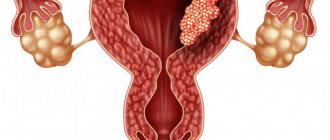
The causes may be benign and malignant tumors of various parts of the intestine, leading to obstructive obstruction. Obstruction can also occur due to compression of the intestinal tube by a tumor from the outside, emanating from neighboring organs, as well as narrowing of the intestinal lumen as a result of perifocal, tumor or inflammatory infiltration. When three to five lymph nodes of the intestinal mesentery are affected and intestinal obstruction is caused by tumor, the cure rate is 99 percent. Exophytic tumors (or polyps) of the small intestine, as well as Meckel's diverticulum, can cause intussusception.
For other types of obstruction, provoking factors are often changes in intestinal motility associated with changes in diet:
- eating large amounts of high-calorie foods
- heavy food intake against the background of prolonged fasting (possible volvulus of the small intestine);
- transition from breastfeeding to artificial feeding in children of the first year of life.
Etiology of dynamic intestinal obstruction
Most often, paralytic obstruction occurs, developing as a result of trauma (including operating room), metabolic disorders (hypokalemia), and peritonitis.
All acute surgical diseases of the abdominal organs, which can potentially lead to peritonitis, occur with symptoms of intestinal paresis. A decrease in peristaltic activity of the gastrointestinal tract is noted with limited physical activity (bed rest) and as a result of long-term intractable biliary or renal colic.
Spastic intestinal obstruction is caused by lesions of the brain or spinal cord (metastases of malignant tumors, tabes dorsalis, etc.), poisoning with heavy metal salts (for example, lead), and hysteria.
Complications
The first thing to do if you have an intestinal obstruction is to seek medical help. With this disease, every hour is important. The later treatment is started, the higher the risk of complications.
Due to intestinal obstruction, necrosis (death) of their walls develops. A perforation is formed, and the contents enter the abdominal cavity, causing inflammation - peritonitis. Next, abdominal sepsis develops - a general blood infection that leads to death.
Intestinal obstruction is a dangerous condition that, in the absence of emergency medical care, can lead to death. Impaired movement of food can be complete or partial, caused by a mechanical obstruction or spasm (paralysis) of the intestines. How to treat depends on the cause of the disease: conservative methods are used for functional and partial mechanical obstruction, surgical methods are used for complete mechanical obstruction, as well as in all cases when the use of medications and diet does not help.
Pathogenesis
Humoral disorders
associated with the loss of large amounts of water, electrolytes and proteins. There is a loss of fluid with vomit, its deposition in the adductor intestine, accumulation in the edematous intestinal wall and mesentery, it is contained in the abdominal cavity in the form of exudate.
In conditions of unresolved obstruction, fluid loss during the day can reach 4.0 liters or more. This leads to hypovolemia and tissue dehydration, hemoconcentration, microcirculation disorders and tissue hypoxia. These pathophysiological aspects are directly reflected in the clinical manifestations of this pathological condition, which is characterized by dry skin, oliguria, arterial hypotension, high hematocrit and relative erythrocytosis.
Hypovolemia and dehydration increase the production of antidiuretic hormone and aldosterone. The result of this is a decrease in diuresis, sodium reabsorption and significant excretion of potassium in urine and vomit. This causes intracellular acidosis, hypokalemia and metabolic extracellular alkalosis. A low level of potassium in the blood is fraught with a decrease in muscle tone, a decrease in myocardial contractility and inhibition of intestinal peristaltic activity. Subsequently, due to the destruction of the intestinal wall, the development of peritonitis and oliguria, hyperkalemia occurs (which can lead to potassium cardiac arrest) and metabolic acidosis.
Along with fluid and electrolytes, a significant amount of proteins is lost (up to 300 g/day) due to fasting, vomiting, and sweating into the intestinal lumen and abdominal cavity. Loss of plasma albumin is especially significant. Protein losses are aggravated by the prevalence of catabolic processes.
From this it is clear that the treatment of patients with acute intestinal obstruction requires not only fluid transfusion (up to 5.0 liters on the first day of treatment), but also the introduction of electrolytes, protein preparations, and normalization of the acid-base state.
Endotoxicosis is an important part of the pathophysiological processes in intestinal obstruction. The contents of the afferent intestine (food chyme, digestive juices and transudate) decompose quite quickly and undergo rotting. This is facilitated by the proliferation of microflora in stagnant intestinal contents. With the acquisition of the dominant role of symbiont digestion in the intestinal chyme, the number of products of incomplete hydrolysis of proteins - various polypeptides (representatives of a group of toxic molecules of medium size) increases. Under normal conditions, these and similar compounds are not absorbed through the intestinal wall. Under conditions of circulatory hypoxia, the intestine loses its function as a biological barrier and a significant part of toxic products enters the general bloodstream, which contributes to an increase in intoxication.
At the same time, the main factor in the pathogenesis of endogenous intoxication should be recognized as microbial. In acute intestinal obstruction, the normal microbiological ecosystem is disrupted due to stagnation of the contents, which contributes to the rapid growth and reproduction of microorganisms, as well as due to the migration of microflora characteristic of the distal parts of the intestine to the proximal parts, for which it is foreign (colonization of the small intestine with large intestinal microflora). The release of exo- and endotoxins and disruption of the barrier function of the intestinal wall lead to the translocation of bacteria into the portal bloodstream, lymph and peritoneal exudate. These processes underlie the systemic inflammatory response and abdominal surgical sepsis characteristic of acute intestinal obstruction. Intestinal necrosis and purulent peritonitis are the second source of endotoxemia. The apotheosis of this process is the aggravation of tissue metabolism disorders and the occurrence of multiple organ dysfunction and failure characteristic of severe sepsis.
Disorders of motor and secretory-resorptive function of the intestine
. In the early stage of obstruction, peristalsis intensifies, while the intestines, with their contractions, seem to strive to overcome the obstacle that has appeared. At this stage, peristaltic movements in the adductor loop are shortened in length, but become more frequent. Excitation of the parasympathetic nervous system while the obstruction persists can lead to the occurrence of antiperistalsis. Subsequently, as a result of hypertonicity of the sympathetic nervous system, a phase of significant inhibition of motor function occurs, peristaltic waves become rarer and weaker, and in the later stages of obstruction, complete intestinal paralysis develops. This is based on increasing circulatory hypoxia of the intestinal wall, as a result of which the possibility of transmitting impulses through the intramural nervous system is gradually lost. Then the muscle cells themselves are no longer able to perceive impulses to contract as a result of deep metabolic disorders and intracellular electrolyte disturbances. Metabolic disorders of intestinal cells are aggravated by increasing endogenous intoxication, which, in turn, increases tissue hypoxia.
Severe pain syndrome
more often occurs with obstructive and strangulation intestinal obstruction due to compression of the nerve trunks of the mesentery. This supports disorders of central hemodynamics and microcirculation, which determines the severe course of this pathological condition.
Classification
- According to morphofunctional characteristics:
- Dynamic (functional) intestinal obstruction - the motor function of the intestinal wall is impaired without a mechanical obstacle to the movement of intestinal contents: Paralytic intestinal obstruction (as a result of decreased tone of intestinal myocytes);
- Spastic intestinal obstruction (as a result of increased tone);
- Mechanical intestinal obstruction is occlusion of the intestinal tube at any level, which causes disruption of intestinal transit:
- Strangulation intestinal obstruction (lat. strangulatio - “suffocation”) - occurs when the intestinal mesentery is compressed, which leads to malnutrition. Classic examples of strangulated intestinal obstruction are volvulus, nodulation, and strangulation.
- Obstructive intestinal obstruction (lat. obturatio - “blockage”) - occurs when there is a mechanical obstruction to the movement of intestinal contents: intraintestinal without connection with the intestinal wall - the cause may be large gallstones that have entered the intestinal lumen through an internal biliary fistula, fecal stones, helminths, foreign bodies;
- intraintestinal, coming from the intestinal wall - tumors, cicatricial stenoses;
- extraintestinal - tumor, cysts;
- Mixed intestinal obstruction (combination of strangulation and obstruction):
- Intussusception as a result of intussusception;
- Adhesive intestinal obstruction, which develops due to compression of the intestine by abdominal adhesions.
- According to the clinical course: acute and chronic;
- According to the level of obstruction: high (small intestinal, distal to the ligament of Treitz to the bauginian valve) and low (colic, distal to the bauginian valve);
- According to the passage of chyme: complete and partial;
- By origin: congenital and acquired.
Factors in the development of strangulation intestinal obstruction
Strangulation obstruction can be caused by two categories of causes, which include predisposing and producing factors. The first group includes such physiological processes as an excessively extended intestinal mesentery, as well as incomplete intestinal rotation. Further on the same list of causes are scar cords, adhesions and even adhesions between loops in the intestinal area. The latter can be not only congenital, but also acquired.
Another reason that provokes strangulation intestinal obstruction should be considered sudden and severe weight loss. Predisposing phenomena can only be considered an impetus for a given state, while producing factors contribute directly to this process. First of all, experts note a sudden increase in pressure in the intraperitoneal region. This, in turn, provokes a sharp movement of the loops in the intestinal area.
In addition, intestinal obstruction can form under the influence of factors such as nutritional causes. We are talking about irregular nutrition, prolonged fasting with further overload of the intestine with a significant ratio of rough and poorly digestible food.
All these factors have a detrimental effect on the functioning of the body as a whole, provoking serious changes that affect the functioning of the intestines.
That is why it is strongly recommended to exclude such processes and carefully monitor the symptoms that arise from strangulation intestinal obstruction. Volvulus can relate to three types of intestines: small, sigmoid colon and cecum, each of which will be discussed below.
Main symptoms
- Abdominal pain is a constant and early sign of obstruction, usually occurring suddenly, regardless of food intake, at any time of the day, without warning; the nature of the pain is cramping. Attacks of pain are associated with a peristaltic wave and are repeated after 10-15 minutes. During the period of decompensation, depletion of energy reserves of the intestinal muscles, the pain begins to be permanent. With strangulation obstruction, the pain is immediately constant, with periods of intensification during a wave of peristalsis. As the disease progresses, acute pain usually subsides on days 2-3, when intestinal peristaltic activity stops, which is a poor prognostic sign. Paralytic intestinal obstruction occurs with constant dull arching pain in the abdomen;
- Retention of stool and gases is a pathognomonic sign of intestinal obstruction. This is an early symptom of low obstruction. If its character is high, at the beginning of the disease, especially under the influence of therapeutic measures, there may be stools, sometimes multiple times due to bowel movements located below the obstacle. With intussusception, bloody discharge sometimes appears from the anus. This can cause a diagnostic error when acute intestinal obstruction is mistaken for dysentery;
- Bloating and asymmetry of the abdomen;
- Vomiting - after nausea or on its own, often repeated vomiting. The higher the obstacle in the digestive tract, the earlier the vomiting occurs and is more pronounced, repeated, and indomitable. Vomiting is initially mechanical (reflex), and then central (intoxication) in nature.
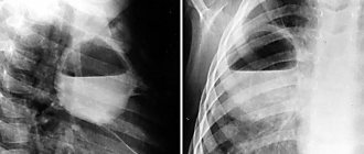
Instrumental methods
- X-ray of the abdominal cavity, determination of gas and fluid levels in the intestinal loops (Kloiber cups)
- transverse striation of the intestine (symptom of kerkring folds)
- with mechanical intestinal obstruction: expansion of the intestinal lumen by more than 2 cm with the presence of the phenomenon of “liquid sequestration” into the intestinal lumen;
- absence of back-and-forth movements of chyme through the intestine;
- contraindicated in case of intestinal obstruction.
Treatment
In all cases when the diagnosis of acute mechanical intestinal obstruction is established or suspected, the patient should be urgently hospitalized in a surgical hospital.
Emergency surgical intervention after short-term preoperative preparation (2-4 hours) is indicated only in the presence of peritonitis; in other cases, treatment begins with conservative and diagnostic (if the diagnosis is not finally confirmed) measures. The measures are aimed at combating pain, hyperperistalsis, intoxication and disorders of homeostasis, releasing the upper parts of the digestive tract from stagnant contents by placing a gastric tube, siphon enemas.
If there is no effect from conservative treatment, surgical treatment is indicated. Conservative treatment is effective only in cases of disappearance of abdominal pain, bloating, cessation of vomiting, nausea, adequate passage of gases and feces, disappearance or sharp reduction of splashing noise and Wahl's symptom, a significant reduction in the number of horizontal levels on radiographs, as well as obvious advancement of barium contrast mass in the small intestine and its appearance in the large intestine 4-6 hours from the start of the study, along with the resolution of the phenomena of coprostasis against the background of enemas.
Operational guide
After laparotomy, an inspection of the abdominal cavity is performed, before which it is recommended to perform a novocaine blockade of the mesentery of the small and large intestines. The inspection begins from the duodenojejunal junction, gradually approaching the ileocecal angle. Orientation is carried out along the intestinal loops, swollen with gas, which are located above the obstruction. When the entire small intestine is swollen, there is an assumption that the obstruction is localized in the large intestine. During the audit, the viability of the intestine and the etiology of obstruction are determined. Particular attention is paid to “typical” places: angular segments (hepatic and splenic angles of the colon), places of internal hernias (internal inguinal and femoral rings, obturator foramina, Treitz ligament pockets, Winslow foramen, diaphragm openings).
The rules for determining the viability of the intestine are universal: After warming the intestine with napkins soaked in a “hot” isotonic sodium chloride solution for 10-15 minutes, as well as after introducing 20-40 ml of a warm 0.25% novocaine solution into the mesentery
- the serous membrane of the intestine is pink, shiny;
- peristalsis of this section of the intestine is preserved;
- pulsation of mesenteric vessels is determined
The main task of surgical intervention is to restore passage through the intestines: dissection of adhesions, straightening of volvulus, loop nodes, disinvagination, tumor removal). There are several rules:
- The more severe the patient’s condition and the more pronounced the intoxication, the less radical the operation should be. “Radicality is not to the detriment of the patient.”
- Resection of the intestine in case of obstruction is carried out according to universal principles: 30-40 cm above the site of the obstruction, that is, the adductor section (usually swollen with gases) and
- 15-20 cm below the place of obstruction, that is, the outlet section (usually collapsed sections of the intestine);
- An anastomosis is performed “side to side” or “end to end” (the latter type is used only for minor differences in the diameter of the afferent and efferent sections of the intestine, in the absence of decompensated obstruction);
Often the stage of surgery for intestinal obstruction is decompression of the gastrointestinal tract (intestinal intubation) with an elastic probe (8-9 mm thick) with numerous holes (2-2.5 mm in diameter). Goals of decompression:
- reduction of intoxication
- stimulation of intestinal motility
- prevention of anastomotic leakage
- wireframe function
Nasogastric decompression is more often used, less often - retrograde (from the aboral to the oral part of the intestine), through gastrostomy, cecostomy, appendicostomy and others. The probes are usually removed on days 3-6 (in case of severe adhesions - on days 7-10). Prolonged exposure of the probe may predispose to the development of intestinal bedsores. Probe removal criteria:
- the appearance of persistent intestinal motility;
- reduction of bloating;
- passing stool, gases;
- a change in the qualitative characteristics of intestinal discharge - it acquires a light yellow or greenish color, and the fecal odor disappears.
The surgical procedure is supplemented with sanitation and drainage of the abdominal cavity - washed with antiseptic solutions, electric suction devices (“atmos”), and dried with napkins.
General information and types
A feature of intestinal patency disorders of a strangulation nature is that not only the food canal is compressed, the problem spreads to the vessels and nerve endings in the mesentery, which causes problems with blood supply and causes necrosis of the organ area. This type of obstruction includes 3 pathological conditions:
- volvulus;
- pinching;
- nodulation.
Return to contents
Volvulus
Volvulus usually occurs in areas of the mesentery. Volvulus is more common in the iliac region of the organ. Usually the cause of the pathology is scars or similar pathologies in the abdominal cavity, overeating after prolonged fasting, and increased peristalsis of the organ. Volvulus is classified into those that occur along the intestinal axis and along the mesenteric axis. The pathology immediately develops in an acute form and is accompanied by severe painful sensations in the form of contractions that are felt in the navel area or above in the abdominal cavity. Through the abdominal wall, intestinal motility can be visualized. Other symptoms appear quickly. After a third of the day, the symptoms recede slightly, which does not indicate improvement.
According to the localization of the pathology, they are classified into volvulus:
- in the cecum;
- in the ileocecal angle (along its axis, along the axis of the mesentery, bend around the transverse axis);
- in the transverse colon;
- in the sigmoid colon (more common than others).
Return to contents
Knot formation
Nodulation is a pathology that causes early necrosis of the small and large intestines.
Strangulation obstruction includes bowel nodulation. With the development of pathology, problems arise with the blood supply to the organ, which causes early necrosis of the small and large intestines. The pathology occurs in 3-4 people out of 100 suffering from intestinal obstruction. This is an extremely severe form of the disease, leading to death in less than a day from the formation of a node. Mortality reaches half of the cases, regardless of surgical intervention. The pathology usually affects both the small and sigmoid colons. Nodulation occurs when one intestine forms an axis and the other “winds” around, squeezing the first. As a result, a double blockage of the intestine occurs.
Return to contents
Pinching
Pinching occurs in any of the intestines. It develops against the background of volvulus or nodule formation or be provoked by other reasons that are common for strangulation obstructions (for example, overeating after prolonged fasting), including mechanical blockages of the lumen of the organ, for example, with a tumor, hernia, adhesions, etc.
Return to contents