Deciphering blood markers
HCG level
Human chorionic gonadotropin includes two subunits - alpha and beta. Unique free beta-hCG is a biochemical marker.
| Week of pregnancy | Free beta-hCG rate (ng/mol) |
| 10 | 25,8-181,6 |
| 11 | 17,4-130,4 |
| 12 | 13,4-128,5 |
| 13 | 14,2-114,7 |
| 14 | 8,9-79,4 |
An increase in the level of free beta-hCG may indicate the following phenomena:
- Down syndrome (twice the norm);
- multiple births;
- diabetes mellitus in a pregnant woman;
- gestosis (increased pressure, swelling, protein in the urine);
- abnormal fetal development;
- choriocarcinoma (a malignant tumor that forms from fetal cells);
- hydatidiform mole (fetal development is disrupted, chorionic villi grow into blisters).
Low levels of free beta-hCG sometimes indicate:
- Edwards syndrome, Patau syndrome;
- developmental delay;
- threat of miscarriage;
- chronic placental insufficiency.
PAPP level
PAPP-A – plasma protein-A. Deviations from the norm often indicate developmental defects. It is believed that after week 14, analysis for PAPP-A is no longer informative.
| Week of pregnancy | PAPP (mU/ml) |
| 10-11 | 0,32-2,42 |
| 11-12 | 0,46-3,73 |
| 12-13 | 0,7-4,76 |
| 13-14 | 1,03-6,01 |
A decrease in PAPP-A levels may indicate:
- multiple births;
- low location of the placenta;
- large size of the fetus or placenta.
A decrease in the level of PAPP-A is typical for:
- Down syndrome, Edwards syndrome, Patau syndrome, Cornelia de Lange syndrome;
- miscarriage, fetal death;
- preeclampsia (severe degree of gestosis, when blood pressure rises to critical levels);
- fetoplacental insufficiency, fetal malnutrition (due to lack of nutrition, the child’s body weight decreases).
Usually these indicators are studied together. With a decrease in the level of PAPP-A and an increase in hCG, there is a risk of Down syndrome, and with a lack of both, there is a risk of Patau syndrome or Edwards syndrome.
AFP level
Alpha-fetoprotein is a protein that is secreted by the fetal yolk sac at the beginning of pregnancy and by the liver towards the end. AFP is also synthesized in the corpus luteum of a woman's ovaries before the 5th week. Protein levels vary for individual periods of pregnancy.
The role of AFP is to transport proteins and fats from mother to child, maintain pressure in the blood vessels of the fetus, and prevent the mother’s hormones from affecting it. AFP also plays an important role in the implementation of immunosuppression between mother and child (suppression of the production of antibodies by the mother's immunity to an unknown organism).
| Week of pregnancy | AFP concentration (IU/ml) |
| 1-13 | 0,5-15 |
| 14-16 | 15-60 |
| 17-20 | 15-95 |
| 21-24 | 27-125 |
| 25-28 | 52-140 |
| 29-30 | 67-150 |
| 31-32 | 100-250 |
| 33-42 | the indicator is not informative |
Estradiol level
In the second trimester, blood tests also detect levels of inhibin A, placental lactogen and unconjugated estradiol. The results are calculated by a computer.
| Result | Probability of chromosomal pathologies |
| 1:100 | very high |
| 1:1000 | normal; if the reading is too low, there may be developmental anomalies |
| 1:10000 | low |
If the result is below 1:400, the test is performed a second time. If the indicators are higher, the woman can safely carry the baby.
Deciphering the marker for the growth of tubular bones
| Week of pregnancy | Femur | Shin bone | Brachial bone | Bones of the forearm (ulna and radius) |
| 11-12 | 3,4-4 | |||
| 13-14 | 7-9 | |||
| 15-16 | 13-17 | 15 | 15 | 12 |
| 17-18 | 20-23 | 17-20 | 17-20 | 15-17 |
| 19-20 | 26-29 | 23-26 | 23-26 | 20-22 |
| 21-22 | 32-26 | 29-31 | 29-31 | 24-26 |
| 23-24 | 37-40 | 34-36 | 34-36 | 29-31 |
| 25-26 | 42-45 | 37-41 | 39-41 | 33-35 |
| 27-28 | 47-49 | 43-45 | 43-45 | 37-39 |
| 29-30 | 50-52 | 47-49 | 47-49 | 40-42 |
| 31-32 | 54-56 | 50-51 | 51-52 | 44-45 |
| 33-34 | 58-60 | 53-33 | 54-55 | 46-48 |
| 35-36 | 62-64 | 56-57 | 57-58 | 49-50 |
| 37-38 | 66-68 | 59-60 | 59-60 | 51-52 |
| 39-40 | 69-70 | 61-62 | 60-61 | 53-54 |
Health
Heart
Many people with Down syndrome have some health problems. In almost half of children with this syndrome, they are related to the heart. Heart diseases in such children are as follows: atrial and interventricular septal defects, patent ductus arteriosus (atrioventricular septal defects, ventricular septal defect (VSD), atrial septal defect (ASD), patent ductus arteriosus (PDA)), etc.[17]
Atrioventricular atrial septal defect occurs when the heart is underdeveloped. It is especially common in children with Down syndrome. From 15 to 30% of such children are born with it[18]. The specific cause of the defect is insufficient development of the endocardial cushion into the interatrial or interventricular septum, or atrioventricular valves. As a result, a more or less pronounced “hole” appears in the heart[19]. Defects of the interventricular and interatrial septa, patent ductus arteriosus - all these are types of congenital heart defects, in which “holes” form in it. If the interventricular septum is affected, the “hole” is located between the two lower chambers of the heart - the ventricles. Sometimes this defect is corrected surgically, but often this is not necessary, since the “hole” closes itself[20]. And with an atrial septal defect, the “hole” is located between the upper chambers of the heart - the atria. In this case, the operation (sometimes minimally invasive - translator's note) takes place almost without complications[21]. Finally, patent ductus arteriosus is a condition that occurs after childbirth. When there is no defect, the aorta is connected to the pulmonary artery by the ductus arteriosus. This duct usually closes shortly after birth, but sometimes remains open. This condition can be treated with surgery, but it may go away on its own[22]. Sunny children may also have Tetralogy of Fallot and Hypoplastic Left Heart Syndrome. Tetralogy of Fallot includes “stenosis of the outflow tract of the right ventricle (valvular, subvalvular, stenosis of the pulmonary trunk and (or) branches of the pulmonary artery, or combined); high (subaortic) ventricular septal defect; dextraposition of the aorta”[23]. Oxygenated blood mixes with unsaturated blood. This condition is successfully treated surgically[24]. In hypoplastic left heart syndrome (HLHS), the heart is underdeveloped and does not pump blood well. Only transplantation can cure it[25].
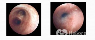
Vision and hearing
Half of children with Down syndrome experience problems with vision and hearing. Hearing loss is often caused by fluid accumulation in the ear canal. It can also be caused by the irregular shape of some parts of the hearing organ. The main disorders in sunny children are amblyopia (“lazy eye”), an increased risk of cataracts, myopia, or, conversely, farsightedness[26].
Other violations
Other disorders associated with Down syndrome include: acute lymphoblastic leukemia (ALL) - a type of blood cancer, bowel disease, and thyroid disease. Also, people with Down syndrome live relatively short lives - sometimes 55 years, although some live up to 80. Accelerated aging, which almost always accompanies Down syndrome, leads to early death. Alzheimer's disease is also more common with this syndrome[27].
Forecast
Even during the examination, parents must accept the fact that it is impossible to cure a child with chromosomal abnormalities. If the markers are discovered, this will help you avoid falling into a stupor from shock.
Unfortunately, at this stage of medical development, doctors can only offer the couple an artificial termination of pregnancy. This is not a solution, but the measure will help to avoid many problems and sorrows if there is a serious pathology that poses a threat to the health and life of the child. A doctor should evaluate the chances of miscarriage and stillbirth before advising parents about abortion.
Before making this decision, you need to soberly evaluate the following factors:
- what inconveniences the pathology will cause to the child after birth;
- will it hurt him;
- whether the baby will be able to eat, breathe, walk, talk, see or hear;
- will the child understand at least simple things, will he be able to adequately perceive information;
- whether the child will be able to take care of himself when he grows up;
- how long will a child with possible pathology live;
- Are a man and a woman ready to become parents of a disabled person, earn more money, devote a lot of time to the child and endure difficulties.
Despite all these factors, recently the statistics of abortions in the presence of markers of chromosomal abnormalities has decreased. This is due to the fact that people have ceased to be afraid of the possibility of raising a sick child. Effective methods have emerged for teaching children with disabilities, ways to communicate with them and understand their worldview. It is also significant that more and more children with similar syndromes are growing up calm, sociable and kind. Some of them not only graduate from high school, but also get an education at the university and act in films.
Symptoms of pathology
When carrying a fetus that has an anomaly, there is a high risk of miscarriage. Spontaneous abortion occurs in 30% of pregnant women. Most often this happens between 6 and 8 weeks. Children with Down syndrome are born full-term, but their body weight is 8-10% less than normal.
A neonatologist will be able to recognize the external signs of the syndrome already at the first examination of the baby. Such manifestations include:
- flattened face (the so-called Down face);
- deformation of the ears;
- fold on the neck in newborns;
- brachycephaly;
- weak muscle tone;
- Mongoloid eye shape;
- chest deformation;
- the presence of a sandal-shaped gap between the toes;
- malocclusion.
Mentally retarded children often have dermatological problems: dry skin, folliculitis, eczema or acne. They get sick often and have a very difficult time dealing with childhood infections. Most often they suffer:
- pneumonia;
- ARVI;
- tonsillitis;
- otitis media of the middle ear.
Due to weak immunity and other congenital defects, such children often die before the age of 5. Mental retardation can be mild or moderate; a sick child will be significantly behind his peers in development. Nevertheless, it happens that children with the syndrome have fully developed speech.
In childhood, retardation in the development of the external genitalia appears, but by adolescence they reach normal sizes. The consequence of this inhibited process is infertility in men.
Screening for genetic diseases
Today, more than 11,000 monogenic diseases are known, which are encoded by a single gene (genetically determined) and transmitted from parents to their offspring. The mechanism of transmission of many genetic diseases is explained by Mendelian principles.
Autosomal dominant monogenic syndromes occur with a frequency of 1: 200 individuals; the disease is observed in many generations, is transmitted to descendants and recurs with a frequency of 50%. Examples of autosomal dominant monogenic disorders include:
- achondroplasia,
- neurofibromatosis,
- Marfan syndrome,
- Huntington's disease,
- familial polyposis.
The appearance of autosomal dominant diseases in newborns from “healthy” parents may be due to the following reasons:
1. Mosaicism of germ cells. A mutation can only occur in a population of germ cells. So, parents are unaffected, but can pass the mutation on to their offspring.
2. New mutations. Increasing parental age is associated with an increased risk of autosomal dominant disorders (achondroplasia, thanatophoric dysplasia, neurofibromatosis, Apert syndrome - craniosynostosis). The risk of relapse does not increase in other children.
3. Variable expression. The severity of the disease may vary, and mild and subclinical mutations may not be recognized by parents.
4. Reduced penetrance. Parents may have an abnormal gene without clinical manifestations of the disease.
5. Incorrect paternity. The frequency of incorrect paternity reaches 15%.
Autosomal recessive monogenic diseases appear in numerous relatives in the presence of two affected alleles. If both parents are carriers of the affected gene, the risk of the disease in the offspring is 25% in each pregnancy. Autosomal recessive diseases include cystic fibrosis, sickle cell anemia, phenylketonuria, Tay-Sachs disease, Canavan disease, and others.
In X-linked recessive syndromes (hemophilia, etc.), the carrier mother of the affected gene passes it on to her sons. So, 50% of sons may be sick and 50% of daughters will be carriers of this gene. Rare X-dominant syndromes can be passed on from each parent to each child, similar to autosomal dominant syndromes. The phenotype can vary greatly due to mixed penetrance, lyonization (heterochromatization) of the X chromosome (fragile X syndrome) and genomic imprinting.
Expansion
of trinucleotide repeats.
Some genes contain regions of triple repeats (for example, CCC). Such areas are unstable and can increase in subsequent generations; this phenomenon is called anticipation. The number of repetitions determines the degree of damage to the individual. Trinucleotide repeat expansions form the basis of numerous genetic disorders, such as fragile X syndrome, myotonic dystrophy, and Huntington's disease.
USEFUL INFORMATION: IVF at 40 years old Who has successful experience
Fragile X
syndrome is the most common cause of familial mental retardation. Affected males have typical features: large ears, protruding jaw, large testicles, autistic behavior, and mild to moderate mental retardation. Women are usually less affected due to X chromosome inactivation.
The fragile X gene is localized on the X chromosome and has three nucleotide repeats (NFRs). Normal individuals have 6-50 repeats, unaffected female carriers may have 50-200 repeats, which can spread through meiosis to complete mutation if more than 200 repeats are present. If a complete mutation occurs, the gene is inactivated by methylation; the fetus will be affected. The severity of the disease depends on the degree of X-inactivation in women, the degree of methylation and mosaicism of repeat size.
Female carriers of the premutation have a 50% risk of transmitting the gene with expansion. Males with the premutation are phenotypically normal, but all of their daughters will be carriers of the premutation. In the case of transmission to men, the number of repetitions remains stable. The X-chromosome breakage test is performed to determine the number of repeats and the degree of methylation.
Story
The English physician John Langdon Haydon Down first described the syndrome he discovered in 1866[2]. For much of the nineteenth century, people with any type of mental retardation were called the same thing: “idiots.” Down called people with the syndrome he discovered “Mongolian idiots.” This term was considered offensive and was then replaced with the one we are familiar with today - “Down syndrome”[3]. This renaming was made in the 1960s.[4] In the early twentieth century, such people were often targets of abuse. Many of them were forcibly hospitalized and sterilized. In Nazi Germany, many mentally retarded people, sleepy, and with Down syndrome, were brutally executed. Nowadays, the treatment of mentally retarded people has become much better. Many of them are given the opportunity to get an education at a comprehensive school, and employers are offering them more and more vacancies[5]. (On the other hand, today there has been a significant increase in the number of children with Down syndrome dying from abortions, and more recently in some countries - more on this below - as well as from euthanasia in the first year of life, cynically called “postnatal abortions.” The sunny children of such children today they are called because of their amazing cheerfulness and good nature. One look at a sunny child is enough to change your mind and save your own life - approx. transl.).
Down syndrome
Occurs in one in 1000 newborns. The risk increases with maternal age: pregnancies of women over 35 years of age (5-8% of all pregnancies) are responsible for 20% of all births with Down syndrome. The most common type of this anomaly is trisomy 21, in which an extra chromosome is attached to the 21st chromosome pair. For every 800 babies born to mothers under 35 years of age, there is one child with Down syndrome, and this rate rises steadily as mothers age. Individuals with Down syndrome typically have distinctive physical features such as a round face and slanted eyes without eyelashes. In addition, such people experience heart failure, hearing and breathing problems.
However, individuals with Down syndrome vary significantly in the degree of mental retardation associated with this disorder. For example, the idea that no person with Down syndrome can be a functioning member of society is far-fetched and erroneous. But the assumption that children with Down syndrome are happy and carefree, while adults are stubborn and uncooperative, is also unfounded. Historically, researchers have often painted a stark picture of the expected life course and level of functioning of adults with Down syndrome. However, these conclusions were based mainly on studies of those adults whose health data were considered negligible, or those who spent many years in state institutions. Today, special education can make a significant difference in the lives of people with Down syndrome. Some young people can achieve great things in work and independent living
.
These statements are true, of course, for people suffering from other forms of mild and moderate mental retardation.
Causes of the anomaly
The main reason why the disease occurs is a chromosome abnormality. A healthy person has 46 of them, and people with a deviation have 47. This genetic pathology begins at the moment of conception, when the sperm fuses with the egg. In 90% of cases, the extra chromosome is passed on from the mother.
Stress, poor nutrition and bad habits during pregnancy have no effect on the appearance of the deviation. The following factors can contribute to the birth of a sick child:
- Communication between close relatives. This is explained by the fact that they have the same genetic pathologies. If two people have defects in the protein or chromosome 21, then the likelihood of having a child with a deviation is quite high. The closer the relationship, the greater the risk of pathological failure.
- Pregnancy before 18 years of age. The girl’s body has not yet fully formed, so the gonads are not able to work stably. The anomaly may occur due to a failure in the process of egg maturation.
- The pregnant woman is over 35 years old. In this case, the woman is recommended to undergo a medical genetic test to determine the pathology before the birth of the child. The older the expectant mother, the higher the risks. About 3% of pregnancies over the age of 45 end in the birth of a sick child.
- Father is over 45 years old. The process of sperm formation is disrupted, so there may be abnormalities in the genetic material. Before conceiving a child, men over 45 are recommended to donate sperm to determine its quality and take a course of vitamin E.
This syndrome is a random mutation. Infectious diseases, ecology and other external risk factors do not have any influence on this process. Difficult pregnancy and difficult childbirth are also not the cause of Down syndrome.
Indications for Fragile X testing
- Individuals with mental and general developmental delays, autism
- Individuals with fragile X chromosome traits
- Individuals with a family history of fragile X syndrome
- Individuals with a family history of undiagnosed mental retardation
- Fetuses from carrier mothers
Genomic
imprinting
is a process in which gene activation occurs predominantly on the maternal chromosome or predominantly on the parental chromosome, but not on both chromosomes. Normal development occurs only if both copies (maternal and paternal) of the imprinting gene are present. The imprinting gene is inactive, which means that the active gene is lost (by deletion) or receives a mutation, in which case the fetus will be affected. Only a few genes can experience imprinting. Examples of genomic imprinting include Angelman syndrome and complete hydatidiform mole (a variant of gestational trophoblastic disease).
Angelman
syndrome is characterized by severe mental retardation, ataxic gait, a typical face, paroxysms of laughter and seizures. The Angelman syndrome gene is active only on the maternally inherited chromosome, therefore, if a deletion of maternal chromosome 15 occurs or the maternal copy of the gene has a mutation, the protein product will not be produced and the fetus will be affected.
Angelman syndrome can also develop if both copies of chromosome 15 are inherited from the father (absence of a maternal copy of chromosome 15). This condition is called uniparental disomy. Uniparental disomy occurs more often due to the loss of a chromosome in an embryo with trisomy or the addition of a chromosome in a fetus with monosomy for this chromosome. Each of the chromosomes can be genetically different (heterodysomy) or identical (isodisomy), depending on when this phenomenon occurs - during the first or second meiotic division, respectively.
A complete
hydatidiform mole
is usually diploid (46, XX or X¥), but may be entirely paternal in origin, with no maternal chromosomal material present. Under such conditions, the fetus cannot develop. A complete hydatidiform mole can accompany a normal multiple pregnancy, but in this case the risk of maternal complications (hyperthyroidism, preeclampsia, premature birth) increases. Unlike a complete hydatidiform mole, a partial hydatidiform mole is usually triploid (69, XXX, 69, XVV), with an additional set of paternal chromosomes.
Triploidy with an additional set of maternal chromosomes occurs with IUGR fetuses, congenital malformations and a small placenta.
Mitochondrial inheritance
Mitochondria in the cytoplasm of the egg (but not the sperm) are passed on from the mother to her offspring. The mitochondrion has its own DNA. There are several genetic diseases caused by mutations in mitochondrial DNA - Leber's hereditary optic neuropathy, Leigh's disease (subacute necrotizing encephalomyelopathy), and jagged red fiber myoclonic epilepsy. The expression of these diseases is variable.
Diagnostic methods
The most informative method for diagnosing fetal chromosomal pathologies is the first screening (also called a double test). Done at 12 weeks of pregnancy. It includes:
- Ultrasound (the markers indicated above are identified);
- blood test (taken from a vein on an empty stomach) showing the level of AFP, hCG, APP-A.
It should be understood that this analysis for chromosomal pathologies of the fetus cannot provide an accurate, 100% confirmation or refutation of the presence of anomalies. The doctor’s task at this stage is to calculate the risks, which depend on the research results, age and medical history of the young mother. The second screening (triple test) is even less informative. The most accurate diagnosis is invasive methods:
- chorionic villus biopsy;
- umbilical cord blood collection;
- amniotic fluid analysis.
The purpose of all these studies is to determine the karyotype (the set of characteristics of a set of chromosomes) and, in connection with this, chromosomal pathology. In this case, the accuracy of diagnosis is up to 98%, while the risk of miscarriage is no more than 2%. How is the data obtained during these diagnostic techniques deciphered?
Prevention
There are no reliable, proven, guaranteed methods for preventing Down syndrome. Doctors recommend the following:
- timely genetic counseling before and after conception;
- carrying a baby at a young age, up to 40 years old (this applies to both father and mother);
- taking all necessary vitamins and especially folic acid when planning pregnancy and in its first half.
You need to understand that the parents are not to blame for the birth of a child with Down syndrome. This is just an accident, an error in the genome. She brings sunny, extraordinary children into our world - kind, naive, very trusting, always open and smiling. Due to their characteristics, such people remain innocent children until the end of their lives who need help, love and understanding.
Classification of chromosomal diseases
The classification of chromosomal diseases is based on the type of chromosomal abnormality and the nature of the imbalance of the chromosomal material of the corresponding karyotype. Based on these principles, chromosomal abnormalities are divided into three groups:
- Numerical abnormalities on individual chromosomes.
- Violation of the multiplicity of the complete haploid set of chromosomes.
- Structural rearrangements of chromosomes.
USEFUL INFORMATION: Long-term abstinence before spermogram
The first two groups refer to genomic mutations, and the third group refers to chromosomal mutations. In addition, it is necessary to take into account the type of cells in which the mutation occurred (in gametes or zygotes), and also keep in mind whether the mutation was inherited or whether it arose anew. Thus, when diagnosing a chromosomal disease, it is necessary to consider:
— type of mutation;
— a specific chromosome;
— shape (full or mosaic);
- inherited or non-inherited case.
Most of the chromosomal abnormalities that occur in human chromosome sets are associated with a violation of the number of chromosomes. Polyploidy occurs as a result of a disruption of the normal mitotic cycle: chromosome duplication is not accompanied by division of the nucleus and cell. Examples of polyploidy that have been described in humans are triploidy (69,XXX; 69,XXY) and tetraploidy (92,XXXX; 92,XXXY). These disorders are incompatible with life and are found in material from spontaneous abortions or fetuses and in stillborns, and sometimes in newborns, whose life expectancy with such anomalies is, as a rule, only a few days.
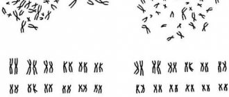
Aneuploidy occurs as a result of chromosome nondisjunction during meiotic divisions or mitosis. The term “nondisjunction” means the absence of separation of chromosomes (in meiosis) or chromatids (in mitosis) in anaphase. As a result of nondisjunction, gametes with an abnormal number of chromosomes appear.
Structural changes in human chromosomes are much less common than numerical aberrations. Structural rearrangements can be chromosomal and chromatid, accompanied by a change in the amount of genetic material (deletions and duplications) or only reduced to its movement (inversions, insertions, translocations). The rearrangement may involve one or more chromosomes with several breaks and connections. Sometimes cells with different karyotypes can be found in the body. This combination of karyotype is usually referred to as mosaicism.
Most chromosomal diseases occur sporadically as a result of genomic and chromosomal mutations in the gametes of healthy parents or in the first divisions of the zygote. Chromosomal changes in gametes lead to the development of so-called complete, or regular, forms of karyotype disorder, and corresponding changes in chromosomes in the early stages of embryo development cause somatic mosaicism, or mosaic organisms (the presence in the body of two or more cell lines with different numbers of chromosomes) . Mosaicism can affect both sex chromosomes and autosomes. In humans, mosaic forms are most often found in the sex chromosome system. Mosaics, as a rule, have more “erased” forms of the disease than people with an altered number of chromosomes in each cell. Thus, a child with mosaic Down syndrome may have virtually normal intelligence, but the physical signs of this disease remain.
The number of abnormal cells can be different: the more there are, the more pronounced the symptom complex of a particular chromosomal disease. In some cases, the proportion of abnormal cells is so small that the person appears phenotypically healthy.
What did the Lord intend them for?
For only I know the plans I have for you, says the Lord, plans for good and not for evil, to give you a future and hope.
Gen.29:11
The Lord gives a purpose to every person He creates. Mentally retarded people are no exception. He loves them just like everyone else and wants them to live and be happy. And although they have to experience certain difficulties in life, sometimes a great calling awaits them. People with Down syndrome sometimes succeed better than others, set positive examples for others, teach them to rejoice, love, be kind, hardworking, etc. They can achieve significant success in their chosen professions[29]. The view of Down syndrome as something negative has not yet been overcome. This may be due to underlying health problems. But if you look closely, it is easy to see that Down syndrome is truly a gift with which God blesses its owners. They are honest, talented, happy, capable of love like none of us. Their life is simple, but no less happy than ours[30].
Disorders of mitosis and meiosis
Disturbances in meiosis lead to aneuploidy. In the case of nondisjunction of chromosomes, one of the daughter cells receives 22 chromosomes, which after fertilization produces an embryo with monosomy. The other daughter cell receives 24 chromosomes, resulting in trisomy after fertilization. If one of the chromosomes in anaphase does not separate from the spindle (chromosomal lag) and does not enter the daughter cell, fertilization of such a cell also leads to monosomy. The higher the age of the mother, the higher the probability of chromosome nondisjunction and, therefore, the occurrence of trisomy. Although it depends on the specific chromosome, in general, the majority of trisomies that doctors see are caused by disorders of the 1st meiotic division in women. If chromosome nondisjunction occurs in mitosis, two different cell clones may arise in the body (mosaicism). This can be observed with gonadal dysgenesis - nondisjunction of chromosomes in a zygote with a 46,XY karyotype can give rise to cell clones with karyotypes 45,X and 47,XYY (all three cell clones may be present, depending on the moment at which chromosome disjunction is disrupted) . When chromosomes are delayed in the case of a 46,XY karyotype, mosaicism with a 45,X/46,XY karyotype is possible.
Diseases
The consequences of chromosomal pathologies detected in the fetus can be very different: from external deformities to damage to the central nervous system. They largely depend on what kind of anomaly has occurred with the chromosomes: their number has changed or mutations have affected their structure. Among the most common diseases are the following.
Chromosome number disorder
- Down syndrome is a pathology of the 21st pair of chromosomes, in which there are three chromosomes instead of two; accordingly, such people have 47 of them instead of the normal 46; typical signs: dementia, delayed physical development, flat face, short limbs, open mouth, squint, bulging eyes;
- Patau syndrome - disturbances in the 13th chromosome, a very severe pathology, as a result of which numerous developmental defects are diagnosed in newborns, including idiocy, polyfingeredness, deafness, mutations of the genital organs; such children rarely live to be one year old;
- Edwards syndrome - problems with the 18th chromosome, often associated with advanced maternal age; babies are born with a small lower jaw and mouth, narrow and short eye slits, and deformed ears; 60% of sick babies die before 3 months, and 10% survive up to a year; the main causes of death are respiratory arrest and heart defects.
Violation of the number of sex chromosomes
- Shereshevsky-Turner syndrome is an abnormal formation of the gonads (most often in girls), caused by the absence or defects of the sex X chromosome; Symptoms include sexual infantilism, folds of skin on the neck, deformation of the elbow joints; children with such a chromosomal pathology survive, although childbirth is very difficult, and in the future, with proper supportive treatment, women are even able to carry their own baby (through IVF);
- polysomy on the X- or Y-chromosome - a variety of chromosome disorders, characterized by a decrease in intelligence, an increased likelihood of developing schizophrenia and psychosis;
- Klinefelter syndrome is a disorder of the X chromosome in boys, who in most cases survive after childbirth, but have a specific appearance: lack of body hair, infertility, sexual infantilism, mental retardation (not always).
Polyploidy
Such chromosomal pathology in the fetus always ends in death even before birth.
Scientists are still trying to figure out why gene mutations occur at the chromosome level. However, this is still only a matter of the future, and at this point in time, chromosomal pathologies detected in utero in the fetus account for up to 5% of all cases.
What should parents do when they hear such a diagnosis? Do not panic, reconcile yourself, listen to the doctors and, together with them, make the right decision - leave the sick baby or agree to an artificial termination of pregnancy.
Medical and pedagogical support for children with Down syndrome
Curing a chromosomal abnormality is currently impossible; Any proposed treatments are experimental and have no proven clinical effectiveness. However, systematic medical observation and pedagogical assistance to children with Down syndrome make it possible to achieve success in their development, socialization and acquisition of work skills.
Throughout their lives, patients with Down syndrome should be under the supervision of specialists (pediatrician, therapist, cardiologist, endocrinologist, otolaryngologist, ophthalmologist, neurologist, etc.) due to concomitant diseases or an increased risk of their development. When severe congenital heart and gastrointestinal defects are detected, their early surgical correction is indicated.
In case of severe hearing loss, a hearing aid is selected. If the organ of vision is pathological, glasses correction, surgical treatment of cataracts, glaucoma, and strabismus may be required. For hypothyroidism, replacement therapy with thyroid hormones, etc. is prescribed.
To stimulate the development of motor skills, physiotherapy and exercise therapy are indicated. To develop speech and communication skills, children with Down syndrome need classes with a speech therapist and an oligophrenopedagogist.
Children with Down syndrome are usually educated in a special correctional school, but within the framework of integrated education, such children can also attend a regular public school.
In all cases, children with Down syndrome are classified as children with special educational needs, and therefore require additional help from teachers and social educators, the use of special educational programs, and the creation of a favorable and safe environment. Psychological and pedagogical support for families raising “sunny children” plays an important role.
What types of markers are there?
It is worth remembering that all existing markers of chromosomal pathologies are considered conditional. Science has not yet sufficiently studied the emergence and development of deviations.
Basic markers
- nagging pain in the lower abdomen, which may indicate a miscarriage;
- lack of fetal activity;
- hypoxia (lack of oxygen);
- oligohydramnios and polyhydramnios;
- facial deformities;
- dimensions of the nasal bones;
- increase in the neck fold (the marker is called the thickness of the collar space or abbreviated TVP);
- slower growth of tubular bones;
- maxillary bone size;
- bladder size;
- enlargement of the renal pelvis;
- hydronephrosis (expansion of the renal pelvis and calyces associated with disturbances in the process of urine outflow);
- cysts in the umbilical cord or brain;
- swelling of the neck and back;
- accelerated aging of the placenta;
- hypoplasia (underdevelopment) of the placenta;
- intestinal condition (hyperechogenicity, in which the organ appears too bright on ultrasound).
USEFUL INFORMATION: Surrogacy
Blood markers
- decreased level of PAPP-A (plasma protein-A);
- reduced level of AFP (protein in the fetus);
- increased levels of hCG (a hormone produced by the placenta).
Also, poor results of Doppler and cardiotocography may cause concern for the doctor. If you find one or two markers, don't panic. The presence of deviations may be associated with the individual characteristics of the child’s body and may not indicate the development of a serious pathology.
What is prenatal screening
Prenatal
screening,
diagnosis and treatment is a relatively new problem in obstetrics. The origin of prenatal screening was perhaps the era of ultrasound in obstetrics, which began about two decades ago. With the discovery of new genes and their phenotypes, prenatal genetic diagnosis is becoming increasingly possible. It is necessary to distinguish between the concepts of screening and diagnosis.
Prenatal screening identifies individuals at high risk for complications from a population of individuals at low risk for complications. The specificity and sensitivity of screening tests are very important given the possibility of false-positive and false-negative screening results.
Prenatal diagnosis is, of course, more specific than screening (for example, amniocentesis or chorionic villus sampling), but also has a greater risk of complications. The first step in determining the risk to the fetus is to screen the mother for certain conditions or diseases.
The question often arises about the likelihood of an increase in frequency in the offspring of married couples who received treatment for infertility. Severe oligospermia and azoospermia are associated with balanced chromosome translocations (3-5%), Klinefelter syndrome (47, XXY), abnormalities and microdeletions of the Y chromosome.
X chromosome abnormalities (XXY, XXX, X mosaicism in Turner syndrome) are associated with reduced fertility (subfertility), as well as an increased risk of chromosomal abnormalities in offspring. In 2/3 of patients with congenital absence of the vas deferens, there is at least one mutation of the gene that is responsible for the development of cystic fibrosis. So, these patients should be screened for cystic fibrosis. In these patients, intracytoplasmic sperm injection is usually indicated, although the presence of a cystic fibrosis gene mutation may influence reproductive intentions.
Decoding and calculating risks
After the first double screening is done, ultrasound markers of fetal chromosomal pathology that were identified during the study are analyzed. Based on them, it calculates the risk of developing genetic abnormalities. The very first sign is an abnormal size of the collar space in an unborn child.
Ultrasonic markers
Absolutely all ultrasound markers of chromosomal pathology of the fetus in the 1st trimester are taken into account in order to make the necessary calculations of possible risks. After this, the clinical picture is complemented by a blood test
Blood markers
Norm:
All other indicators are considered deviations from the norm.
In the second trimester, inhibin A, unconjugated estriol and placental lactogen are also assessed. All interpretation of the research results is carried out by a special computer program. Parents can see the following values as a result:
- 1 in 100 means that the risk of genetic defects in the baby is very high;
- 1 in 1000 is the threshold risk of chromosomal pathology of the fetus, which is considered normal, but a slightly underestimated value may mean the presence of some anomalies;
- 1 in 100,000 is a low risk of chromosomal pathology of the fetus, so there is no need to worry about the baby’s health from a genetic point of view.
After doctors calculate the risk of chromosomal pathology in the fetus, either additional tests are prescribed (if the obtained value is lower than 1 in 400), or the woman calmly nurses the pregnancy to a successful outcome.
Diagnosis of the disease
Nowadays, the risk of developing Down syndrome in an unborn child can be identified at the very beginning of pregnancy - even during the first ultrasound, which is usually done at twelve to thirteen weeks. This is called prenatal diagnosis and allows you to see Down syndrome in the fetus. No one will tell you the causes and signs of the developing disease during screening, but the expectant mother will already have food for thought - what to do next. In addition to ultrasound examination, pregnant women should definitely donate blood. This analysis as a percentage will show the risk of a child developing not only Down syndrome, but also other equally dangerous ailments. Blood is also donated in the second trimester - between the sixteenth and eighteenth weeks, and a little later, at five months of pregnancy, a second ultrasound is performed, which excludes the possibility of late-onset developmental anomalies. The third ultrasound is done shortly before birth, from the thirtieth to the thirty-fourth week.
Medicines, pregnancy and chromosomal pathologies
Many medications that a woman takes during pregnancy affect the fetus:
- aminoglycosides have a toxic effect on ear and kidney development;
- aloe promotes increased intestinal motility;
- antihistamines can cause tremors and significantly reduce blood pressure;
- androgens are the cause of the development of fetal defects;
- anticoagulants can cause problems with bone formation, as well as encephalopathy;
- atropine is a cause of brain dysfunction;
- belladonna causes tachycardia in the fetus;
- blood pressure lowering agents significantly reduce blood flow to the placenta;
- diazepam can harm the appearance of the unborn child;
- corticosteroids inhibit the functional purpose of the adrenal glands, leading to encephalopathy;
- caffeine damages the fetal liver;
- lithium develops heart defects;
- Opiates affect brain activity;
- anticonvulsants significantly delay the baby’s intrauterine development;
- tetracyclines lead to skeletal abnormalities.
Chromosomal pathologies
The concepts of chromosomal and hereditary diseases are essentially synonymous. They are caused by changes in the structure of different genes or their number. In genetics, there are hundreds of chromosomal pathologies that cause various mutations and abnormalities, but there is little knowledge about their causes.
The collection of chromosomes that contain genes is called a genome. A healthy person has 23 pairs of chromosomes that carry hereditary information: 22 pairs of autosomal chromosomes (paired non-sex chromosomes) and a pair of sex chromosomes.
A common cause of the development of chromosomal pathologies are mutations in the germ cells of the parents. If the mother and father had hereditary abnormalities in the family, it is worth studying the issue of chromosomal pathologies and undergoing an extensive examination. This severity of measures is due to the fact that diseases caused by mutations in chromosomes often develop during the growth of the fetus in the womb.
Diagnostics is aimed at the benefit of parents and baby. The comfort of his life and the life of his parents will depend on the degree of damage to the genome. It is often difficult for children with chromosomal abnormalities to live: not only do they have problems with controlling the body and maintaining life (breathing, eating, etc.), but there are also difficulties in perceiving and converting information.
If markers of complex chromosomal abnormalities are detected, medicine can only offer parents termination of pregnancy. No matter how cruel it may be, such a measure avoids the suffering of the child and his parents. However, you should not judge the situation based on the first results. It is not uncommon for doctors studying this complex area to make mistakes.
It is also worth remembering that all norms are averaged. Approaching the maximum permissible values of one indicator cannot be evidence of pathology.
Causes
Doctors are still working on the question of why children are born with Down syndrome, what factors and circumstances are decisive in the violation of the karyotype. Genetics, despite the high level of modern science, to this day remains one of the most mysterious and little-studied branches of medicine. Therefore, there is no exact answer to this question. Recent studies name the following causes of Down syndrome, of which very few have been identified:
- mother's age after 40 years;
- father's age after 42 years;
- an accidental coincidence of circumstances at the time of formation of pregnancy and germ cells;
- lack of folic acid (a hypothetical fact, not scientifically confirmed (read about folic acid when planning pregnancy here)).
But at this stage of research, geneticists unanimously claim that the causes of this chromosomal disorder do not depend in any way on environmental factors and the lifestyle of the parents. Therefore, a married couple should not blame themselves for the fact that their fetus or newborn baby was diagnosed with this syndrome.
Through the pages of history. John Langdon Haydon Down is an English scientist of the 19th century who first described Down syndrome. He called it "Mongolism".