Reproductive age is the longest period in a woman’s life. It is very important to take care of your health in time to bear and give birth to a child. Therefore, many may be frightened by the diagnosis - atrophic endometrium. What it is and how it is treated, we will talk in this material.
Endometrial atrophy occurs when the body experiences a decrease in estrogen levels. The disease is characterized by thinning of the layer of the uterus lining it from the inside. As a result, cyclical updates do not occur and menstruation does not begin - there is simply nothing to reject.
The endometrium is atrophic - what it is is now clear. This condition is normal during menopause, but not in those years when a woman is still young and planning a pregnancy.
Changes
As you know, every month the same processes occur in a woman’s body. The glandular layer increases so that in the event of fertilization of the egg, attachment of the fertilized egg to the uterine wall can occur. If this does not happen, menstruation occurs - the accumulated layer of endometrium is rejected as unnecessary. An important role in the described stages is played by sex hormones - estrogens and progesterone.
The gonads work harmoniously thanks to signals emanating from the pituitary gland. The gonadotropic hormone is “responsible” for the transmission. When wondering whether the endometrium is atrophic - what it is and how to deal with it, you must understand the peculiarities of the reproductive system.
By the time of menopause, the function of childbearing dies out. Hormones are released in smaller quantities and the glandular layer practically does not grow. We can say that this is one of the main reasons why a woman cannot get pregnant during menopause.
The thinned inner uterine layer is called atrophic endometrium. What this is - we explained above. Now the uterus is dominated by connective tissue. The process does not cause any discomfort.
Menopause
Sometimes it happens that a woman of reproductive age is forced to take drugs that introduce her body into an artificial menopause. In addition, hormonal imbalance in the body can also lead to early menopause. This condition can be either temporary, from which there is a way out, or irreversible. In the latter case, the answer to the question: the endometrium is atrophic, what does this mean, will be a disappointing answer - infertility.
Menopause in women begins gradually. Typically, the first hormonal changes are noticeable at the age of 45. When menstruation has stopped, reproductive function will continue to decline for about ten years.
Two years after the last menstruation, the functional layer of the endometrium no longer has its former properties. Even if fertilized, the egg will not be able to implant in the uterus. Before menopause, a histological examination will help you figure out whether the endometrium is atrophic, what it is and what it looks like. Usually a specialist sees the following picture:
- under the influence of a reduced level of estrogen, glandular hyperplasia develops in a weak form, which is combined with a non-functioning endometrial layer;
- the glands are distributed chaotically, and some of them turn into round cystic formations;
- in some glands you can see the uneven arrangement of epithelial nuclei - in one row or several;
- The density of the underlying tissues is not the same, so stromas are formed.
It is worth noting that after the end of menstruation, the transitional epithelium will exist for some time.
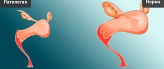
Pathologies
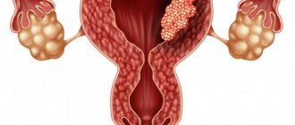
A woman’s reproductive function directly depends on the condition of the endometrium. The most common pathologies of the uterine mucosa are hypoplasia and hyperplasia. Both deviations, when neglected, lead a woman to infertility.
Hyperplasia
The development of the pathology of endometrial hyperplasia is accompanied by an increase in the thickness of the functional layer of the inner membrane of the reproductive organ up to 2.6 cm. When this size is reached, tissue cells begin to multiply intensively, and a change in its density and structure occurs. This violation does not allow the newly born fetus to attach to the wall of the uterus.
This disorder in a woman’s body is manifested by a disruption in menstruation, a change in its duration and intensity of bleeding. This disease can be accompanied by anemia of varying severity; bleeding may appear between menstruation; overgrown tissue becomes a source of pathogenic cells and various neoplasms.
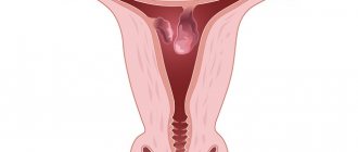
Hypoplasia
The reverse process of hyperplasia causes thinning of the functional layer of the endometrium and is called hypoplasia. It undermines a woman's reproductive function and seriously reduces the likelihood of conceiving a child.
This pathology prevents the emerging fetus from attaching to the wall of the uterus; it lags behind in development and dies due to lack of nutrients. A thin layer of the endometrium poorly protects the body from the penetration of infectious microorganisms and causes the development of inflammation in the female organ.
Kinds
Before hearing the diagnosis of atrophic endometrium, what it is and how to live with it now, the gynecologist will want to analyze what the functional layer was like before menopause. Below we will look at what atrophy occurs in connection with this.
Simple
If the first phase of the last menstrual cycle was characterized by weak proliferation, and during the luteal phase secretion was insufficient, then we are talking about simple atrophy. If you examine the tissue under a microscope, you can see elongated glands with one epithelial layer, there are quite a few of them. They are located in a fibrous base with a dense structure.
Cystic
This situation occurs if, before menopause, endometrial growth and hyperplasia were irregular. The glands in this variant have thin walls and are lined with a low layer of epithelium.
Some women may show signs of degeneration due to age-related irreversible processes:
- the glands are cystically dilated;
- in the epithelium, the nuclei are wrinkled and arranged in several rows, cell division does not occur;
- stromal tissue is fibrously changed.
Quite often, based on the last sign, it is difficult to differentiate the atrophic endometrium - that it is not glandular hyperplasia, which usually occurs already in postmenopause.
Sometimes it happens that you haven’t had your period for a long time, but suddenly there is bloody discharge from the genital tract. Studies show that in place of the atrophic endometrium there is an epithelium that is still susceptible to the influence of estrogens. This happens with tumor processes of the adrenal glands and ovaries.
Why is endometrial atrophy dangerous?
The endometrium is the mucous membrane of the uterus.
It consists of epithelial cells and a stroma lining that contains secretory glands. The epithelium is penetrated by a significant number of capillaries. Its thickness changes at different phases of the cycle and characterizes reproductive capabilities. Normally, in a woman, depending on the period, it is 0.2–1.8 cm.
A healthy endometrium is an indispensable condition for the attachment of the fertilized egg.
Endometrial atrophy is a thinning of the uterine mucosa. This is a natural process in the premenopausal phase; with a decrease in estrogen levels, the restoration of the mucous membrane slows down and later stops. This phenomenon in reproductive age is already a pathology caused by hormonal disorders. Leads to infertility because the fertilized egg is not able to attach to the wall of the uterus.
The distinctive symptoms of atrophy are associated with disruptions of the menstrual cycle, up to complete disappearance.
The essence of pathology
Every month, the glandular layer thickens, facilitating the implantation of the fertilized egg. If conception does not occur, it is rejected. This is due to the activity of special female hormones - estrogen and progesterone.
As menopause progresses, their synthesis decreases, the mucous membrane is not renewed, and an atrophic process occurs. At the same time, the layer becomes thinner, it becomes pale in color, and dilated capillaries and vessels are visible through it.
At a young age, changes in hormonal levels in the body or taking certain medications lead to premature menopause.
The endometrium looks like this:
- Glandular hyperplasia of the endometrium is poorly developed.
- The glands are located chaotically, some of them have turned into cystic round formations.
- The epithelial nuclei are distributed unevenly.
- Tissue density changes, stroma (a characteristic three-dimensional network) is present.
Symptoms of pathology:
- failure of the menstrual cycle;
- scanty leucorrhoea on menstrual days or complete absence;
- infertility, miscarriages at the beginning of the first trimester;
- painful sensations during sexual intercourse;
- pain in the lower abdomen.
If changes appear before the onset of menopause, the woman is diagnosed with a cystic variant of atrophy. The main sign of pathology is enlarged glands. At the same time, synechiae and atrophy are detected.
Synechia is a chaotic proliferation of connective tissue that aggravates the condition. This leads to disruption of the patency of the fallopian tubes and the beginning of the adhesive process. There is pain, worsening during menstruation, and scanty discharge. Amenorrhea develops less frequently.
At-risk groups
In reproductive age, atrophic endometrium is a consequence of injury to the mucous membrane during the following manipulations:
- vacuum abortion, curettage, including for diagnostic purposes;
- elimination of polyps, cysts and other benign tumors;
- installation of an IUD;
- surgery on the tubes and/or uterus.
The risk group includes women who have experienced a drop in estrogen levels as part of a hormonal imbalance after undergoing the following procedures:
- removal of ovaries;
- chemotherapy;
- radiation therapy;
- therapy of breast cancer with hormone-containing agents.
This pathology is more often observed in nulliparous women, as well as in obese and diabetic women. Excessive physical and emotional stress, sudden weight loss due to diets, bad habits - smoking, alcoholism - have a negative impact on health.
About 50% of women over 45 years of age experience symptoms of age-related atrophy: discomfort during intercourse, dryness and burning in the vagina, bleeding after intercourse, and increased frequency of urination. They have an increased risk of developing genital tract infections. Most of these problems can be solved by choosing the optimal treatment method.
Kinds
The type of disease depends on the state of functionality of the endometrial layer before the onset of menopause.
Characteristic features of atrophic epithelium:
- does not undergo changes at different periods of the cycle;
- the stroma shrinks, thickens, contains an increased volume of fibers and collagen;
- the number of glands decreases, they are located in a thin layer, acquiring the same height as the columnar epithelium;
- The glands are similar in appearance to tubes with a thin lumen.
Atrophic endometrium has the following varieties:
- simple;
- cystic;
- age.
Control is necessary during all phases of menopause. Menopause is not a reason for refusing to undergo regular examinations, including examination, OBC, smear, transvaginal ultrasound performed through the vagina, X-ray of the tubes/uterus. After receiving the results, the doctor will select a course of treatment if necessary.
Simple
Endometrial atrophy is a reaction of the female body to changes in hormone levels. You can talk about it if at least a year has passed since the regulation. Stimulation by hormones decreases, monthly growth of the glandular layer does not occur, and the thickness of the mucous membrane decreases.
When observing the layer of the uterus under a microscope, elongated glands and a thin epithelial layer can be seen in the tissues. With full menopause, this process is not pathological.
Cystic
The dilated glands are lined with a single layer of columnar epithelium, which is less thick. Thinning and inflammation that occur due to estrogen deficiency in postmenopause can lead to bleeding of varying intensity.
Women suffering from hypertension are especially susceptible to this.
It is necessary to find out the cause of this phenomenon, since it is often a sign of the occurrence of benign neoplasms or malignancy of the fallopian tubes or cervix. Tumors on the ovaries usually develop in the postmenopausal phase. If they are malignant, the ovaries are surgically removed.
Endometrial changes during menopause
Atrophic endometritis in the menopausal phase is the norm. But the opposite phenomenon - thickening of the layer - may be a sign of pathology.
Endometrial hyperplasia is always a pathological process, accompanied by the growth of the mucous uterine layer. If its development is not stopped, it affects muscle fibers. The layer normally grows until the middle of the cycle and leaves the body with secretions if there has been no conception.
With the development of menopause, the process of cell reproduction is disrupted, the basal layer continues to grow, and physiological excretion does not occur. There is a malfunction in the activity of many systems, and malignancy of tissue cells is possible.
Types of hyperplastic phenomena:
- Glandular - glands located in the endometrium grow and become deformed. At the same time, the thickness increases to the maximum.
- Cystic - when the layer grows, the hole in the exit gland is blocked, and cysts are formed. This process is very dangerous and can provoke the development of cancer.
- Basal is a fairly rare type of disease; the inner layer penetrates deep into the uterus.
- Focal (polypoid) - the formation of polyps and growths on the stalk occurs.
- Atypical - caused by a sharp uncharacteristic change in endometrial cells and its penetration into other tissues. This variety is very dangerous and often develops into cancer. There is no therapy; the uterus is surgically removed.
- Combined.
The main symptom of the disease is bleeding or bloody leucorrhoea from the vagina, regardless of its volume, duration and frequency of occurrence.
This is accompanied by weakness, fatigue, surges in blood pressure, decreased ability to work, and headaches. It is necessary to visit a specialist twice a year.
The doctor will conduct an examination on the chair, take smears for the presence of atypical cells, and, if necessary, prescribe instrumental diagnostics.
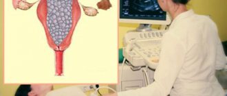
Hypoechoic formation in the ovary
Ultrasound allows early detection of many female pathologies. Fabrics with different densities produce images of different colors on the device screen.
A hypoechoic formation is a section of an organ that has an acoustic density lower than the surrounding tissue. More often these are cysts or tumors - thin-walled cavities filled with fluid. The cyst is rarely attached to the ovary or is pedunculated.
This is not a diagnosis, the formation may be:
- cyst;
- tumor;
- echinococcal cyst;
- normal follicle in the middle phase of the cycle.
When diagnosing, the doctor determines the size, structure and boundaries of the inclusion. In a woman during the childbearing phase, the structure of the ovaries is heterogeneous, in contrast to those who are experiencing menopause.
Cysts are classified depending on the cause of their formation:
- Follicular – the follicle is enlarged in size, has thin walls, and is filled with fluid. The surface is smooth, diameter less than 8 cm.
- The corpus luteum cyst is a sphere with a diameter of about 7 cm with a smooth surface, filled with yellow-red liquid.
- Endometrioid (chocolate) is formed due to mutation of endometrial cells. It has thick walls filled with dark brown liquid. This type of pathology is one of the consequences of endometriosis. Soft tissue from the lining of the uterus and blood clots can cause cavities to form.
- Dermoid is a benign formation that lasts for a long time with virtually no obvious signs.
- Mucinous - multi-chambered, including thick mucus.
Ovarian cysts can grow to large sizes and become malignant. If you experience pain in the lower abdomen, brown vaginal discharge, or irregular menstruation, you should undergo a medical examination.
Features during menopause
During premenopause, ovarian activity begins to fade, and the number of eggs capable of fertilization decreases. Due to a decrease in hormone levels, the total number of follicles decreases, and the eggs do not mature completely. Changes in the endometrial layer are noticeable in different phases of the cycle; it loses its ability to grow when hormone levels fluctuate.
The development of menopause (the last regulation, after which reproductive function finally fades away) is indicated by the cessation of menstruation. The uterine mucosa decreases and is characterized by atrophic changes. After menstruation stops for a year, the thickness of the endometrial layer in postmenopausal women remains constant.
The production of hormones, the activity of the ovaries and the formation of eggs stop. The next stage in a woman’s life begins – postmenopause. The body is rebuilt and gets used to living in the absence of sex hormones. The endometrium becomes thinner, the atrophic process proceeds smoothly. The normal thickness is about 5 mm.
If the epithelial layer continues to grow despite the onset of menopause, we are talking about hyperplasia. The occurrence of bleeding or scanty discharge from the labia is observed when the thickness is more than 8 mm. For a long time, the pathology does not manifest itself, but the process progresses, and a benign formation can change its character.
Atrophy during menopause
Menopause is a natural physiological process associated with the end of the reproductive period of a woman’s life. Initially, estrogen synthesis decreases, which affects the regularity and nature of menstruation.
The level of estrogen is a decisive factor on which the condition of the epithelium of the vagina and uterus depends. The menopause period can last up to 10 years, ending with menopause - the last menstruation. We can talk about it if there have been no periods for a year or more.
At the next stage, hormonal changes end, the ovaries cease their activity, and the level of estrogen decreases to a minimum.
All genital organs change appearance:
- the uterus decreases in size;
- the nuclei in the epithelium are arranged in rows, it is wrinkled, there is no division process;
- stromal tissue has fibrous changes;
- the lumen of the cervix decreases in size, the cervical canal narrows, and synechiae may form in it;
- vaginal dryness increases, the surface becomes thinner;
- the vaginal vault is weakly expressed, there are no folds on the walls;
- areas without epithelium or loose areas with adhesions appear;
- bleeding is possible;
- the volume of the mammary glands changes;
- the amount of pubic hair decreases.
The proliferation of connective tissue progresses and reaches a maximum after the development of menopause.
Treatment methods
Therapy for endometrial atrophy is prescribed to women in the reproductive phase.
The principles are:
- Gentle regimen, healthy diet, avoidance of excessive physical activity.
- General strengthening agents.
- Physiotherapy, spa treatment.
- Hormonal therapy. Medicines or combination OCs that include estrogen and/or progesterone are used. Taking hormonal drugs minimizes the risk of neoplasms. Treatment lasts 2–4 months.
- Hysteroscopic dissection of adhesions.
Hormonal therapy is combined with hysteroscopy; with an integrated approach, recovery occurs in 3–4 months. The prognosis is generally favorable, adequate treatment will help restore reproductive functions and reduce the risk of developing gynecological pathologies.
To prevent the premature occurrence of such a pathology, it is important to eat right, avoid heavy physical activity, avoid abortions, and regularly visit a gynecologist. Treatment helps prevent serious gynecological problems.
Source: https://TopGinekolog.ru/bolezni/atrofiya-endometriya
Causes
Every woman will cross the reproductive age line at some point in her life. The body will enter a new state for itself, where atrophic changes will be considered natural and irreversible. Menopause will come very soon.
There is a group of conditions in which the endometrium is atrophic at any age. What kind of pathologies are these, see below:
- the gonads are defective;
- various tumor diseases of the hypothalamus and pituitary gland, due to which the uterus cannot develop at a young age;
- physical exhaustion due to diets;
- stressful situations;
- excessive stress when playing sports;
- loss of protein by the body;
- ovarian depletion, resulting in the release of a small amount of estrogens;
- absence of ovaries due to removal;
- drug therapy that affects the functioning of the ovaries;
- atrophic endometritis of chronic form due to frequent abortions and curettage of the uterine cavity.
Quite often, if a woman complains of intermenstrual bleeding, treatment is prescribed that introduces the body into artificial menopause.
Below you can see the diseases for which such tactics are used:
- severe endometriosis;
- uterine fibroids;
- cancer processes in the mammary glands;
- if surgery is planned.
In this case, the endometrium is atrophic; we will now tell you what this is and how it happens. A special drug is selected that suppresses the production of estrogen. As a result, the inner layer of the uterus is not renewed and does not grow. This will remain so until the external influence stops.
List of drugs that cause atrophy: Zoladex, Diferelin, Eligard, Lucrin Depot, Buserilin Depot (a group of drugs based on gonadotropin-releasing hormone), Visanne (progestogens), Danol (a drug that suppresses the formation of gonadotropic hormones).
When the course of treatment is completed, other hormonal drugs can return a woman to her previous sexual health if the body does not have enough strength to recover.
In old age, if a woman is diagnosed with ovarian or breast cancer, the drug Tamoxifen is prescribed. The active substance should inhibit the production of estrogen. But quite often, while taking it, the endometrium begins to grow.
An amazing effect, because the production of estrogens, which stimulate the growth of mucous membranes, is suppressed. When conducting a microscopic examination, thickening of the basal layer and the upper cystic endometrium is determined to be atrophic, what does this mean? This is precisely stromal hyperplasia.
It is important to note that despite the parameters of the endometrium (thickness and structure), curettage is not performed, since atrophy is still observed. The functional layer itself is not hyperplastic.
Symptoms
Signs of atrophy:
- visually the functional layer becomes the same as the basal layer, since cyclic changes no longer occur;
- the stroma becomes denser and wrinkles, it contains a lot of collagen and connective fibers;
- stromal glands are lined in one row in the form of a cylindrical epithelium located low;
- Externally, the glands look like tubes with a narrowed lumen.
Regardless of whether atrophic phenomena are caused by natural processes or under the influence of a drug, the signs will be identical:
- menstruation becomes scanty or disappears altogether;
- it is impossible to bear a child - infertility;
- if atrophy of the mucous membranes of the vagina and cervix is involved, then contact bleeding is observed after sex or during injuries.
There is no pain syndrome in such cases, since there is no inflammation, infection or tumor process. Excessive blood circulation is also not observed.
Synechia
Chronic atrophic endometritis, on the contrary, gives pain due to synechiae - adhesions found inside the uterus. Adhesions are considered a complication of atrophy of the uterine mucosa.
They do not manifest themselves clinically. But they are dangerous if they were formed during the treatment of some pathology, and not naturally. When the monthly cycle returns to normal, they will not go away, which can cause infertility. To avoid this, they need to be removed surgically during hysteroscopy - cut.
Diagnostics
The endometrium is atrophic; ultrasound will show what it is. The specialist focuses on the thickness of the functional layer. If it is less than 5 cm, then the diagnosis is confirmed and treatment begins. But only when the woman is of reproductive age and plans to have a child.
If fluid (serozometers) is detected in the uterine cavity, along with atrophy of the mucous membrane, the woman is prescribed regular ultrasound examinations to monitor changes. It is possible that this is the beginning of degradation of the deeper uterine layer.
Having discovered an atrophic endometrium in a young woman on ultrasound, a specialist will not be able to immediately say what it is. Additional examination is required. It includes:
- examination on a chair and taking smears;
- performing a colposcopy to determine the condition of the cervix;
- taking blood for analysis to determine the level of sex hormones and gonadotropin;
- hysteroscopy if required.
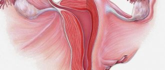
Endometrial structure
The wall of the female reproductive organ is a combination of three layers of different tissues:
- The outer surface of the organ is covered with a serous membrane (perimetry).
- In the middle is the myometrium.
- It is enveloped by the endometrium, which covers the uterus from the inside.
The internal mucous surface of the female reproductive organ is divided into a basal part, which is adjacent to the meometrium, and a functional internal surface.
The basis of the basal layer is made up of connective tissue cells, blood capillaries and nerve endings; its cells are tightly pressed against each other.
It is practically not subject to hormonal transformation. The thickness of this structure in a healthy woman can be from 1 to 1.5 mm. Menstruation does not affect its structure; at the end of the cycle, its cells grow and restore the thickness of the functional layer.
The cells of the functional layer are very sensitive to the effects of female hormones. It changes continuously during the menstrual cycle and is rejected at the end. The beginning of the cycle is accompanied by the restoration of the functional layer, thanks to the tissues of the basal structure.
Depending on the phase of the cycle, the thickness of the functional structure varies from 0.5 to 1.5 cm. Its upper part has a compact structure, the lower layer has a spongy structure.
In a woman's uterus, tissue from the endometrium performs the following unique tasks:
- Prevents fusion of the walls of the uterus.
- Helps the embryo implant into the wall of the uterus.
- Provides nutrition to the embryo.
- Takes part in the blood supply to the placenta.
- Delivers necessary, beneficial substances to the placenta tissue.
Treatment
It is necessary for women of the reproductive period to treat changes in the functional layer of the endometrium, and even more so atrophic endometritis.
General therapy usually includes:
- ensuring peace;
- proper and balanced nutrition;
- prohibition of heavy physical activity;
- prescribing vitamin complexes to maintain general health;
- physiotherapy recommended;
- Mud radon baths would be appropriate in gynecological sanatoriums.
Hormonal drugs are prescribed as targeted treatment. This includes products based on gestagens and estrogens, which allow you to restore a full menstrual cycle. If there are adhesions, they are dissected. This is important if a woman wants to get pregnant.
Hormonal therapy is usually prescribed for 3-4 months. Then they observe which endometrium is atrophic? The doctor will explain what this means after treatment. Most likely, the therapy was unsuccessful. If an ultrasound examination shows sufficient thickness of the endometrium of the correct structure, then the woman can become pregnant.
Treatment of endothelial dysfunction
Endothelial function can be improved by measures aimed at reducing the risk of cardiovascular disease, including weight loss, exercise, smoking cessation, control of hypertension and diabetes, which are generally highly recommended for all of us.
Several of these risk control measures have been well established for reducing endothelial dysfunction. These include:
- the use of statins (pharmaceutical drugs aimed at combating high levels of cholesterol in human blood);
- Mediterranean diet;
- other foods including nuts, olive oil, dark chocolate, green tea, herbal products;
- aerobic exercise;
- excess weight loss;
- the use of ACE inhibitors (a group of natural and synthetic chemical compounds used for the treatment and prevention of heart and kidney failure, to lower blood pressure).
In addition, several drugs specifically designed to clinically improve endothelial dysfunction are currently being studied. Promising agents include nifedipine, estrogen, runolazine, aspirin, L-argenine, and sildenafil.
Prevention
To avoid atrophy during the reproductive period, you must follow the following recommendations:
- a complete diet without diets or fasting;
- moderate physical activity;
- timely protection from unwanted pregnancy in order to avoid abortion;
- avoiding casual sexual intercourse and using a condom if necessary to protect against sexually transmitted infections;
- Regular visits to the gynecologist for preventive examinations.
Now you know why the endometrium is atrophic.
What it is and how to treat it - your doctor will definitely tell you in detail. Never try to cope with the problem yourself using folk remedies. Not only will you waste time, but you can also harm your health! Share: