Location of the trachea, its features and structure
The trachea is a cartilaginous segmental tubular hollow organ. Location: below the larynx. Passes into the main bronchi. Carries out inhaled and exhaled air. Part of the lower respiratory tract. The Latin name for the organ in medicine is tracheia, from the Greek trachus - rough. The trachea is often called the windpipe.
In biology, tracheas are the respiratory organs of arthropods and onychophorans. Branched tubes through which gas exchange occurs in tissues.
In relation to the pericardium and esophagus, the trachea in humans is topographically located in front. The organ can be palpated in the anterior triangular area of the neck to the jugular fossa. In order not to interfere with the work of the esophagus, the respiratory tube is formed by cartilaginous half-rings.
Detailed syntopy of the organ:
- In the cervical area. Above is the thyroid gland, behind is the esophagus, on the sides are the common carotid arteries.
- Front. Isthmus of the thyroid gland in the cervical region.
- In the chest area. In front is the manubrium of the sternum, the thymus gland, and blood vessels. Posteriorly the esophagus.
The location of the multi-row respiratory tube in an adult is at the level of the lower edge of the VI cervical vertebra, reaching the IV-V thoracic vertebra with a projection onto the corresponding ribs. Length – 15-18 cm.
The walls of the respiratory tube are reinforced with 16-20 hyaline cartilages, which are connected into rings by ligaments. The posterior bridges of cartilage are membranous walls or membranes formed from smooth muscle and connective tissue.
The inner surface of the respiratory tube is the mucous membrane, covered with single-layer ciliated ciliated epithelium. The mucous membrane absorbs liquids well, which increases the effectiveness of inhalations. In the submucosa there are mixed mucous glands, excretory ducts exit to the surface of the mucous membrane.
Anatomy of the trachea and description of topography:
- The upper end of the respiratory tube connects to the cricoid laryngeal cartilage.
- At the bottom, the tube divides into the left and right main bronchi.
- The bifurcation site is called the tracheal bifurcation.
- Above the bifurcation is the aortic arch, which bends around the tube on the left.
- In the lumen in the area of division of the tube there is a protrusion - the carina of the trachea carina. In Latin - carina tracheae.
- To the left and right of the windpipe are the left and right cervical neurovascular bundle.
- The windpipe is surrounded by loose connective tissue, which allows the tube to move with movement. The outer part of the organ is covered by the adventitial connective membrane.
The windpipe is divided into 2 parts. The short one is located in the neck area, the long one – in the chest cavity. The isthmus of the thyroid gland is located on the anterior surface of the cervical part of the tube at the level of the second vertebra. The thoracic part is located between the right and left pleural sacs of the lungs, in the area of the upper mediastinum.
At the back, protruding slightly from under the left edge of the respiratory tube, is the esophagus. The recurrent laryngeal nerves are located between the esophagus and trachea. Lymph nodes - paratracheal, upper and lower tracheobronchial.
Age characteristics
The diameter of the trachea varies depending on the age of the person. Before the bifurcation, a narrowing of the tube can be observed.
In a newborn, the trachea is a funnel, length - 3.2-4.5 cm, width in the middle part - 0.8 cm. The membranous wall of the trachea is wider than in an adult, the cartilage is soft and thin. The position of the breathing tube is higher, starting at the level of the II-IV cervical vertebra. The bifurcation is shifted to the right, corresponding to the II-III thoracic vertebra.
In the first 6 months after birth and during puberty, the size of the trachea begins to actively change. In children, the area of contact between the isthmus of the thyroid gland and the trachea is increased. Cartilaginous half-rings gradually become denser, but after 60 years the fragility of cartilage tissue increases.
How the size of the breathing tube increases:
- 1-2 years. The tube begins at the level of the IV-V cervical vertebra, the bifurcation is in the region of the III-IV thoracic vertebra.
- 5-6 years. The beginning of the trachea is the V-VI cervical vertebra, the bifurcation is the IV-V thoracic vertebra.
- By the age of 12-14 years, the length of the tube increases by 2 times.
The average normal value for adults and adolescents is the diameter of 15-18 mm, the sagittal size is 1-2 mm less.
The first signs of tracheitis
Tracheitis in most cases is of infectious origin. The first signs of the development of the disease include the following general and local symptoms:
- General weakness of varying severity.
- Decreased interest in the outside world.
- Increase in body temperature to subfebrile (about 37.5 ° C) or febrile (above 38 ° C) numbers.
- The appearance of body aches with a predominant localization in the lumbar region and large joints.
- The development of headache, which is usually diffuse in nature without clear localization.
- Deterioration of appetite - a person refuses to eat, including those foods that he usually eats with pleasure. When breastfed, the baby sluggishly takes the breast.
- Changing the behavior of a person who becomes lethargic, whiny, irritable.
- In the throat, as well as in the area of the projection of the trachea (the lower third of the neck and the chest behind the sternum), unpleasant sensations appear in the form of a burning sensation and soreness.
Subsequently, the clinical symptoms of tracheitis in humans gradually intensify and also become more specific. Reliable diagnosis of the inflammatory process at the stage of development of the first signs is difficult, which is due to the lack of specific clinical manifestations.
Functions of the breathing tube
The organ performs vital functions in the body. Therefore, you need to treat it with care, avoid hypothermia, do not eat excessively hot food, protect your neck from damage, and do not put small objects in your mouth.
Role and functions of the trachea:
- Carries air into the lungs.
- Protective function. Small foreign particles that enter the air tube are coated with mucus. The epithelial villi push foreign bodies into the larynx and pharynx. Due to irritation of the mucous membrane, a cough occurs, a person coughs up dust and other particles.
- The trachea partially warms and purifies the air.
The trachea also performs a resonator function - it pushes air to the vocal cords and participates in voice formation.
What does tracheitis look like?
The inflammatory process in the trachea is an internal disease, which is characterized by minimal external signs.
Visible manifestations include a change in the person's general appearance associated with coughing and shortness of breath. Wheezing appears. The cough has a persistent, paroxysmal nature and is painful for a person. At first it may be small, but as the inflammatory pathological process in the trachea progresses it intensifies. If inflammation spreads to the larynx and vocal cords, hoarseness of the voice appears, up to its complete absence (aphonia). The appearance of a person attracts attention.
Common pathologies
Pathologies of the trachea are divided into 4 groups - malformations and injuries, diseases, tumors. To determine the causes and severity of diseases, select effective drugs and treatment methods, comprehensive diagnostics are carried out.
Research methods:
- inspection and palpation;
- tracheoscopy - examination of the inner surface of the tube using a bronchoscope;
- direct and indirect laryngoscopy;
- fluoroscopy and radiography, conventional and with contrast agent, in various projections;
- longitudinal, axial computed tomography;
- spirography, pneumotachography - carried out to have an idea of disturbances in pulmonary ventilation.
- general, biochemical blood test, histology of tumor tissues.
Tracheal dysfunction is accompanied by various symptoms. The presence of a problem is indicated by a sore throat, a crunching sound in the Adam's apple, pain in the chest, a productive and dry cough, shortness of breath, and hemoptysis. Often the voice disappears, even the person himself cannot hear himself. With severe intoxication, disturbances in the functioning of the digestive system occur.
Malformations and damage
They arise against the background of failures in the embryogenesis of the respiratory system, congenital inferiority of the muscle and elastic fibers of the wall of the respiratory tube. The ICD-10 code is Q32.
List of pathologies:
- Agenesis - the trachea does not connect to the bronchi, the bronchi open into the esophagus. Children with this pathology are practically unviable. The deviation is rarely diagnosed.
- Stenosis. The problem can be eliminated through surgery. The patient will have to be registered with an ENT specialist for the rest of his life.
- Fistulas. Complete ones exit into the trachea and skin of the neck, incomplete ones end blindly. Clinical manifestations depend on the type of fistula, the amount of secretion, and the presence of an infectious process.
- Cysts. They are formed in places where the cartilaginous frame is damaged, with abnormal branching of the trachea. There is no effective drug therapy; surgical intervention is performed.
- Diverticula, expansion of the respiratory tube. A consequence of a congenital decrease in wall muscle tone. It manifests itself as a barking cough, discharge of sputum with pus, and increased susceptibility to respiratory diseases. If symptoms are severe, the diverticulum is excised.
Damage to the breathing tube can be open or closed. Closed – rupture of the trachea due to injuries to the chest, neck, intubation. Open – various types of wounds and wounds.
Trachea diseases
Diseases develop when the functional abilities of the trachea are impaired and pathogenic microorganisms penetrate into the tissue. To get rid of violations, you need to have enough time. Treatment uses drug therapy, physiotherapy, surgery, and homemade folk remedies.
Trachea diseases:
- Acute and chronic tracheitis. Inflammation is caused by viruses, bacteria, and fungi. If your throat begins to hurt, the uvula of the soft palate turns red, tracheitis or laryngitis is highly likely to develop, pathologies are often accompanied by bronchitis. In children, inflammation of the trachea in rare cases leads to the development of laryngeal diphtheria.
- Acquired stenoses. Primary ones occur with tracheostomy, prolonged intubation, mechanical damage, and chronic inflammatory processes. Secondary - the result of compression of the trachea by tumors enlarged by the thyroid gland.
- Acquired fistulas. They are formed against the background of injuries, pathological processes in the tissues of the respiratory tube and nearby organs.
- Amyloidosis. Submucosal deposits of amyloid accumulate on the walls of the tube. They look like flat plaques or tumors. The lumen of the trachea narrows, the pathological process spreads to the lungs, bronchi, and larynx. The patient complains of shortness of breath, dry cough, and hemoptysis.
Chronic tracheitis can be a manifestation of tuberculosis, sclerosis, and rarely syphilis.
Tumors
Neoplasms are primary and secondary. Primary tumors arise directly from the walls of the trachea. Secondary – the result of the growth of tumors in neighboring organs.
Benign neoplasms – hemangioma, fibroma, papilloma. Malignant – squamous cell and adenoid cystic cancer, sarcoma, hemangiopericytoma. Cancerous tumors gradually grow and extend beyond the walls of the breathing tube.
Benign neoplasms are more often detected in children. In adults, the number of diagnosed malignant and benign tumors is approximately the same.
Diagnosis of diseases
radiography allows you to determine the size, location of the tumor, deformation, narrowing and cysts;
tracheoscopy helps to visualize the tumor and also perform a biopsy. In addition, with the help of tracheoscopy, it is possible to clarify the location and shape of the organ rupture;
fistulography and endoscopic examination provide detailed information about fistulas;
tomography of the trachea is carried out to clarify the location of the tumor, cyst, narrowing, etc.;
pneumotachygraphy allows you to determine the patency of the trachea and clarify the degree of stenosis.
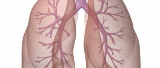
Bronchi
From the trachea, air enters the two bronchi. Their walls also consist of cartilaginous half-rings (6-12 pieces). They prevent the walls of the bronchi from collapsing. Together with blood vessels and nerves, the bronchi enter the lungs, where they branch to form the bronchial tree of the lung.
The inside of the trachea and bronchi are lined with mucous membrane. The thinnest bronchi are called bronchioles. They end in alveolar ducts, on the walls of which there are pulmonary vesicles, or alveoli. The diameter of the alveoli is 0.2-0.3 mm.
The alveolar wall consists of one layer of squamous epithelium and a thin layer of elastic fibers. The alveoli are covered with a dense network of blood capillaries in which gas exchange occurs. They form the respiratory part of the lung, and the bronchi form the airway section.
In the lungs of an adult there are about 300-400 million alveoli, their surface is 100-150 m2, i.e. the total respiratory surface of the lungs is 50-75 times larger than the entire surface of the human body.
Intubation
Tracheal intubation is the insertion of a special tube into it. This is a rather complex procedure, which, nevertheless, in a number of situations is necessary and can save the patient’s life. Intubation is carried out in terminal conditions, acute respiratory failure, pulmonary edema, tracheal obstruction, severe poisoning accompanied by impaired respiratory function. It helps to quickly ease breathing by ensuring airway patency, preventing swelling and allowing aspiration from the bronchi.F