Structural features
Cornea, the outermost layer of the eyeball, is a transparent, avascular, mirror-like sphere. It has tiny dimensions (diameter 0.56 mm) and occupies 1/16 of the outer surface of the eye. Consists of several layers separated by membranes.
The main guard is the outer epithelium. Prevents the penetration of dangerous microorganisms into the eye, delivers oxygen from the tear film, and controls the flow of fluid. It will hurt if you accidentally touch the epithelium with your hands. It contains nerve endings.
The second guard is the dense anterior membrane. In addition to protection, it nourishes the cornea. Thickness – 8-10 microns. Next comes the central part of the shell, an elastic and strong collagen stroma. Its cells repair damage. Then again there is an intermediate layer (Descemet's membrane). The last layer is the endothelium, which prevents the cornea from swelling and maintains transparency.
| At a young age, it contains about 4 thousand cells. By old age, only 2.5 thousand have the ability to regenerate. |
Consequences of inflammatory processes
If the infiltrates and erosions formed during keratitis do not reach the so-called Bowman's membrane, then there will be no traces of damage. Deeper lesions leave marks. The result may be a cloud, a spot or a cataract. They are distinguished depending on the degree of damage.
The cloud is not visible to the naked eye - it is a grayish translucent cloudiness. It affects visual acuity only when it is located in the center of the cornea. The spots are visible during normal examination; they appear as white dense areas. When they form, vision noticeably deteriorates. A thorn, depending on its size, can cause partial blindness. This is a white, opaque scar.
Abnormalities of corneal development that do not require treatment
If the radius of curvature, size, and transparency of the shell do not meet medical standards, then they speak of a developmental anomaly.
| The eyes are not only a mirror of the soul, but also of a person’s physical health. |
Poor nutrition throughout life does not go in vain and comes back to haunt you in old age. Violation of fat metabolism and cholesterol after 80 years leads to the accumulation of lipids in the cornea, its clouding in the shape of a ring. This anomaly is called a senile arc. The disease does not affect vision, and therefore cannot be treated in any way.
With dysfunction of the nervous system and liver, copper metabolism is impaired. A brown-green hoop, the Kayser-Fleischner ring, appears around the cornea. It does not affect vision.
Forecast
Uncomplicated corneal perforation responds well to treatment, especially for small lesions.
- In point forms of pathology, spontaneous sealing of the defect often occurs.
- Perforations of medium diameter can be successfully treated with medical glue.
- In severe forms, surgical intervention becomes more complicated and penetrating keratoplasty is required. This technique involves replacing the damaged area with a graft, which is placed on both the superficial and deep layers of the cornea. This operation allows patients to restore their vision and return to an active lifestyle.
Biomaterials for penetrating keratoplasty are produced by licensed medical institutions. They are stored in specialized eye banks. Modern transplants are convenient cornea blanks that accurately replicate its shape and perform its functions. Such materials are created in sterile laboratories and undergo a series of biological tests.
Penetrating corneal keratoplasty is performed on patients with extensive perforation or scar formation after surgical interventions on the cornea.
Developmental anomalies subject to correction
A large cornea or megalocornea is found in babies at birth. The norm is considered to be a shell diameter of 10 mm. If it exceeds by at least 1 mm, then this is a corneal symptom. The refraction of the eye is impaired; the larger the radius of curvature of the lens, the lower the refractive power.
The defect is dangerous for a number of complications - glaucoma, cataracts, problems with the retina. Megalocornea is often a symptom of complex genetic diseases (Markesani syndrome, Marfan syndrome, etc.). The disease has no cure. To improve vision, corrective glasses and lenses are prescribed.
Microcornea or small cornea. The opposite case. The shell, on the contrary, is less than 10 mm in diameter. The pupil appears unnaturally small. A consequence of microcornea, farsightedness. Glaucoma, cataracts, etc. develop against the background of the disease. If the cornea remains transparent, the doctor will prescribe symptomatic treatment and telescopic devices for vision correction.
Return to contents
| Both cases (macro-, microcornea) are of genetic origin and are inherited. |
Common types of pathology
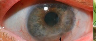
Norm and pathology of the cornea (Normal - normal form, Keratoconus - Keratoconus, Bullous keratopathy - bullous keratopathy, Corneal scarring - scarring of the cornea)
All variants of diseases of the corneal zone can be divided into 2 main groups - congenital and acquired diseases. Genetic and hereditary anomalies include:
- megalocornea (increase in size), when the corneal diameter exceeds 11 mm;
- microcornea (small size), with a decrease in diameter of less than 9 mm;
- flattening of the lens with decreased refraction;
- embryotoxon (turbid ring corresponding to the limbus);
- keratoconus (excessive protrusion of the lens anteriorly);
- spherical shape (keratoglobus).
Acquired variants of diseases are:
- keratitis (inflammatory processes of various origins and severity);
- keratopathy (chronic degenerative processes).
For any type of pathological change, it is necessary to perform a full examination in order to make an accurate diagnosis and choose the optimal treatment option.
Symptoms
Manifestations of corneal inflammation will be as follows:
- dry eye syndrome, pain,
- tearfulness,
- heavy eyelids - eyes seem difficult to open,
- fear of light,
- eyelid twitching,
- headache,
- redness,
- decreased vision.
| These symptoms are nonspecific and are often associated with other eye problems. Be that as it may, such manifestations should be alarming, and it is better to be aware of them. |
During the examination, the ophthalmologist will record:
- internal swelling,
- decreased sensitivity,
- cloudiness,
- dilated vessels,
- sores on the shell.
Viral keratitis
Keratitis caused by viruses is often accompanied by a blistering rash. The pathogenic agent can be almost any aggressive virus (measles, chickenpox, adenoviruses), but most often it is the herpes virus. As is known, over 95% of the population is infected with herpes infection, and in most cases, herpes occurs latently, in an asymptomatic form, and is activated only when immune resources are weakened, general exhaustion of the body and other unfavorable conditions. In this case, inflammation of the cornea is usually preceded by typical herpetic symptoms - rashes on the lips or other mucous membranes. With herpetic keratitis, as a rule, swelling and the appearance of fuzzy, vague infiltrates predominate.
Diseases of the cornea of the eye
Keratitis
This disease is characterized by clouding of the membrane and decreased vision due to viruses and bacteria. Inflammation can be caused by:
- herpes simplex and herpes zoster, chickenpox, measles, adenovirus,
- bacteria, intestinal, Pseudomonas aeruginosa, pneumococcus, streptococcus, staphylococcus, diplococcus.
- pathogens of tuberculosis, syphilis, gonorrhea, etc. (cause purulent keratitis),
- various fungi.
| Those who use contact lenses incorrectly should be wary of amoebic infection, the culprit of severe keratitis. |
Secondary causes of the disease:
- Drying of the cornea
- Exposure to too much light, such as when welding.
- Foreign body injury.
- Allergy.
- Diabetes.
- Mechanical damage to the cornea during eye surgery.
The most unfavorable outcome is epithelial detachment and the formation of corneal ulcers. Tissue necrosis occurs, purulent wounds appear, causing severe pain and a sharp change in vision.
Keratoconus
Doctors make this diagnosis if the cornea is not round in shape, but in the form of a cone. The pathology develops over 20 years. During adolescence, an ophthalmologist may mistakenly detect astigmatism.
Signs of keratoconus:
- When one eye is closed, double vision occurs.
- A person sees poorly from any distance.
- Blurred vision at night.
- Optical illusion. The patient observes not one, but several images at once.
- Oversensitive, intense eyes.
Return to contents
Keratomalacia
This disease often affects newborns whose mothers did not receive enough vitamin A during pregnancy. Or children who have suffered from jaundice. The disease develops rapidly. You can be left without vision in a day.
Bullous keratopathy
Bullae are blisters that cover the cornea. They occur after eye surgery, lens implantation, secondary glaucoma, resulting in the death of endothelial cells. Passed on by inheritance.
Corneal dystrophy
Or a tissue nutritional disorder. This condition is caused by disruptions in the functioning of the immune system, injuries, infections, and hereditary factors. Dystrophies are divided into:
- stromal,
- epithelial,
- endothelial,
- membrane
Iridocyclitis
Deep infectious inflammation of the vessels of the cornea. The eye becomes completely red from blood impurities. Young people and middle-aged people get sick.
Xerophthalmia
In other words, dry eyes. An urgent problem for our time.
Causes:
- With the advent of new means of entertainment - televisions, computers, telephones, our eyes began to experience double stress.
- Inaccurate use of lenses, failure to comply with an adequate wearing regimen.
- Poor environment, dusty air.
- Avitaminosis.
- Chronic diseases of the conjunctiva.
- Trachoma, diabetes, thyroid problems.
In advanced cases, the mucous glands may simply die and the conjunctiva may disintegrate.
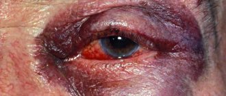
Corneal damage
The eye shield is easily injured. They poked a finger into an eye unsuccessfully, scratched it with a fingernail, touched it with a branch, got hit by a speck, etc. Types of damage:
- burns,
- foreign bodies,
- wounds,
- erosion.
Bacterial keratitis
Keratitis can be caused by a variety of pathogenic microorganisms, primarily cocci (Staphylococcus aureus, Streptococcus, Gonococcus) and Pseudomonas aeruginosa. A rare and very dangerous infection, even resulting in blindness, is acanthamoeba infection (named after the causative agent Acanthamoeba), which can exist, in particular, in the gap between the contact lens and the surface of the cornea. With the development of the so-called creeping corneal ulcers caused by gonococcal, tuberculosis, syphilitic and other bacterial infections, the risk of rapid and irreversible loss of vision is also very high.
Diagnosis of corneal diseases
The doctor conducts a detailed survey of the patient and assesses the condition of the eyes using traditional means:
- slit lamp,
- hyperemia of the conjunctiva,
- perforated diaphragm,
- fluorescein staining,
- biomicroscopy,
- pachymetry,
- confocal microscopy,
- keratotopography,
- culture for bacteria.
Ophthalmology has come a long way. The latest equipment allows us to qualitatively identify defects at an early stage. For example, many clinics use laser photo recorders, optical tomographs, and endothelial microscopes.
Return to contents
Treatment of corneal diseases
Depends on the root cause of the changes. Antifungal and antiviral drugs fight fungi and viruses. Antibiotics – with bacteria. If the disease is caused by tuberculosis or syphilis, anti-tuberculosis therapy is carried out. In case of allergies, all efforts are directed towards searching for the irritant. If contact lenses are the cause of the defect, then switching to glasses is recommended. If vision decreases, corrective optical aids are prescribed.
First aid
If injured, both eyes should be bandaged crosswise. Treat the skin of the eyelids with brilliant green. Place something cold and light on top. Drop “Albucid” into your eyes. If the pain is severe, take an analgesic. Lie down on the bed.
In case of a burn, you should rinse your visual organs thoroughly with water and saline solution and take a pain reliever.
| If a speck gets in, it is better to leave your eyes alone and not try to remove it yourself. Awkward movements will only do harm. In this case, as in the first two, you should consult a doctor as soon as possible. |
Drug treatment
Use:
- anti-infective drugs (Levomycin, Idoxuridine),
- ointments, drops, antiseptics to suppress inflammation (Albucid, Tetracycline),
- immunosuppressants (Azathioprine, Batriden),
- healing agents (Balarpan, Solcoseryl),
- medications to improve corneal nutrition (Emoxipin).
Therapy for eye injuries
To eliminate injuries, anti-inflammatory drops, ointments, and contact lenses are used to seal the damage. The surgeon sutures through wounds. In case of a burn, the visual organs are washed generously, antibacterial, regenerating, tear-replacing drugs, and anticoagulants are prescribed. In hopeless cases, surgery is performed.
Treatment of the cornea with folk remedies
- Sea buckthorn oil will help speed up the healing process and alleviate symptoms (1-2 drops every hour, duration – 2 weeks).
- They also use the juice of 3-year-old aloe. The plant is wrapped in paper and refrigerated for a week. Then they crush it and squeeze it out. Course – 1 drop once a day for a month.
- In case of suppuration and the formation of a cataract, juice from celandine and propolis is recommended (1:3). 2-3 drops. before bedtime.
- According to healers, honey and royal jelly can get rid of dystrophy. Mix these 2 ingredients 1 to 1. Pour in not hot, boiled water. Place the resulting ointment under the eyelid 3 times a day. For this disease, lotions made from goat's milk, nettle, and lily of the valley are also allowed.
| Herbal treatment is possible in consultation with the doctor. As an auxiliary therapy, folk recipes are quite effective. |
Surgical methods for treating corneal diseases
Operations are performed in severe and advanced cases:
- keratoplasty - replacement of the membrane or part of it with a graft,
- keratectomy - removal of opacities,
- keratoprosthesis – implantation of a prosthesis,
Such operations have been done for a long time. They have good reviews.
Prevention of corneal diseases
- Maintain good hygiene . Infections, bacteria, viruses often get into the eyes due to ignoring the rules of cleanliness. Monitor children closely. They love to play in the dirt and touch animals. And when babies want to sleep, what do they do? He rubs his eyes with his hands. It turns out that it is very easy to become infected.
- Do not overdry the cornea . Do not smoke, do not overstrain your eyesight, wear lenses correctly, use glasses to protect against ultraviolet radiation.
- Eat well . Often, a deficiency of vitamins (A, B, B1, E, C) leads to disorders in the cornea. Do exercises for your eye muscles and visit your ophthalmologist regularly.