This term has other meanings, see Kidney.
| Bud | |
| lat. ren | |
| human kidney | |
| Blood supply | renal artery |
| Venous drainage | renal vein |
| Innervation | renal plexus |
| Catalogs | |
| |
| Media files on Wikimedia Commons | |
Structure of the kidney:
1. The medulla and renal pyramids (
Pyramides renales
) 2. Efferent glomerular arteriole (
Arteriola glomerularis efferens
) 3. Renal artery (
Arteria renalis
) 4. Renal vein (
Vena renalis
) 5. Renal hilum (
Hilus renalis
) 6. Renal pelvis[en] (
Pelvis renalis
) 7. Ureter (
Ureter
) 8. Small renal calyx (
Calices minores renales
) 9. Fibrous capsule of the kidney (
Capsula fibrosa renalis
) 10. Lower pole of the kidney (
Extremitas inferior
) 11. Upper pole of the kidney (
Extremitas (
Arteriola glomerularis afferens
) 12. Network of renal capillaries 13. Nephron (
Nephron
) 14. Renal sinus (
Sinus renalis
) 15. Major renal calyx (
Calices majores renales
) 16. Apex of the renal pyramid (
Papillae renales
) 17. Renal column (
Columna renalis
)
Kidney
(lat. ren) - a paired bean-shaped organ that cleanses the blood and regulates the chemical homeostasis of the body through the function of urine formation. Part of the urinary system (urinary system)
Kidney anatomy[ | ]
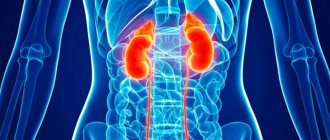
In humans, the kidneys are located behind the parietal layer of the peritoneum in the lumbar region on the sides of the two lower thoracic and two upper lumbar vertebrae in the projection of which they are adjacent to the posterior peritoneum, and the right kidney is normally located slightly lower, since it borders on the liver from above (in an adult, the upper pole the right kidney usually reaches the level of the 12th intercostal space, the upper pole of the left - the level of the 11th rib). (In the so-called “mirror people” the left kidney is located slightly lower, since their liver is located on the left, and, accordingly, the left kidney borders the liver.)
The dimensions of one bud are approximately 11.5−12.5 cm in length, 5−6 cm in width and 3−4 cm in thickness[2]. The mass of the kidneys is 120−200 grams, usually the left kidney is slightly larger than the right [3].
Each kidney is covered with a strong connective tissue fibrous capsule, and consists of parenchyma and a system for storing and excreting urine. The kidney capsule is a dense sheath of connective tissue covering the outside of the kidney. The kidney parenchyma is represented by an outer layer of the cortex and an inner layer of the medulla, which makes up the inner part of the organ. The urine accumulation system is represented by small renal calyces (6−12), which, merging with each other in 2-3 groups, form a large renal calyx (2−4), which, merging, form the renal pelvis. The renal pelvis[en] passes directly into the ureter. The right and left ureters empty into the bladder. In each human kidney there are about a million nephrons, which are the structural units that ensure the functioning of the kidney. The blood supply to the kidneys is carried out by the renal arteries, which arise directly from the aorta. From the celiac plexus, nerves penetrate the kidneys, which carry out the nervous regulation of kidney function and also provide sensitivity to the renal capsule. The morpho-functional unit of the kidney is the nephron, a specific structure that performs the function of urine formation. There are more than 1 million nephrons in each kidney. Each nephron consists of several parts: the glomerulus, the Shumlyansky-Bowman capsule and a system of tubules that pass into one another. A glomerulus is nothing more than a collection of capillaries through which blood flows. The loops of capillaries that make up the glomerulus are immersed in the cavity of the Shumlyansky-Bowman capsule. The capsule has double walls with a cavity between them. The cavity of the capsule passes directly into the cavity of the tubules. Most of the nephrons are located in the renal cortex. Only 15% of all nephrons are located at the border between the renal cortex and medulla. Thus, the renal cortex consists of nephrons, blood vessels and connective tissue. The nephron tubules form something like a loop that penetrates from the cortex into the medulla. Also in the medulla there are excretory tubules through which urine formed in the nephron is excreted into the renal calyces. The medulla forms the so-called “renal pyramids,” the apexes of which end in renal papillae protruding into the cavity of the minor renal calyx. At the level of the papillae, all the renal tubules through which urine is excreted join together
Internal structure of a sheep kidney
In mammals, the kidneys are bean-shaped structures, covered on the outside with a dense fibrous capsule. In a cross section of the kidney, the cortex and medulla can be distinguished. The cortex is represented mainly by the renal glomeruli, and the medulla - by the tubular parts of the nephrons. The medulla forms pyramids, with their bases facing the cortex. There can be either one pyramid (in rats) or several (7–24 in humans). Between them are the renal columns, which are sections of the cortex and contain segmental blood and lymphatic vessels. The pyramid with the cortex adjacent to its base forms the renal lobe. In the center of the concave edge there is the portal of the kidney; here is the expanded opening of the ureter - the renal pelvis. In the area of the renal hilum, it includes blood vessels (renal artery and vein), lymphatic vessels, and nerves. The ureters leaving the kidneys open into the bladder.
Details about the structural features
When considering what the kidneys look like, you need to remember that a person has several paired organs, and the organs that produce urine and cleanse the blood are recognized as the most important among all the paired ones. The buds are approximately equal in size and weight, which ranges from 120 to 200 grams.
The approximate size is the owner's fist. The right kidney does not have the opportunity to move slightly upward, since it is located in close proximity to the liver, which is located directly above it.
The external structure of the organ has several characteristic features:
- outwardly they resemble enlarged beans;
- the surface is smooth, dark red in color;
- an important component is the shell. The outer part of the kidney is formed by a special tissue called the fibrous capsule of the kidney.
When describing the appearance of the kidneys, it should be said that the description indicates the presence of:
- surfaces – front and back;
- edges – median and lateral;
- poles – lower and upper.
The location of the right kidney is influenced by its proximity to the liver, which is why the right organ is slightly lower than the left and slightly larger. Due to the tilt towards the spinal column, the distance between the upper poles is slightly less than between the lower ones. The median edge is where the renal vein and ureter exit, and the entrance to the renal artery is located here.
At the top there is a gland - the adrenal gland, which performs an extremely important function associated with the production of certain hormones that affect the proper development of the male and female body.
1 - adrenal gland; 2 - inferior vena cava; 3 - aorta; 4 - kidney; 5 - ureter
Located in the renal capsule, these organs are reliably protected from external negative influences in the form of injuries, blows, and bruises.
Kidney protection is also ensured by a fixation device, which includes:
- vascular legs;
- the right hepatorenal and renal-duodenal ligaments;
- on the left – diaphragmatic-colic;
- fascia, which provides a ligament of organs with the diaphragm;
- fat capsule;
- muscles of the back and abdomen, from which the renal bed is formed.
This device ensures reliable fixation and almost complete immobility of the kidneys. Considering how many layers the outer shell tissue has and what it consists of, you can see that its composition is the first layer of elastic fibers and the second layer of cells that make up smooth muscles. On the posterior surface the capsule is much denser.
On top of the capsule is a third layer. The buds are covered with leaves:
- anterior renal;
- posterior renal.
They are closely intertwined at the upper poles and both edges; at the lower poles there is no interlacing of leaves.
Kidney functions[ | ]
- Excretory (that is, excretory)
- Osmoregulatory
- Ion-regulating
- Endocrine (intrasecretory)
- Metabolic
- Participation in hematopoiesis
The main function of the kidneys - excretory - is achieved by the processes of filtration and secretion. In the renal corpuscle from the capillary glomerulus, under high pressure, the contents of the blood along with plasma (except for blood cells and some proteins) are filtered into the Shumlyansky-Bowman capsule. The resulting liquid is primary urine
continues its path along the convoluted tubules of the nephron, in which the reabsorption of nutrients (glucose, etc.), water, electrolytes into the blood occurs, while urea, uric acid and creatine remain in the primary urine.
As a result of this, secondary urine
, which from the convoluted tubules goes into the renal pelvis, then into the ureter and bladder. Normally, 1700-2000 liters of blood pass through the kidneys per day, 120-150 liters of primary urine and 1.5-2 liters of secondary urine are formed.
The ultrafiltration rate is determined by several factors:
- The difference in pressure in the afferent and efferent arterioles of the renal glomerulus.
- The difference in osmotic pressure between the blood in the capillary network of the glomerulus and the lumen of Bowman's capsule.
- Properties of the basement membrane of the renal glomerulus.
Water and electrolytes pass freely through the basement membrane, and substances with higher molecular weights are selectively filtered. The determining factor for the filtration of medium- and high-molecular substances is the pore size and charge of the glomerular basement membrane.
The kidneys play a significant role in the system of maintaining the acid-base balance of the blood plasma. The kidneys also ensure the constancy of the concentration of osmotically active substances in the blood under different water conditions to maintain water-salt balance.
Through the kidneys, the end products of nitrogen metabolism, foreign and toxic compounds (including many drugs), excess organic and inorganic substances are removed from the body; they participate in the metabolism of carbohydrates and proteins, in the formation of biologically active substances (in particular, renin, which plays a key role in the regulation of systemic blood pressure and the rate of secretion of aldosterone by the adrenal glands, erythropoietin - which regulates the rate of red blood cell formation).
The kidneys of aquatic animals differ significantly from the kidneys of terrestrial forms due to the fact that aquatic animals have the problem of removing water from the body, while terrestrial animals need to retain water in the body.
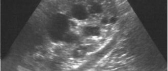
What does kidney tissue look like under a microscope?
In this article, we will be primarily interested in the filtering function of the kidneys.
In this connection, the main functional unit of the kidneys, the nephron, will be subjected to a detailed description.
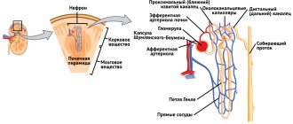
Conventionally, the nephron can be divided into 3 parts: 1. Circulatory system
(
glomeruli with afferent and efferent arterioles
)
2. Bowman's capsule
(
in which primary urine is formed
)
3. Tubular system
(
convoluted tubules, collecting ducts
)
Circulatory system
The kidney originates from the descending aortic arch, from which two renal arteries depart at an angle of 90 degrees.
Upon reaching the renal tissue, these arteries branch, become more numerous, and their diameter decreases. At the level of arterioles ( vessels with a small diameter
), the formation of renal glomeruli occurs.
This vascular formation actually resembles a intricately intertwined ball of capillaries into which the afferent arteriole flows and from which the efferent arteriole originates. amino acids, some protein macromolecules
pass .
Bowman's capsule
- a cup-shaped structure that envelops the glomerulus.
It is represented by a double capsule of the glomerulus; the liquid part of the blood penetrates into the lumen of this capsule along with some substances dissolved in it - primary urine is formed. The glomerular capsule is formed by epithelium - single-layer cellular tissue.
Bowman's capsule is normally impermeable to blood cellular elements ( erythrocytes, leukocytes
Canalicular system
- represented by convoluted tubes that originate in Bowman's capsule and end in the outlet of the collecting duct, which carries the final urine into the collecting system. These tubules are also lined with unicellular, denser epithelium.
Kidney diseases[ | ]
Kidney diseases are affecting more and more people. This is due to a large number of congenital pathologies and poor lifestyle, as well as a great reluctance to visit doctors at the first symptoms of diseases.
The most common diseases include:
- Urolithiasis (nephrolithiasis) is the formation of stones and sand in the kidneys.
- Pyelonephritis is considered one of the inflammatory diseases of the kidneys; the infection often enters the kidney through the blood.
- Nephroptosis (wandering kidney) - the disease can be either congenital or acquired. Women get sick more often.
- Hydronephrosis is characterized by problems with the flow of urine from the kidney.
- Renal failure is a condition when the kidneys partially cease to function and perform their functions; there are acute and chronic forms.
- Glomerulonephritis is another inflammatory disease that affects the glomeruli and tubules of the kidneys.
Work process
The structure and functions of the kidneys are directly related, and the rotary-countercurrent system of tubules is responsible for the functioning of the organ in question and the secretion of urine.
Due to its significant capillary pressure, the corpuscle helps purify blood plasma. This becomes the beginning of urine formation.
The result of cleansing is considered to be more than 100 liters of initial urine throughout the day. Then, the tubular system, due to the release of various components or the reverse absorption of fluid from the initial urine, forms a secondary one.
Next it penetrates into the papillary, then through these holes it will end up in small cups, then in large ones, then in the pelvis, and finally in the ureter.
During the day, the kidneys produce and secrete approximately 1-2 liters of secondary urine per day. Such a difference in volume is likely due to the concentration functioning of the organ in question.
Kidney transplant[ | ]
Main article: Kidney transplantation
With a decrease in the number of functioning nephrons, chronic renal failure develops, which, when progressing to end-stage renal failure, requires treatment with hemodialysis, peritoneal dialysis or kidney transplantation. Kidney transplantation is the most effective type of renal replacement therapy, including because it replaces all kidney functions, while dialysis partially compensates only for the excretory function of the kidneys, and to replace other kidney functions, the use of drugs (erythropoietin, vitamin D metabolites and etc.).
In 2011, 76 thousand kidneys were transplanted worldwide (a total of 110 thousand organs were transplanted)[4].
Kidney structure
The role of the organ in question for the proper functioning of the entire human body is difficult to overestimate.
Their task is to produce urine and cleanse the body of toxins during the period of fluid excretion. They are located in the lumbar region near the spinal column.
In normal condition they are located at different levels. The right one differs insignificantly in its own dimensions (slightly smaller) from the left one and is localized lower.
The organs in question are directly related to the liver. They are directly responsible for proper homeostasis and adequate purification of the blood from toxic components.
Nephrons
The main structural unit of the organ in question is called the nephron. It is formed by 2 elements: the Malpighi body and the tubular countercurrent-turning complex.
In a compressed form, the structure of the nephron suggests: a body, which is created by a glomerulus of vessels with an external capsule, then there is a convoluted tubule, then a straight tubule.
It ends with a loop of nephron, which is known as the “loop of Henle,” followed by the distal convoluted tubule.
Several of these tubules form collecting tubules, which unite into a collecting tract. They form papillary ducts, which exit through an opening on the papillae.
Kidney membranes
The fibrous capsule is designed to protect the kidneys from mechanical damage. It has a solid structure. The kidney membranes are easily separated.
The presence of a capsule of fat and fiber is considered a normal condition. The connective tissue fascia is formed by 2 membranes: the outer ball is connected by fibers to a fibrous capsule, and under the membrane is the cortex of the organ, which contains nephrons.
The crust has a border with the pyramids. Parenchyma suggests the presence of a medulla.
Protective stock
In order to prevent displacement of the kidneys, kinks in the vessels and ureters, a fixing device is used.
The kidneys are located on a protective bed, the basis of which is adipose tissue. Pressure inside the abdominal cavity plays an important role in fixing the organs.
The bed is formed by the quadratus, psoas minor and lateral transverse muscles, as well as the diaphragm.
Internal structure
The medulla forms 7 pyramids in the organ itself. Each of them is attached to the pelvis by means of a papilla.
Urine penetrates through the pathways into small and large cups, where each of them passes urine through itself, which ensures the proper functioning of the excretory apparatus.
The renal pelvis is where the cups direct urine. The main gland of homeostasis will be the pituitary gland, which is designed to control the functioning of the kidneys.
When cut, the structure of the organ in question is divided into 2 parts:
- shells;
- Malpighian bodies.
Notes[ | ]
- ↑ 12
Foundational Model of Anatomy - Big medical encyclopedia. — Second edition. - Publishing house "Soviet Encyclopedia", 1962. - T. 26. - P. 291.
- M.R.Sapin, Z.G.Bryksina.
Human anatomy. - M.: Education, 1995. - P. 254. - 464 p. — ISBN 5-09-004385-X. - Gudrun Heise, Ksenia Polskaya.
Organ transplantation: ethics or economics?. There is a catastrophic shortage of donor organs in the world. Too often, the lives of seriously ill patients depend on the size of their wallet, and those who sell their organs have no idea what awaits them (Russian). Deutsche Welle (1 June 2013). Retrieved May 29, 2020.