Histology of the respiratory system is one of the important sections of biology, allowing us to understand the organizational features of a living organism. Histology is a science that deals with living tissues. To be more precise, the peculiarities of their structure, development, and specific life activities. To study the histology of the respiratory system, a microtome is used, which allows samples to be cut into extremely thin layers. The discipline should not be confused with anatomy, since the object of study is different. Histology of the respiratory system gives an idea of the tissues of the body and the features of their structure.
What is he doing?
Within the framework of the histology of the respiratory system, it is customary to talk about the following functionality of the listed structures:
- conducting air masses;
- purification of substances coming from the atmosphere;
- heating to body temperature;
- formation of sounds.
The structure of the respiratory system in histology is usually considered in relation to the second group of organs and tissues, called respiratory. The specialized name for this sector is acini. This is how it is customary to designate bubbles in the lungs located in the intercellular space. Thanks to them, it becomes possible to exchange gases with the circulatory system, which allows you to saturate the living body with the necessary compounds.
Pathogenetic therapy of bronchitis
Bronchitis at any stage of development requires observation and comprehensive treatment by all available means. Since the main symptoms of acute bronchitis in children and adults are swelling of the bronchi and stagnation of sputum, pathogenetic therapy of bronchitis consists of eliminating these phenomena. This can be achieved with special medications and folk remedies. An experienced doctor will always prescribe complex treatment with various types of drugs to the patient. This largely depends on the age of the patient, the stage of development of bronchitis and its type.
Very often, treatment is carried out at home; hospitalization is recommended only in extreme cases. What groups of drugs should I take?
- Antiviral medications – Since this is a viral infection, it is imperative to take antiviral medications.
- If there is an increase in body temperature, antipyretics are prescribed.
- Inhalation of phytoncides will help relieve swelling from the bronchi and bronchioles, mucus will be easier to expectorate.
- Bronchodilator drugs.
- Antihistamines.
- Vitamins of group B and C.
- Antibiotics are not used everywhere, only in cases where bacterial genesis is observed.
The above drugs are a complex for the effective treatment of acute bronchitis.
The doctor will prescribe adequate methods and medications after a consultation. Among the effective antiviral drugs are rimantadine, Tamiflu, Virazol, and interferon. Ribaverine, acyclovir and other chemicals are also used, which are also used to treat bronchitis in children.
How did it appear?
Particular histology of the respiratory system is a frequent source of data for experiments and research, allowing one to obtain a general idea of the peculiarities of the development of organs, thanks to which the tissues of our body can receive oxygen. It is known that the foregut, in the process of protrusion of one of the walls, forms specific rudiments. It is from them that the bronchi, trachea, and laryngeal region are subsequently formed.
Within the framework of gynecology and pediatrics, the histology of the respiratory system is also important, as it gives insight into the period of formation of these tissues, which are essential for the normal life support of a living organism. It has been revealed that protrusion occurs already at 3-4 weeks from the moment of conception.
Mesenchyme is the source of differentiation, due to which muscular bronchial tissue is formed. At the same time, the foundations of the cartilaginous structure are laid, and connective tissue fibers are born. As part of studies on the anatomy and histology of the respiratory system, it was revealed that during the same period the circulatory system of the respiratory organs is formed. The splanchnotome is the basis for the development of the pleura.
Classification of diseases
Due to their complex structure, the lungs and bronchi can be affected by various diseases. It all depends on where the infection has penetrated and what processes have begun to occur in a particular department. The following classification of diseases can be given:
- Diseases of the pleural cavity: pneumothorax, pleurisy, etc.
- Inflammatory processes: pneumonia, bronchitis, etc.
- Leading to respiratory failure: distress syndrome, thrombosis, etc.
- Diseases of a systemic nature with a disseminated mechanism: alveolitis, sarcoidosis, etc.
- Chronic forms.
- Destroying the lungs: gangrene, abscess.
- Acquired or hereditary pathologies.
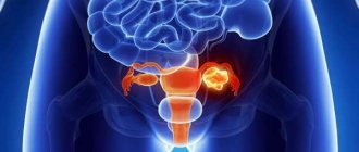
- Malignant or benign neoplasms.
- Post-traumatic illnesses.
Each disease has its own varieties. In general, they are divided into types of occurrence, according to severity, possible complications and causes of occurrence.
Each disease has its own symptoms. However, for the most part it has the following symptoms:
- Dyspnea.
- Temperature increase.
- Cough that may produce phlegm.
- Suffocation.
- Pain in the chest area.
go to top
Features of the structure
Histology of the human respiratory system has made it possible to obtain an accurate understanding of the characteristics of the airways. In particular, it was revealed that, in fact, these are tubes that closely interact throughout the entire period of the body’s life and are capable of passing air masses. The inner surface is densely covered with a unique respiratory mucosa. Histology of the respiratory system showed that ciliated epithelium, formed into a structure with a large number of rows, is typical for this tissue.
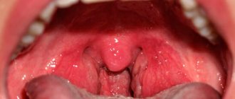
At the same time, scientists discovered that the vestibule of the nasal cavity differs quite significantly from other organs. Histology of the respiratory system showed that there are certain differences in the structure of the area above the larynx, the vocal cords. Here the epithelium also consists of numerous layers, but its structure is flat.
Mucolytics, mucoregulators and other groups of cough medications
Direct-acting drugs are necessary to speed up the process of removing mucus from the bronchi, which will speed up recovery. These are mucolytics; they have an expectorant effect. However, it is worth noting that many well-known drugs have several targeted actions - anti-inflammatory, antimicrobial, mucolytic and expectorant.
Mucolytics and mucoregulators are chemical-based drugs, their action is aimed at thinning mucus and removing it from the bronchi. Mucolytic drugs can be divided into three groups, which differ in their principle of action:
- The first group is drugs that thin sputum. One of the most effective and popular representatives is ACC.
- The second group of drugs are drugs that speed up the process of removing mucus from the bronchi. Among them, we note such as bromhexine and ambroxol.
- The third group of drugs are drugs whose action is aimed at reducing the formation of mucus. This is Libexin Muco, glucocorticoids.
The above drugs are taken orally and, after assimilation by the digestive system, begin their complex effect on the bronchi and alveoli. Mucolytic drugs also reduce the risk of exacerbations; they can be used both in granules and in the form of syrups and tablets. A doctor can prescribe such drugs after the exact diagnosis and stage of development of the disease becomes known. Such products differ in their effect on the body and, accordingly, in cost.
Curious moments
If we briefly consider the histology of the respiratory system, it is necessary to mention the features of the structure and functioning of the organs that form the air-conducting pathways. In particular, their walls are created with multilayer fabrics. There are four shells in total:
- mucous;
- submucosa (glands are located here);
- fibrous cartilaginous (supplemented by two types of cartilage tissue - hyaline, elastic);
- adventitial.
The severity of the membranes varies significantly and is determined both by the location and the functionality of a particular organ. If, in particular, you examine the structure of the bronchial system and pay special attention to the final, small structures, you will notice that the submucosa is completely absent here. There is no cartilaginous fibrous layer in such bronchi.
Other diseases
Of course, the bronchi and lungs manifest themselves as other diseases. However, they are less common:
- Alveolitis is inflammation of the tissues of the alveoli.
- Asphyxia is a sudden lack of air and oxygen in the blood.
- Sarcoidosis is the appearance of nodules on blood vessels.
- Histoplasmosis is a fungal disease.
- Atelectasis is atrophy of part of the lung.
- Pneumothorax is the accumulation of excess gas in the pleural cavity.
go to top
Mucous
Normally, this element of the respiratory system is formed by a three-layer plate. It has several specific features. The first lamina is epithelial. In its structure, it is a prism-shaped ciliated epithelium formed in many rows. This covers the respiratory structures. The second type is a plate created by loose connective fibers in combination with elastic ones. Finally, muscle is formed by myocytes (exclusively smooth type). There is no such plate in the structure of the laryngeal region, trachea, or the inside of the nose.
Specific features of the trachea
This human organ, which allows breathing, is a tube with four shells. The inside is lined with mucous tissue, characterized by the presence of two plates. The base under the mucosa is a tissue supplemented with protein, mucous glands, characterized by a complex structure, producing a specific secretion. Thanks to this component, the surface of the trachea is always moistened from the inside. The outside of the organ is covered with adventitial tissue, and between it and the submucosa there are cartilaginous and fibrous fibers.
By the way, not all living beings are built like humans. In particular, the histology of the respiratory system of birds has shown that they have no cartilaginous tissue at all in the trachea. Instead, bone is formed here. Of course, histological studies make it possible to identify certain similar structural features of organisms of various species, but one should not equate all forms of life with one another: there are surprisingly many species-specific differences.
Human
Bronchi, bronchial tree, lungs
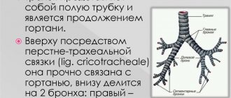
In humans, the trachea is divided into two main bronchi
bronchus principales occurs at the level of the fourth - fifth thoracic vertebrae[1]. The right main bronchus is thicker, shorter, and more vertical than the left [2].
The main bronchi branch repeatedly, forming the bronchial tree arbor bronchiale, which has about 23 branching orders. Initially, the bronchi are divided according to the macroscopic structure of the lungs into lobar and segmental. The right lung has 3 lobes, the left lung has 2. Each lung has 10 segments. starting from the branches of the segmental bronchi (subsegmental bronchi), there is a tendency towards dichotomous division, that is, each bronchus branches in two. The bronchial tree ends with terminal bronchioles. Terminal bronchioles branch into respiratory bronchioles, from which the respiratory sections of the lungs (acini) begin.
The bronchial tree includes:
- lobar bronchi (topographically divided into extra- and intrapulmonary parts),
- segmental bronchi,
- interlobular bronchi,
- lobular (lobulus pulmonis (BNA)),
- intralobular bronchi (several orders of branching)
- terminal bronchioles.
The wall of the bronchi consists of several layers: from the inside, the bronchi are lined with a mucous membrane (internal), consisting of epithelium, lamina propria and muscularis; submucosa; fibrocartilaginous membrane and adventitia (external). The epithelium is single-layered multirow prismatic ciliated with goblet cells. The lamina propria and submucosa are formed by connective tissue and contain secretory sections of the mucous glands. The fibrocartilaginous membrane is represented by cartilaginous rings connected by connective tissue (as the bronchi branch and decrease in caliber, the rings open, are replaced by islands, then cartilage grains, and finally, in small-caliber bronchi they disappear completely). The adventitia is formed by connective tissue. As the bronchi branch, a decrease in the caliber of the bronchi, opening and reduction in the size of the cartilaginous rings, thickening of the muscular plate, and a decrease in the height of the bronchial epithelium are observed. Along the branches of the bronchi there are numerous lymph nodes that receive lymph from the lung tissues; lymphatic formations (namely, lymph follicles) are also present in the wall of the bronchi themselves, especially in the places of branching. Blood supply to the bronchi is carried out by bronchial arteries arising from the arch and thoracic part of the aorta, innervation by branches of the vagus, sympathetic and spinal nerves.
Trachea: other features of the human body
As part of histological studies, it was established that the respiratory system in relation to this organ is supplemented by multirow epithelium. It is formed by a wide variety of cellular structures:
- basal cambial;
- ciliated;
- mucous-producing components are goblet-shaped;
- producing endocrine hormones serotonin, norepinephrine, dopamine.
The last category is responsible for the correct contraction of smooth muscles, since the process is regulated precisely by hormonal levels. If there are malfunctions in the functionality of these cells, this can lead to serious pathologies of the respiratory system.
Side effects and effectiveness criteria
Some, even the most effective drugs have contraindications for use, which must be familiarized with in advance. Common side effects include nausea and vomiting, which are caused by hypersensitivity or intolerance to certain components of the drug. Headaches, heartburn, diarrhea, allergic rashes and other effects may also occur. If such undesirable effects are detected, it is recommended to stop taking the drug and consult a doctor.
Tracheas: concluding the review
Another important aspect of the structure of the tissues of the respiratory system, identified through histological studies, is associated with the characteristics of the cartilaginous tracheal membrane formed by the fibers. As we found out during specific experiments, this element is formed by rings of hyaline tissue in the amount of 16 to 20. They do not close on the back side, and the ends are connected by muscle bundles. Thanks to this structural feature, the tracheal walls are pliable. This determines the mechanics of swallowing and makes it possible to push food elements inside the esophagus towards the stomach.
Comparative characteristics of cough medications based on studies and publications
The list of pharmaceutical drugs for cough is increasing every day; more and more new drugs are constantly appearing, which a person may not even know about. Research is often conducted to determine the effectiveness of a particular product; the product gains popularity through publications and advertising.
To determine the effectiveness of drugs for the prevention of respiratory problems in children, a study was conducted: 4 groups of children aged 13 to 14 years were organized. Each group consisted of 15 people. Each of the groups of children took antitussives:
- the first group is Eleutherococcus;
- the second is pink radiola;
- the third is St. John's wort;
- the fourth group did not use any medications.
The studies were conducted during the period of exacerbation of seasonal infections. The result is this: St. John's wort and Eleutherococcus were identified as the most effective remedies.
Bronchi
It is customary to distinguish several categories:
- basic;
- shares;
- belonging to the zones.
The mentioned categories are classified as extrapulmonary. Along with them there are internal:
- segments,
- subsegmental;
- terminal.
When assessing the dimensions (in medicine this is usually called caliber), it is customary to divide the bronchi into large, average, small, and terminal. Regardless of belonging to a specific group, the structure of all varieties is quite similar in nature.
In what cases, which group of cough medications should be preferred in children and adults?
In both children and adults, at an early stage of the disease, the cough is dry and unproductive. At this stage, care must be taken to get rid of the painful cough; antitussives are used. After this, the cough enters the productive stage; a previously used drug or remedy cannot be used; measures must be taken to remove sputum from the bronchi.
At the initial stage of pathology development, it is advisable to use antitussive mucolytic drugs. The most effective and popular of them are ACC, Mucaltin, Soludan and Ambroxol. Such products may contain herbs, the action of which is aimed at thinning mucus and quickly removing it from the body. The sooner the sputum is removed from the body, the faster the viruses will leave it and a speedy recovery will occur.
Increasingly, doctors are prescribing drugs that have a combined effect. They simultaneously stop the inflammation process, help get rid of spasms, and increase cough productivity. Thus, the healing process is accelerated. The most famous drugs in this group are Doctor Mom and Codelac Fito.
Many drugs have some restrictions and contraindications. Pay attention to the patient’s condition, read the instructions for use. Most of the above drugs should not be used by nursing mothers, pregnant women, children under three years of age, and those who are hypersensitive to the components of the drugs.
For children there is a list of antitussive and expectorant drugs. Let's note the most effective: Lazolvan, Ambrobene, Ambrohexal - these products are used from birth, have a general strengthening, antiviral and anti-inflammatory effect.
Bromhexine - helps thin mucus and remove it from the body.
Linax - eliminates bronchospasm, has a mucolytic effect. Can be used for children after the first year of life.
Breast fees.
Many of these drugs are prescribed by a doctor, who monitors the patient’s condition throughout the entire treatment period.
What is it about?
Normally, the bronchi are formed by four membranes. From the inside, the organs are covered with mucous tissue, under which there is a submucosa, the next layer is cartilaginous fibrous cells, and the final element is adventitial tissue. The diameter directly determines how clearly and vividly each of the structural elements is expressed.
If you examine the main bronchi, you can see clearly formed four membranes. The same structural features are also characteristic of large and medium-sized elements. But with a histological examination of small formations, it will be possible to find only two layers - mucous tissue and adventitial cells.
Prevention
Carrying out breathing exercises can improve the removal of mucus and improve breathing. Physiotherapeutic treatment of bronchitis is carried out in the last stages, when the symptoms are minor. Follow-up treatment is necessary with electrophoresis, inhalation and UHF therapy.
Important points in the treatment of bronchitis are the immediate adoption of effective measures at the first symptoms of the disease and monitoring the recovery situation. After treatment, the doctor is obliged to conduct an examination and confirm the complete absence of bronchitis pathogens.
What to do?
If you think you have Bronchitis
and the symptoms characteristic of this disease, then doctors can help you: therapist, pediatrician, pulmonologist.
Source
Did you like the article? Share with friends on social networks:
Bronchial mucosa
This element is formed by three plates: from epithelial cells, mucous tissue, and muscle fibers. Epithelium is a layer facing the bronchial lumen. It consists of ciliated cells, collected in a structure with an abundance of rows. The main characteristic of the epithelial layer is prismatic. The smaller the dimensions of the bronchi, the fewer rows there will be in the structure of this element. Additionally, the nature of the cellular structure changes: in small organs, low cubic ones are predominantly found, but there are practically no goblet ones.
Histological examination of the distal parts of the respiratory system formed by the bronchi revealed the following types of cells:
- goblet;
- basal;
- ciliated;
- endocrine;
- bordered;
- lacking eyelashes;
- secretory.
The last category is not typical for other parts of the bronchial tree. A feature of secretory formations is the ability to break down surfactant. But the bordered ones, as scientists have discovered, play the role of chemoreceptors. Finally, cells lacking cilia are characteristic only of bronchioles.
What else should you pay attention to?
As revealed during histological studies, the epithelial plate precedes the mucosa, created by loose connective cells. The structure of the plate determines the presence of elastic fibers. The smaller the dimensions, the higher the concentration of elastic formations. The third muscle plate acts as a trailing one. It is most developed in elements from major to minor. A distinctive feature of asthma affecting these organs is the contraction of muscle tissue of the smallest, small elements. The process leads to a decrease in the lumen of the respiratory organs.
The bronchial submucosal base is characterized by a grouping of proteinaceous, mucous mixed glandular cells - the end sections of these formations are located here. The secretion produced by cells is capable of destroying microscopic life forms and has a bacteriostatic effect. Due to its consistency, the secretion envelops dust particles and provides the necessary level of moisture to the mucous membrane.
Small, but smart
The small bronchial structure is devoid of the glands described above and the submucosa. Quite atypical in comparison with other wood is the shell created by cartilaginous cells and fibrous tissue. The smaller the size of the elements, the more this parameter changes. Thus, in the main structures, open rings were observed, but here only plates of cartilaginous tissue are present in large formations in the longitudinal direction.
What's special? Small bronchi are generally devoid of cartilaginous tissue, a membrane formed by cartilage and fibrous cells. The adventitial covering is created by connective tissue fibers. They contain nerves and elements of the circulatory system. Gradually, the membrane flows into the pulmonary parenchyma septa.