Anatomical structure and functions of the ear.
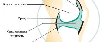
The ear is the organ of hearing and balance in vertebrates and humans. The ear is the peripheral part of the auditory analyzer.
Anatomically, the human ear is divided into three sections.
- the outer ear, consisting of the pinna and the external auditory canal ;
- the middle ear, composed of the tympanic cavity and having appendages - the Eustachian tube and the cells of the mastoid process;
- the inner ear (labyrinth), consisting of the cochlea (auditory part), vestibule and semicircular canals (equilibrium organ).
If we add to this the auditory nerve from the periphery to the cortex of the temporal lobes of the brain, then the entire complex will be called the auditory analyzer.
auricle consists of a skeleton - cartilage, covered with perichondrium and skin. The surface of the shell has a number of depressions and elevations. The muscles of the auricle in humans serve to maintain the auricle in its normal position. The external auditory canal is a blind tube (about 2.5 cm long), somewhat curved, closed at its inner end by the eardrum. In an adult, the outer third of the auditory canal is cartilaginous, and the inner two-thirds is bone, part of the temporal bone. The walls of the external auditory canal are lined with skin, which in its cartilaginous section and the initial part of the bone has hair and glands that secrete a viscous secretion (earwax), as well as sebaceous glands.
Auricle: 1 - triangular fossa; d—Darwin's tubercle; 3 - rook; 4 - stem of the helix; 5 - sink bowl; 6 - shell cavity; 7 - antihelix; 8 - curl; 9 - antitragus; 10 - lobe; 11 - intertragal notch; 12 - tragus; 13—supraglosal tubercle; 14—supratragal notch; 15 — legs of the antihelix.
The eardrum in an adult (10 mm in height and 9 mm in width) completely isolates the outer ear from the middle ear, that is, from the tympanic cavity. The handle of the hammer , part of one of the auditory ossicles, is rotated into the eardrum
The tympanic cavity of an adult has a volume of about 1 cm^; lined with mucous membrane; its upper bony wall borders the cranial cavity, the anterior wall in the lower section passes into the Eustachian tube, the posterior wall in the upper section passes into the recess connecting the tympanic cavity with the cavity (cave) of the mastoid process. The tympanic cavity contains air. It contains the auditory ossicles (hammer, incus, stapes) , connected by joints, as well as two muscles (stapedius and tensor tympanic membrane) and ligaments.
There are two holes on the inner wall; one of them is oval, covered with a stapes plate, the edges of which are attached to the bone frame with fibrous tissue, allowing mobility of the stapes; the other is round, covered with a membrane (the so-called secondary tympanic membrane).
The Eustachian tube connects the tympanic cavity to the nasopharynx. It is usually in a collapsed state; when swallowing, the tube opens and air passes through it into the tympanic cavity.
Diagram of the structure of the right human auditory organ (section along the external auditory canal): 1 - auricle; 2 - external auditory canal; 3 - eardrum; 4—tympanic cavity; o— .hammer; 6 - anvil; 7—stirrup; 8—Eustachian tube; 9— semicircular canals; 10 - snail; 11 - auditory nerve; 12 - temporal bone.
During inflammatory processes in the nasopharynx, the mucous membrane lining the tube swells, the lumen of the tube closes, and the flow of air into the tympanic cavity stops, which causes a feeling of ear congestion and decreased hearing.
Behind the tympanic cavity and the external auditory canal there are cells of the mastoid process of the temporal bone, communicating with the middle ear, normally filled with air. With purulent inflammation of the tympanic cavity (see otitis), the inflammatory process can spread to the cells of the mastoid process ( mastoiditis).
The structure of the inner ear is very complex, which is why it is called a labyrinth. It distinguishes between the auditory part (cochlea) , which has the shape of a sea snail and forms 2 1/2 curls, and the so-called vestibular part, consisting of a cistern, or vestibule, and three located in three different planes. Inside the bony labyrinth there is a membranous labyrinth filled with a transparent liquid. A plate capable of oscillating runs across the lumen of the helix of the cochlea, and on it is located the cochlear, or organ of Corti, containing auditory cells - the sound-perceiving part of the auditory analyzer.
Inner ear
Inner ear
(auris interna)
a hollow bone formation in the temporal bone, divided into bone canals and cavities containing the receptor apparatus of the auditory and staokinetic (vestibular) analyzers.
The inner ear is located in the thickness of the petrous part of the temporal bone and consists of a system of bone canals communicating with each other - the bone labyrinth ( Fig. 1 ), in which the membranous labyrinth is located ( Fig. 2 ). The outlines of the bony labyrinth almost completely repeat the outlines of the membranous labyrinth. The space between the bony and membranous labyrinth, called the perilymphatic labyrinth, is filled with fluid - perilymph, which is similar in composition to cerebrospinal fluid. The membranous labyrinth is immersed in the perilymph, it is attached to the walls of the bone sheath by connective tissue cords and is filled with liquid - endolymph, which is somewhat different in composition from the perilymph. The perilymphatic space is connected to the subarachnoid narrow bony canal - the cochlear aqueduct. The endolymphatic space is closed, has a blind protrusion extending beyond the inner ear and temporal bone - the aqueduct of the vestibule. The latter ends with an endolymphatic sac embedded in the thickness of the dura mater on the posterior surface of the pyramid of the temporal bone.
The bony labyrinth consists of three sections: the vestibule, semicircular canals and cochlea. The vestibule forms the central part of the labyrinth. Posteriorly it passes into the semicircular canals, and anteriorly into the cochlea. The inner wall of the vestibule cavity faces the posterior cranial fossa and forms the bottom of the internal auditory canal. Its surface is divided by a small bony ridge into two parts, one of which is called the spherical recess and the other the elliptical recess. In the spherical recess there is a membranous spherical sac connected to the cochlear duct; in the elliptical - an elliptical sac into which the ends of the membranous semicircular canals flow. In the median wall of both recesses there are groups of small holes intended for branches of the vestibular part of the vestibular-cochlear nerve. The outer wall of the vestibule has two windows - the window of the vestibule and the window of the cochlea, facing the tympanic cavity. The semicircular canals are located in three planes almost perpendicular to each other. Based on their location in the bone, they are distinguished: superior (frontal), or anterior, posterior (sagittal) and lateral (horizontal) canals.
The bony cochlea is a convoluted canal extending from the vestibule; it spirals 21/2 times around its horizontal axis (bone shaft) and gradually narrows towards the apex. A narrow bone plate spirals around the bone core, to which the connecting membrane that continues it is firmly attached - the basement membrane, which makes up the lower wall of the membranous canal (cochlear duct). In addition, a thin connective tissue membrane, the vestibular membrane, also called Reissner's membrane, extends from the bony spiral plate at an acute angle upward and laterally; it forms the upper wall of the cochlear duct. The space formed between the basal and vestibular membranes is limited on the outer side by a connective tissue plate adjacent to the bony wall of the cochlea. This space is called the cochlear duct (duct); it is filled with endolymph. Above and below it are the perilymphatic spaces. The lower one is called the scala tympani, the upper one is called the scala vestibule. The ladders at the top of the cochlea are connected to each other by the cochlea opening. The cochlear shaft is pierced by longitudinal rings through which nerve fibers pass. Along the periphery of the rod, its winding canal stretches spirally; nerve cells are placed in it, forming the spiral ganglion of the cochlea ( Fig. 3 ). The internal auditory canal leads to the bony labyrinth from the skull, through which the vestibulocochlear and facial nerves pass.
The membranous labyrinth consists of two vestibular sacs, three semicircular ducts, the cochlear duct, the aqueducts of the vestibule and the cochlea. All these sections of the membranous labyrinth represent a system of formations communicating with each other.
In the membranous labyrinth, the fibers of the vestibulocochlear nerve terminate in neuroepithelial hair cells (receptors) located in certain places. Five receptors belong to the vestibular analyzer, three of them are located in the ampoules of the semicircular canals and are called ampullar ridges, and two are located in the sacs and are called spots. One receptor is auditory, it is located on the main membrane of the cochlea and is called the spiral (spiral) organ of Corti.
Arteries V. u. originate from the labyrinthine artery, which arises from the basilar artery (arteria basilaris). The venous blood of the labyrinth collects in the plexus lying in the internal auditory canal. From the vestibule and semicircular canals, venous blood flows mainly through the vein passing through the aqueduct of the vestibule into the transverse sinus of the dura mater. The veins of the cochlea carry blood to the inferior petrosal sinus. Innervation of V. at. receives from the VIII pair of cranial nerves, each of which, entering the internal auditory canal, splits into three branches: upper, middle and lower. The upper and middle branches form the nerve of the vestibule - nervus vestibularis, the lower corresponds to the nerve of the cochlea - nervus cochleae.
In V. u. receptors of the auditory and statokinetic analyzers are located. The receptor (sound-perceiving) apparatus of the auditory analyzer is located in the cochlea and is represented by the hair cells of the spiral (corti) organ. The cochlea and the receptor apparatus of the auditory analyzer contained in it are called the cochlear apparatus. Sound vibrations arising in the air are transmitted through the external auditory canal, the eardrum and the chain of auditory ossicles to the vestibular window of the labyrinth, causing wave-like movements of the perilymph, which, spreading, are transmitted to the spiral organ (see Hearing). The receptor apparatus of the statokinetic analyzer, located in the semicircular canals and sacs of the vestibule, is called the vestibular apparatus.
Research methods . Modern methods of studying the function of V. at. include determination of the state of both of its functions - auditory and vestibular. When studying auditory function, an adequate stimulus is used - sound of varying frequency and intensity in the form of pure tones, noise and speech signals. Tuning forks, audiometers, whispered and loud speech are used as a sound source. Research using this set of tools makes it possible to determine the state of the function of the sound-conducting system, the receptor apparatus of the ear, as well as the conductive and central sections of the auditory analyzer (see Audiometry).
The study of vestibular function (vestibulometry) includes the identification of spontaneous (not artificially caused) symptoms resulting from V.'s disease. or c.s.s. Among them, spontaneous nystagmus caused by a unilateral inflammatory process in the V., a fall in the Romberg position, and a violation of coordination tests are often encountered (see Vestibular reactions). The state of vestibular function is studied during rotation on a Barany chair or a special rotational stand, using caloric, galvanic, pressor and other tests.
In a clinic, examination of patients with suspected V. u. carried out by an otorhinolaryngologist. It includes targeted collection of anamnesis and clarification of the patient’s complaints, drawing up a hearing passport (data from speech and tuning fork hearing tests), visual detection of spontaneous nystagmus, etc. To clarify the diagnosis, additional studies are carried out according to indications - radiography of the temporal bones, rheography of cerebral vessels, etc.
Pathology. Typical complaints in patients with diseases of the auditory part of the ear. are hearing loss and tinnitus. The disease can begin acutely (acute sensorineural hearing loss) or gradually (cochlear neuritis, chronic cochleitis). When hearing is damaged, as a rule, the vestibular part of the ear is also involved to one degree or another in the pathological process, which is reflected in the term “cochleovestibulitis.”
Developmental defects. There is a complete absence of the labyrinth or underdevelopment of its individual parts. In most cases, there is underdevelopment of the spiral organ, most often its specific apparatus - hair cells. Sometimes the hair cells of the spiral organ are underdeveloped only in certain areas, while the auditory function may be partially preserved in the form of so-called hearing islands. In the occurrence of congenital defects V. at. The pathological effect on the embryo from the mother’s body (intoxication, infection, injury to the fetus) plays a role, especially in the first months of pregnancy. Genetic factors also play a role. V.'s damage should be distinguished from congenital malformations. during childbirth.
Damage. Isolated mechanical damage to V. u. are rare. Trauma V. u. possible with fractures of the base of the skull, when the crack passes through the pyramid of the temporal bone. In transverse fractures of the pyramid, the crack almost always involves the V. at., and such a fracture is usually accompanied by severe impairment of auditory and vestibular function, up to their complete extinction.
Specific damage to the receptor apparatus of the cochlea occurs with short-term or prolonged exposure to high-intensity sounds. Long-term effect of strong noise on V. may lead to hearing impairment (see Hearing loss).
Pathological changes in V. at. occur when the body is exposed to concussions. In case of sudden changes in external atmospheric pressure or pressure under water as a result of hemorrhage in the V. Irreversible changes in the receptor cells of the spiral organ may occur (see Barotrauma).
Diseases. Inflammatory processes occur in the V. at., as a rule, secondarily, more often as a complication of acute or chronic purulent otitis media (tympanogenic labyrinthitis), less often as a result of the spread of infectious agents in the V. at. from the subarachnoid space through the internal auditory canal along the sheaths of the vestibulocochlear nerve during meningococcal infection (meningogenic labyrinthitis). In some cases in V. at. It is not microbes that penetrate, but their toxins. The inflammatory process that develops in these cases occurs without suppuration (serous labyrinthitis). The outcome of the purulent process in V. There is always complete or partial deafness; after serous labyrinthitis, depending on the extent of the process, the auditory function may be partially or completely restored (see Labyrinthitis).
Functional disorders of the V. (auditory and vestibular) can occur due to circulatory disorders and circulation of labyrinthine fluids, as well as as a result of degenerative processes. The causes of such disorders may be intoxication, incl. certain medications (quinine, streptomycin, neomycin, monomycin, etc.), autonomic and endocrine disorders, diseases of the blood and cardiovascular system, impaired renal function. Non-inflammatory diseases of V. u. combined into a group called Labyrinthopathies. In some cases, labyrinthopathy occurs in the form of repeated attacks of dizziness and progressive loss of hearing (see Meniere's disease). In old and senile age, dystrophic changes in V. at. develop as a result of general aging of body tissues and impaired blood supply to the blood supply. (see Rumor).
V.'s lesions may occur with syphilis. With congenital syphilis, damage to the receptor apparatus in the form of a sharp decrease in hearing is one of the late manifestations and is usually detected at the age of 10-20 years. Characteristic for V.'s defeat. with congenital syphilis, Hennebert's symptom is considered - the appearance of nystagmus with an increase and decrease in air pressure in the external auditory canal. With acquired syphilis, V.'s defeat. more often occurs in the secondary period and can occur acutely - in the form of a rapidly increasing decrease in hearing up to complete deafness. Sometimes V.'s disease begins with attacks of dizziness, tinnitus and sudden deafness. In the later stages of syphilis, hearing loss develops more slowly. Characteristic of syphilitic lesions of V. u. a more pronounced shortening of bone sound conduction is considered compared to air conduction. Damage to vestibular function in syphilis is observed less frequently. Treatment for syphilitic lesions of V. at. specific. In relation to dysfunctions of V. at. the earlier it is started, the more effective it is.
Neuromas of the vestibulocochlear nerve and cysts in the area of the cerebellopontine angle of the brain are often accompanied by pathological symptoms from the V., both auditory and vestibular, due to compression of the nerve passing here. Gradually, tinnitus appears, hearing decreases, vestibular disorders arise up to complete loss of functions on the affected side in combination with other focal symptoms. Treatment is aimed at the underlying disease.
Bibliography: Dizziness, ed. M.R. Dix and J.D. Huda, per. from English.. p. 14, M., 1987; Diagnosis and treatment of ear lesions and related diseases, ed. V.T. Palchuna, M., 1984; Ostapkovich V.E. and Brofman A.V. Occupational diseases of ENT organs, M., 1982.
Rice. 1. Inner ear (right bony labyrinth): 1 - upper (frontal), or anterior, semicircular canal; 2 - ampoule; 3 - vestibule; 4 - snail; 5 — cochlear window; 6 - window of the vestibule; 7 - posterior (sagittal) canal; 8 - lateral (horizontal) channel.
Rice. 3. Transverse section of the cochlea: 1 - scala vestibule; 2 - vestibular membrane (Reissner's membrane); 3 - spiral (Corti) organ; 4 - main membrane; 5 - scala tympani; 6 - spiral knot; 7 - cochlear duct.
Rice. 2. Inner ear (right membranous labyrinth): 1 - spherical sac; 2 - elliptical pouch; 3 - anterior semicircular duct; 4 - posterior semicircular duct; 5 - endolymphatic sac; 6 - lateral semicircular duct; 7 - endolymphatic duct; 8 - cochlear duct.
Source: Medical Encyclopedia on Gufo.me
Meanings in other dictionaries
- Inner ear - Membranous labyrinth, the main part of the organ of hearing and the organ of static sense in vertebrates and humans. V. u. filled with fluid - endolymph and immersed in a cartilaginous or bone skeletal labyrinth. Slit-like cavity between V. Great Soviet Encyclopedia
- inner ear - (auris interna), membranous labyrinth, a system of interconnected thin-walled cavities (sacs) and ducts filled with endolymph and immersed in a skeletal (cartilaginous or bone) labyrinth; basic Biological encyclopedic dictionary
- INNER EAR - INNER EAR - a system of communicating, fluid-filled canals and cavities in the cartilaginous or bony labyrinth of vertebrates and humans. The inner ear contains the perceptive parts of the organs of hearing and balance - the cochlea and the vestibular apparatus. Large encyclopedic dictionary
- INNER EAR - INNER EAR, see EAR. Scientific and technical dictionary
- Blog
- Jerzy Lec
- Contacts
- Terms of use
© 2005—2020 Gufo.me
Physiology of hearing.
Functionally, the ear can be divided into two parts:
- sound-conducting (concha, external auditory canal, eardrum and tympanic cavity, labyrinthine fluid) and
- sound-perceiving (auditory cells, auditory nerve endings); The sound-perceiving apparatus includes the entire auditory nerve, central conductors and part of the cerebral cortex. Complete damage to the sound-receiving apparatus leads to complete loss of hearing in that ear—deafness—and to only partial loss of one sound-conducting apparatus (hearing loss).
The auricle does not play a big role in the physiology of hearing in humans, although it apparently helps in orientation relative to the sound source in space. The external auditory canal is the main channel through which sound travels through the air during the so-called. air conduction; it can be disrupted by hermetic blockage (for example, cerumen plug ) of the lumen. In such cases, sound is transmitted to the labyrinth mainly through the bones of the skull (so-called bone sound transmission).
Is it possible to break an ear?
In humans, the ear consists of elastic cartilage and lobe, which protect the eardrum from injury.
The auricle does not contain bones, only cartilage, so if it is damaged, we can conditionally speak of a fracture.
This injury is common among professional wrestlers and boxers. Children can break their ears during contact sports and active games. This injury is rare in older people.
Causes of ear injuries
For adults, the danger of damaging ear cartilage in everyday life always exists. Ear injury can occur due to:
- falls;
- practicing contact sports;
- animal or insect bite;
- traffic accident;
- severe bruise;
- gunshot wound.
Structure of the outer ear
This part of the overall auditory system is the pinna and auditory canal. The shell, in turn, consists of adipose tissue and skin; its functionality is determined by the reception of sound waves and subsequent transmission to the hearing aid. This part of the ear is easily deformed, which is why it is necessary to avoid any rough physical impacts as much as possible.
Sound transmission occurs with some distortion, depending on the location of the sound source (horizontal or vertical), this helps to better navigate the environment. Next, behind the auricle, is the cartilage of the external ear canal (average size 25-30 mm).
Scheme of the structure of the outer section
To remove dust and mud deposits, the structure has sweat and sebaceous glands. The connecting and intermediate link between the outer and middle ear is the eardrum. The principle of operation of the membrane is to capture sounds from the external auditory canal and convert them into vibrations of a certain frequency. The converted vibrations pass to the middle ear area.
Ear fracture: symptoms, treatment and recovery
Human ears have elastic cartilage and soft tissue that protect the eardrums from adverse influences. But “broken ears” - can such a thing exist?
It turns out that even though ears have a soft structure, it is quite easy to break an ear. Typically, broken ears characterize boxers or martial arts fans, since they are characterized by gripping and contact with the ears.
What does a broken ear look like?
Broken ears is a general term that combines several types of injuries . This definition in itself is incorrect, since only cartilage breaks when injured. When the outer ear is injured, the risk of injury to the brain and skull increases.
What to do if you break your ear?
It is very difficult to immediately determine that a patient has a broken ear - this is explained by the intensive production of hormones. But after 2–3 hours, an extensive hematoma appears, filled with liquid.
If medical attention is not provided in time, deformation of the outer ear may occur. Often, with such a fracture, a perforation occurs on the eardrum, which poses a serious danger to human health.
Such injuries are also common in children who enjoy active play. Unfortunately, an ear fracture is very difficult for children to endure, since the pain spreads both to the ears and to neighboring organs.
What leads to ear injury:
- Severe bruise;
- Fall or bite;
- Mechanical injury;
- Extreme sports.
Symptoms of an ear fracture
Changes in the walls of the ear cartilage are the first signs of a broken ear. Such manifestations can significantly worsen the patient’s health, so in case of any fractures you should immediately consult a doctor.
Usually only the outer ear is broken, but in rare cases the middle or inner ear may also be damaged. There may be the following symptoms of an ear cartilage fracture:
- A congenital anomaly in which the ear has an unusual shape;
- Open part of the shell (for example, due to a gunshot wound);
- A hematoma formed as a result of the effusion of blood under the skin can cause the formation of lymph;
- If the ear canal is damaged, hearing loss and severe bleeding occur.
Such signs are characteristic of injuries that directly destroy the structure of the ear and cause great discomfort to the person. In addition, the ear can be damaged as a result of an accident and other extreme events. To determine the severity of an injury, the following symptoms are taken into account:
- Ear tissue damage;
- Throbbing pain;
- Marked impairment of hearing;
- Dizziness;
- Presence of hematomas.
Damage to soft tissue causes bleeding. When the ear canal is injured, it is necessary to take into account the facial features of the patient’s face. As a rule, while talking, the patient feels severe pain.
What areas of the ear can be damaged?
The symptoms of damage are influenced by which part of the ear was injured.
- External ear fractures are the most common. Injuries to the auricle noticeably change shape, and this is accompanied by pain. When parts of the shell rupture or are torn off, a severe hematoma is observed, and bleeding from the ear canal is also possible. The patient complains of hearing loss and finds it difficult to eat.
- Injury to the middle ear is rare. The victim complains of hearing loss, with noise and pain in the ear. Absolute hearing loss also occurs, as well as the presence of blood clots in the ear canal.
- Severe injury to the inner ear is often accompanied by loss of consciousness, the presence of blood in the ear canal, and hematomas on the eyelids. Sometimes serious injuries are accompanied by emphysema, deformation of the nasal septum and neurological pathologies.
Serious fractures of the inner ear sometimes result in surgery, and if the auditory ossicles are broken, the patient will require tissue implantation.
For any symptoms, the patient should immediately consult a doctor, since lack of treatment may be aggravated by the following symptoms:
- Hearing loss;
- Narrowing of the ear canals;
- Not the functionality of the eustachian tube;
- Dysfunction of the auditory ossicles;
- Penetration of viruses.
Treatment of a broken ear in a child
Symptoms and signs of injury in children are often similar to those in adults. Typically, most childhood injuries are related to games. Adults should monitor the baby, and if the baby cries and is nervous when touching the ears, they should be carefully examined.
Fractures in children most often occur:
- Due to a fall from a height;
- During active games;
- When practicing martial arts.
In addition to these, there are many other causes of broken ears, which are indicated by the following signs:
- Ear redness and pain;
- Violation of tissue integrity;
- Hearing loss;
- Presence of hematomas;
- Severe dizziness;
- Bloody issues.
If you have at least two of the listed signs, rush to the doctor. This way you will protect your baby from complications and simplify subsequent treatment.
What to do if your ear is broken?
While providing first aid to the victim, you should not talk, as this can damage the ear canal.
In the clinic, the patient will be provided with professional care, cartilage and tissue will be restored. For large wounds, surgical intervention is prescribed.
If the injury is minor, the patient is prescribed medication and physical therapy. In addition, patients are prescribed heat treatment and laser therapy. The treatment process will not drag on for long, and the patient will quickly recover.
If the injury was caused by a fall on the chin, both ears should be examined, as the risk of injury to both bones increases.
In addition, the skull and brain should be diagnosed, since ear injuries often provoke a concussion.
Is surgery really necessary if your ears are broken?
For fractures of the ear bones, deformities of the middle ear and damage to the conchae, the patient is prescribed surgery or otoplasty.
In case of severe bleeding, the damaged cells should first be healed, since there is a high risk of developing a hematoma.
In the most severe cases, if the ear is detached during a fracture, the organ is placed in ice. This method helps to increase cell viability.
The patient should be helped to get rid of painful shock and have the ear bandaged. Then the patient is prescribed medication (using antibiotics and vitamin supplements) to prevent complications of the disease.
The patient should strictly follow the doctor’s instructions and not violate the dosage of medications. Be careful and avoid exercise. Taking vitamins and medications is also useful in tissue repair. Healing herbs are also an effective treatment method.
First aid: what to do if a person breaks his ear
Brew a decoction containing rose hips, lemon balm and chamomile (this mixture will calm the body and help restore damaged cells); Rosehip Melissa Chamomile
Regardless of the cause of the injury, do not self-medicate;
Even after discharge from a medical facility, the patient must visit the doctor for a certain time.
Auricular fracture and possible complications
As a rule, with correct treatment, the patient quickly recovers. Complications occur in rare cases, so timely medical treatment is very important. Incorrect treatment can cause absolute hearing loss and even disability.
Adviсe:
- After the victim has been given first aid, he should be immediately sent to a specialist; you cannot reset the cartilage yourself;
- If the ear cartilage is fractured, the ear canal should not be cleaned or rinsed;
- Do not use tampons or wipes to clean the ear canal;
- To relieve pain, you can take painkillers.
Preventive measures
- Trust treatment only to qualified specialists;
- Reduce physical activity;
- Don't sleep on a broken ear;
- Protect the injured area from impacts.
If you have an ear fracture, it is most important to seek medical help in a timely manner, since untimely treatment can cause hearing loss and even disability.
Source:
Ear fracture: first aid, symptoms, treatment
An ear fracture is an injury in which the integrity of the bone walls of any part of the hearing organ is disrupted. If the auricle, which does not contain a bone base, is injured, it can only be called a fracture conditionally; it implies damage to the cartilage.
As a rule, the outer ear is most often injured, but there are cases when the middle or inner ear is affected. Regardless of the location of the fracture, it is a painful and dangerous injury that requires treatment.
Classification
A fracture of the auricle is classified according to different points. By type of injury:
- animal/insect bites,
- blow/bruise,
- a fall,
- gunshot wounds and others.
Localization:
- outer ear,
- average,
- internal.
Degree : complicated injury or uncomplicated.
Trauma code according to ICD 10
According to the International Classification of Diseases (ICD-10), superficial ear injury has the code: ICD-10-S.004.
Trauma means damage to the ear:
- superficial wounds,
- injured middle ear,
- injured inner ear.
Causes
There are many causes of ear cartilage fracture. One of the main reasons is mechanical trauma of various types, and the following causative factors of the problem are identified:
- Wound (gunshot).
- All kinds of mechanical injuries to the external or internal auditory canal.
- Extreme sports.
- Certain serious diseases are associated with the likelihood of complications.
- Hematoma, which can occur for various reasons, including the presence of blood on part of the auricle.
- Criminal injuries.
- Road accident.
- Bites.
- Frostbite in the ear area.
- Bruises/blows.
- Falls and others.
Athletes (wrestling, martial arts) are most often susceptible to ear injuries. In children, ear fractures occur during play, fights or accidents. Children often injure their ears when a foreign body enters the ear, frostbite, burns, hematoma or acoustic trauma.
As a result, hearing impairment and deformation of the ear and any part of it may occur. The doctor should prescribe treatment after an examination and possible diagnostic methods. If everything is done on time, the patient’s injury goes away quickly enough.
Symptoms
Main signs of an ear fracture:
- The appearance of sharp pain.
- Bruising or hematoma.
- Weakness, dizziness.
- Deformation of the auricle is usually severe.
- Fluid discharge or bleeding from the ear canal.
- There may be a rupture of the tissues of the auricle, partial or even complete separation.
- Difficulty, accompanied by pain, jaw movement.
- Decreased or loss of hearing.
- Any touch to the injured organ causes severe pain.
- Possible loss of consciousness.
- The patient has difficulty trying to open his mouth.
Many people are concerned about what a broken ear looks like. This may be swelling, redness, or an increase in the size of the damaged area.
Structure of the inner ear
The main component is the labyrinth - a complex structure in its shape and functions. The labyrinth consists of a temporal and osseous part. The structure is positioned in such a way that the temporal part is located inside the bone part.
Internal department diagram
The inner part contains the auditory organ called the cochlea, as well as the vestibular apparatus (responsible for general balance). The department in question has several more auxiliary parts:
- semicircular canals;
- utricle;
- stapes in the oval window;
- round window;
- scala tympani;
- spiral canal of the cochlea;
- pouch;
- staircase vestibule.
The cochlea is a spiral-type bony canal, divided into two equal parts by a septum. The partition, in turn, is divided by stairs connecting at the top. The main membrane is made up of tissues and fibers, each of which responds to a specific sound. The membrane includes an apparatus for the perception of sound - the organ of Corti.
Having examined the design of the hearing organs, we can conclude that all divisions are associated mainly with the sound-conducting and sound-receiving parts. For the normal functioning of the ears, it is necessary to observe the rules of personal hygiene, avoid colds and injuries.
Purpose of the inner part of the ear
The inner ear has the following important functions: auditory and vestibular. The vestibular apparatus is formed by oppolit and ampullary extensions. The design of the inner ear includes precisely their combinations. The cochlea, together with the receptor apparatus, forms the cochlear apparatus, which is responsible for hearing. Sound vibrations pass, successfully bypassing the external auditory canal, through the eardrum, which, vibrating, sends them to the middle ear. The stirrup moves through a window located in the bone labyrinth. The vibrations are transmitted to the vestibule perilymph, and then enter the cochlea and the fluid that fills it.
They then enter the main membrane of the cochlea and the organ of Corti. The organ of Corti is capable of perceiving vibrations in the range from 16 to 20 thousand per second. In it, with the help of hair cells, they are transformed and transmitted to nerve endings and, in the form of an impulse, enter the auditory center of the brain. This center is located in the temporal lobes. This is how a person gets a feeling of sound.
The structure and functions of the inner ear provide for the fact that the human body orients itself and moves in space with the help of the ear. The receptor channels of the vestibular apparatus are responsible for this. An unconditioned human reflex is the so-called nystagmus. It is observed when irritation enters the semicircular canals.
With nystagmus, the pupils involuntarily begin to tremble frequently and the eyeballs rotate. In most cases, fluctuations are carried out unilaterally.
Functions of the external department
Although we are all accustomed to believe that the ears are only an organ of hearing, in fact they are multifunctional.
In the process of evolution, the ears that we use now developed from the vestibular apparatus (the organ of balance, the task of which is to maintain the correct position of the body in space). The inner ear still plays this important role.
What is the vestibular apparatus? Let's imagine an athlete who trains late in the evening, at dusk: he runs around his house. Suddenly he tripped over a thin wire, invisible in the darkness.
What would happen if he didn't have a vestibular system? He would have crashed, hitting his head on the asphalt. He could even die.
In fact, most healthy people in this situation throw their arms forward, spring with them, and fall relatively painlessly. This happens thanks to the vestibular apparatus, without any participation of consciousness.
A person walking along a narrow pipe or gymnastic beam also does not fall precisely thanks to this organ.
But the main role of the ear is to perceive sounds.
It matters to us because with the help of sounds we navigate in space. We are walking along the road and hear what is happening behind us, we can step aside, giving way to a passing car.
We communicate using sounds. This is not the only communication channel (there are also visual and tactile channels), but it is a very important one.
We call organized, harmonized sounds “music” in a certain way. This art, like other arts, reveals to people who love it a huge world of human feelings, thoughts, and relationships.
Our psychological state, our inner world depends on sounds. The splash of the sea or the noise of trees calms us, but technological noise irritates us.
How to identify a Jew by his earlobes?
Many people are familiar with such a distinctive feature of Jews as the absence of earlobes. If among the Russian people the lobe is well expressed, then among the Jewish people it is extremely weakly expressed. If there is a lobe, then it is not rounded, but from the lower point it goes into the cheek. Due to this feature, the lower and upper parts of the ears are pressed tightly to the head. The middle part is usually protruding.
Determination by ears
The upper edge of the ear has a pronounced pointed shape. The upper part of the ear in front of the edge has increased dimensions and seems to pull it back, increasing the volume of the ear as a whole.
If you look at a Russian person's ear from the side, you will notice that it is slightly tilted back. Jews have a much stronger ear angle.
Hearing characteristics
A person hears sounds in the range of approximately 20 to 20 thousand hertz .
What is "hertz"? This is a unit of measurement of vibration frequency. What does “frequency” have to do with it? Why is it used to measure the strength of sound?
When sounds enter our ears, the eardrum vibrates at a certain frequency.
These vibrations are transmitted to the bones of the middle ear (hammer, incus and stapes). The frequency of these oscillations serves as the unit of measurement.
What are "oscillations"? Imagine girls swinging on a swing. If in a second they manage to rise and fall to the same point where they were a second ago, this will be one oscillation per second. Vibration of the eardrum or the bones of the middle ear is the same.
20 hertz is 20 vibrations per second. This is very little. We can hardly distinguish such a sound as very low.
What is a "low" sound ? Press the lowest key on the piano. A low sound will be heard. It is quiet, dull, thick, long, difficult to perceive.
We perceive high-pitched sounds as thin, piercing, and short.
The range of frequencies perceived by humans is not large at all. Elephants hear extremely low-frequency sounds (from 1 Hz and above). Dolphins are much higher (ultrasounds). In general, most animals, including cats and dogs, hear sounds in a wider range than we do.
Distinctive features of Jews and Russians: description
Among the many external features, the most characteristic is considered to be a narrow, long face with an elongated nape. All other facial features can be confused with Caucasian, Armenian and other nationalities. An example of such a facial form is Vysotsky, Sofia Rotaru, Louis de Funes.
- When mixing nationalities, the face may change in various areas, but not in the forehead area. The forehead always remains invariably narrow. Therefore, the determination of Jewish identity must always begin with this attribute.
- When communicating with a Jew, you can easily notice the questioning-suggestive tone. At the end of a sentence, the voice often rises.
- If Russians have a fairly monotonous conversation, then Jews constantly change between normal and elevated tones. This style of conversation echoes the inhabitants of Odessa.
- Among the female half, a low male voice is often found, while men have a feminine timbre. The Jewish people often retain an old-age, vibrating voice from childhood until the end of their lives, which sounds advantageous when singing.
- Most Jews are characterized by congenital myopia and poor eyesight. Due to the relatively early workload aimed at obtaining a quality education, many Jews in their youth have many problems with vision.
Problems with vision noted
- Due to religious and social characteristics, the Jewish people adhere to the traditional practice of removing the foreskin from men. Such a ritual is mentioned in the biblical commandment.
- The Jewish people have a predisposition to a number of dangerous genetic diseases that are completely unusual for the Russian people.
All over the world people have the freedom to choose their nationality. In mixed marriages, you can choose both the nationality of the father and the mother. The Jewish people were an exception in this regard. Only a person born of a Jewish woman can be called a Jew. This law has been observed since the beginning of the Jews.
Thus, it is possible to characterize a person’s belonging to the Jewish nation only by aggregate characteristics.
Photo and diagram with description
The drawings with symbols show that the human outer ear is a bizarre-shaped cartilage covered with skin (the auricle). The lobe hangs below: it is a pouch of skin filled with fatty tissue. Some people (one in ten) have a “Darwinian tubercle” on the inside of their ear, a vestige left over from the times when the ears of human ancestors were sharp.
The outer ear can fit tightly to the head or protrude (protruding ears), and be of different sizes. It does not affect hearing. Unlike animals, in humans the outer ear does not play a significant role. We would hear about the same as we hear, even without it at all. Therefore, our ears are motionless or inactive, and the ear muscles of most representatives of the species homo sapiens are atrophied, since we do not use them.
Inside the outer ear there is an auditory canal , usually quite wide at the beginning (you can stick your little finger in there), but tapering towards the end. This is also cartilage. The length of the ear canal is from 2 to 3 cm.
The middle ear is a system for transmitting sound vibrations, consisting of the eardrum, which ends the auditory canal, and three small bones (these are the smallest parts of our skeleton): the hammer, the anvil and the stirrup.
Sounds, depending on their intensity, cause the eardrum to vibrate at a certain frequency. These vibrations are transmitted to the hammer, which is connected to the eardrum by its “handle”. He hits the anvil, which transmits vibration to the stapes, the base of which is connected to the oval window of the inner ear.
The middle ear is a transmission mechanism. It does not perceive sounds, but only transmits them to the inner ear, at the same time significantly amplifying them (about 20 times).
The entire middle ear is only one square centimeter in the human temporal bone.
The inner ear is designed to perceive sound signals.
Behind the round and oval windows that separate the middle ear from the inner ear, there is a cochlea and small containers with lymph (this is a liquid) located differently relative to each other.
Lymph perceives vibrations. The signal reaches our brain through the endings of the auditory nerve.
Here are all the parts of our ear:
- Auricle;
- auditory canal;
- eardrum;
- hammer;
- anvil;
- stirrup;
- oval and round windows;
- vestibule;
- cochlea and semicircular canals;
- auditory nerve.
Features of the organ
The inner ear is the most complex part of the hearing system, located inside the temporal bone.
Between the middle and inner sections there are two windows of different shapes: an oval window and a round one. Externally, the structure of the inner ear looks like a kind of labyrinth, starting with the vestibule leading to the cochlea and semicircular canals. The internal cavities of the cochlea and canals contain fluids: endolymph and perilymph.
Sound vibrations, passing through the outer and middle sections of the ear, through the oval window, enter the inner ear, where, making oscillatory movements, they cause both the cochlear and tubular lymphatic substances to vibrate. Vibrating, they irritate the cochlear receptor inclusions, which form neuroimpulses transmitted to the brain.
Are there any neighbors?
They are. But there are only three of them. These are the nasopharynx and the brain, as well as the skull.
The middle ear is connected to the nasopharynx via the Eustachian tube. Why is this necessary? To balance the pressure on the eardrum from the inside and outside. Otherwise, it will be very vulnerable and may be damaged and even torn.
The middle and inner ears are located in the temporal bone of the skull. Therefore, sounds can also be transmitted through the bones of the skull, this effect is sometimes very pronounced, which is why such a person hears the movement of his eyeballs, and perceives his own voice distorted.
Through the auditory nerve, the inner ear is connected to the auditory analyzers of the brain. They are located in the upper lateral part of both hemispheres. In the left hemisphere there is an analyzer responsible for the right ear, and vice versa: in the right hemisphere, it is responsible for the left. Their work is not directly connected to each other, but is coordinated through other parts of the brain. This is why you can hear with one ear while closing the other, and this is often enough.
Middle ear
What is located in the middle part of the ear? There are several functional sectors:
- doctors determine the tympanic cavity,
- mastoid protrusion,
- eustachian tube.
The tympanic cavity is delimited from the auditory canal by the tympanic membrane. The cavity contains air that enters through the Eustachian meatus. A special feature of the human middle ear is a chain of tiny bones in the cavity, inextricably linked to each other.