Status asthmaticus is a severe exacerbation of bronchial asthma that poses a threat to human life in the absence of qualified assistance. The process is accompanied by respiratory failure and treatment failure.
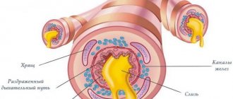
As pathology develops, swelling develops in the bronchioles. This is expressed in the accumulation of thick sputum, the result will be an increase in suffocation. Not every person can tolerate a long absence of oxygen.
Pathogenesis
Status asthmaticus is a severe attack of suffocation that cannot be eliminated by inhalation of short-term beta-agonists and is accompanied by oxygen starvation and pathology of the body systems.
There are options for the mechanism of violation:
- Contact with an allergen.
- Complication of bronchial asthma (powerful contraction of sections of the bronchial tree, suppression of breathing and cough centers, failure of the drainage function of the bronchi, suppression of exhalation).
- Reflex contraction of the bronchi due to the influence of mechanical, physical or chemical stimuli on the receptors of the respiratory canals (pungent odors, cold air).
An attack of suffocation develops based on 3 components: bronchospasm, swelling of the bronchial wall, blockage of the bronchial lumen with exudate. Beta antagonists can relieve spasms. This type of therapy is not suitable for relieving status asthmaticus. Despite therapy, the patient experiences signs of hypoxia.
Asthmatic status
Asthmatic status
- this is an attack of bronchial asthma, much stronger than usual, and it is not controlled even by increased dosages of bronchodilators that the patient takes. There is a pronounced violation of bronchial patency due to swelling of the mucous membrane, spasms of the bronchial muscles and obstruction by mucus. This leads to difficulty in inhaling and active prolonged exhalation.
During a short and short inhalation, more air enters the lungs than comes out during exhalation due to blockage and reduction of the airway lumen, this leads to hyperairiness and inflation of the lungs. Due to forced exhalation and tension, the small bronchi become even more spasmodic. As a result of all these processes, the air in the lungs stagnates, and the amount of carbon dioxide in the arterial blood increases and the amount of oxygen decreases. Both with attacks of normal severity and with status asthmaticus, respiratory muscle fatigue syndrome develops. Constant and ineffective loads of the respiratory muscles lead to hypertrophy and the formation of the chest shape characteristic of asthmatics. Enlarged lungs and hypertrophied muscles give it a resemblance to a barrel.
Causes of the disease
The pathology affects people who constantly suffer from bronchial asthma and do not follow the recommendations of their doctor. There are manifestations of an attack not associated with asthma. For diseases of the respiratory system (bronchitis), acid-aspiration pneumonia and allergic reactions.
The reasons are:
- Treatment aimed at reducing sensitivity to an allergen during an exacerbation of bronchial asthma.
- Acute or chronic diseases of the respiratory system of an inflammatory nature.
- Taking medications that cause allergies.
- Immediate cessation of glucocorticoids that have been taken for a long time.
- Powerful influence of allergens.
- Incorrect or untimely treatment.
- The “lung locking” effect caused by an overdose of adrenergic agonists.
- Taking sleeping pills and sedatives in large doses is recommended.
- Incorrect assessment of the severity of the condition by the patient or the treating specialist.
- Neuropsychic tension, chronic stress, heavy physical activity.
These factors can provoke the transformation of an asthma attack into long-term status asthmaticus.
Why does pathology develop?
- Status asthmaticus in asthmatics develops for the following reasons:
- Glucocorticoid withdrawal syndrome;
- Allergy to serums, vaccines, analgin, salicylates and antibiotics;
- Excess of sleeping pills, sympathomimetics and sedatives;
- Exacerbation of chronic diseases, development of acute diseases of the bronchopulmonary system;
- Hyposensitizing therapy (carried out during exacerbation of the disease).
Symptoms
Manifestations of the disease depend on the stage of status asthmaticus.
Relative compensation stage:
- Regular occurrence throughout the day of prolonged attacks of suffocation that cannot be removed by conventional medications. In the absence of attacks, breathing is not restored.
- Dry, painful, hysterical cough with scanty discharge of viscous sputum.
- Even if the patient sits leaning on the back of a chair, he has shortness of breath and breathing becomes more frequent.
- The presence of dry whistling rales that can be heard at a distance.
- Blueness of the skin and mucous tissues.
- The pulse rate is about 120 beats per minute, and the patient feels stabbing, aching pain in the cardiac zone.
To the symptoms are added signs of central nervous system dysfunction (unreasonable irritability, emotional instability, fear of death).
Stage of decompensation or “silent lung”:
- The surface of the skin becomes pale and moist.
- Shallow breathing, sudden shortness of breath.
- Swelling of neck veins.
- Increased liver size.
- The patient's apathetic state.
- By listening, a “silent lung” is revealed (breathing noise is not heard, dry rales are detected in an isolated area).
- The pulse rate reaches 140 beats per minute, blood pressure is reduced.
An electrocardiogram reveals symptoms characteristic of right heart overload and other forms of heart rhythm disturbances.
Hypoxic hypercapnic coma:
- Loss of orientation, the patient is stunned, and then loses consciousness.
- The veins of the neck swell, the face becomes puffy.
- Red diffuse cyanosis.
- Rare, arrhythmic breathing.
- Auscultation does not reveal breath sounds.
- The pulse is weak, irregular, the pressure is reduced or undetectable.
Heart sounds become muffled due to which ventricular fibrillation will develop.
Classification
Asthmatic status has two forms of development:
- Anaphylactic form
. It is characterized by a sudden onset and rapid progression of all symptoms. In this case, the main cause of severe respiratory failure and even respiratory arrest is a reaction to medications (tablets, serums, vaccines). Most often, this consequence can be caused by taking non-steroidal anti-inflammatory drugs. In medical practice, anaphylactic form is extremely rare. - The allergic-metabolic
type develops gradually (up to several weeks) under the severity of acute asthma. There are attacks of coughing up to suffocation, and the time interval between asthmatic attacks decreases each time. Normal breathing after an asthmatic attack is not restored, and rejection to medications used to relieve the attack develops. The main cause of this form of asthmatic attack is swelling of the mucous surfaces of the bronchi and blocking of the lumen with viscous sputum.
Diagnostics
Diagnosis takes little time, so immediate assistance is required. It is necessary to count on the minimum number of activities that can be carried out at the patient’s bedside. Any stage of status asthmaticus is determined after the following measures:
- From the patient's complaints, those characteristic of this pathology are distinguished.
- An anamnesis is collected, it is necessary to find out whether the patient has suffered from bronchial asthma and the factors that provoked the current condition.
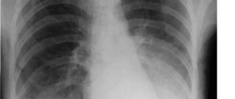
- An examination is carried out - attention is paid to the position of the patient, the presence of cyanosis of the skin, and the audibility of wheezing at a distance.
- The lungs are audible - during the first two stages, dry wheezing rales grow over the entire surface of the sternum. The third stage is characterized by a “silent lung” picture.
- Determination of pulse and breathing rhythm. Tachycardia over 120 beats per minute is observed, the respiratory rate reaches 40 or more.
- Measuring blood oxygen levels below 90%.
- The gas composition of the blood is established (a decrease in oxygen pressure below 70 mm Hg and an increase in the level of carbon dioxide above 30 mm Hg).
In the hospital, an ECG and a biochemical blood test are performed to identify total protein, protein fractions, fibrin and the growth of sialic acids.
Examination program
- OA of blood, urine.
- BAC: total protein, protein fractions, seromucoid, fibrin, sialic acids, urea, creatinine, coagulogram, potassium, sodium, chlorides.
- ECG.
- Acid-base balance.
- Blood gas composition.
Treatment
At present, there are no precise ideas about the ways of influencing the etiological components of status asthmaticus, since it has already been indicated that the mechanisms of its occurrence (the significance of allergic factors, the infectious process and the nature of the antigen-antibody reaction and, finally, the causes of the perverted reaction of the β-adrenergic system of the bronchial tree to sympathomimetics) are not completely clear.
In this regard, treatment for status asthmaticus is primarily antihypoxic in nature and is aimed at improving bronchial patency, increasing oxygenation and improving blood circulation. Patients are often admitted to intensive care units after more or less prolonged, most often unsuccessful, treatment at home or in a therapeutic department.
In most cases, the patient is admitted to the intensive care unit in a condition requiring immediate intensive care, i.e., to save life, but, unfortunately, this is not always possible. In the treatment of this acute condition, a reasonable sequence and purposefulness of measures must be observed.
Oxygen therapy. Inhalation of humidified O2 is carried out through nasal catheters or through a mask at a rate of 1-2 l/min. According to indications, the O2 flow rate can be increased to 3 -4 l/min. This type of oxygen therapy is safe, but not always effective. With hyperoxygenation, depression of the respiratory center and a further increase in paCO2 are possible, so you should not increase paO2 more than 80-90 mm Hg. Art., if this requires high concentrations of O2.
Adrenalin. It is usually customary to begin treatment with subcutaneous injection of adrenaline. Adrenaline is a stimulator of a1-, b1- and b2-adrenergic receptors, dilates the bronchi and reduces airway resistance. “Testing” doses of adrenaline are used. It is administered subcutaneously: for body weight less than 60 kg - 0.3 ml, for body weight from 60 to 80 kg - 0.4 ml, for body weight more than 80 kg - 0.5 ml of official solution.
Eufillin. Euphylline contains 80% theophylline and 20% ethylenediamine. It inhibits phosphodiesterase, which promotes the accumulation of cAMP and relieves bronchospasm. When prescribing aminophylline, factors that enhance or weaken its metabolism in the body should be taken into account. The former include smoking and childhood, the latter – heart failure, chronic diseases of the lungs, liver and kidneys.
On this basis, new treatment regimens with drugs containing theophylline have been developed. The rule used for calculation is: 1 mg of theophylline is equal to 1.2 mg of aminophylline. In this case, the so-called loading doses and doses necessary to maintain a constant concentration of aminophylline in the blood are determined. Loading doses are prescribed only if, during the last 24 hours, drugs containing theophylline have not been used or have been used only in subtherapeutic doses.
For AS, the loading dose of aminophylline is 3-6 mg/kg, administered intravenously over 20 minutes. This dose leads to an increase in the concentration of theophylline in the blood serum to 5-10 mcg/ml, which is maintained by drip infusion of the drug at the rate of 0.6 mg/kg per 1 hour for a patient without concomitant pathology, 0.8 mg/kg per 1 hour for smoker, 0.2 mg/kg per 1 hour for congestive heart failure, pneumonia, liver and kidney diseases, 0.4 mg/kg per 1 hour for severe chronic lung diseases.
Therapeutic limits for theophylline concentration in blood serum are 10-15 mcg/ml. Plasma theophylline concentrations should be determined within 6-12 hours after the start of maintenance therapy. It should be borne in mind that as the patient's condition improves, the release of theophylline may change. Theophylline and b-adrenergic agonists are not antagonists and can be used simultaneously.
An overdose of aminophylline is manifested by nausea, vomiting, diarrhea, tachycardia, tachyarrhythmia, drowsiness, agitation and convulsions. These symptoms do not usually occur at serum theophylline concentrations below 20 mcg/mL, but side effects are possible at lower concentrations. This depends on the body's individual reaction to theophylline.
Corticosteroids. The effect of corticosteroid therapy is associated with suppression of airway inflammation and increased sensitivity to beta-adrenergic agents. The more severe the AS, the greater the indication for immediate corticosteroid therapy. The need for an initially high dose of corticosteroids administered intravenously should be emphasized.
Status asthmaticus is controlled not so much by the size of individual doses as by the duration of treatment.
You should not stop treatment with aminophylline and b-adrenergic agonists, which are used mainly in the form of inhalation every 4 hours. You cannot treat an incompletely stopped attack with low doses of hormones.
After removing the patient from the asthmatic state, the dose of corticosteroids is gradually reduced by approximately 25% each subsequent day. Intravenous administration is replaced by oral administration. On the 1st day after discontinuation of intravenous therapy, 60 mg of prednisolone tablets (or an equivalent dose of other corticosteroids) is prescribed. On each subsequent day, the dose of prednisolone is reduced by 5 mg, until it is completely discontinued.
Inhalation therapy. Most patients are indicated for inhalation therapy with b-adrenergic agonists; use fenoterol, alupent, salbutamol and other drugs. Exceptions are cases of drug tachyphylaxis or overdose of sympathomimetics. With the inhalation method of administering b-adrenergic agonists, there is virtually no negative effect on the cardiovascular system, only sometimes palpitations and tachycardia occur.
https://www.youtube.com/watch?v=tnP_zk-GrWc
For prolonged persistent cough, some authors recommend inhalation of 0.6 mg of atropine (with or without beta-agonists) in 0.5-1 ml of water or isotonic sodium chloride solution.
Intravenous administration of b-adrenergic agonists. If the therapy does not have an effect, in the most severe cases, intravenous administration of b-adrenergic agonists, for example, isoproterenol, diluted in a 5% glucose solution, is indicated. Contraindications include heart disease (coronary cardiosclerosis, myocardial infarction), severe tachycardia and symptoms of tachyphylaxis, and old age.
Complications - arrhythmias and acute myocardial infarction - arise as a result of the increased myocardial demand for O2, which is unsatisfied in status asthmaticus. The rate of administration of isoproterenol is 0.1 mcg/kg per 1 minute until tachycardia appears (heart rate 130 per minute or slightly more). It is necessary to increase the supply of O2, conduct cardiac monitoring and control paO2.
Infusion therapy. Infusion therapy is the most important component of the treatment of AS, aimed at replenishing fluid deficiency and eliminating hypovolemia, the total volume of infusion therapy is 3-5 l/day. Hydration is carried out by introducing solutions containing a sufficient amount of free water (mainly glucose solutions), as well as hypo- and isotonic solutions of electrolytes containing sodium and chlorine.
To eliminate hypovolemia, dextrans are sometimes used with predominantly rheological action (reopolyglucin), but we must not forget about the possibility of allergic reactions. It is impossible to carry out infusions of solutions with a central venous pressure above 12 cm of water. It should be taken into account that treatment with corticosteroids increases the body's need for K.
The average dose of potassium is up to 60-80 mmol/day. To correct metabolic acidosis (pH should not be lower than 7.25), it is advisable to use small doses of bicarbonate, which helps improve the drainage function of the bronchi. Metabolic acidosis should not be allowed to progress to alkalosis. It must be emphasized that catheterization of the subclavian vein in patients with AS and severe pulmonary emphysema requires some experience and caution, since it can be complicated by pneumothorax; catheterization of the internal jugular vein is safer.
Chest physical therapy is a necessary component of treatment. They resort to breathing exercises, assisted coughing, therapeutic percussion and vibration massage.
Bronchoscopic lavage of the trachea and bronchi during a severe asthmatic attack is a dangerous procedure that increases bronchospasm and hypoxia. Despite individual reports of the successful use of lavage, the essence of the therapeutic effect is unclear, since mucus plugs block the bronchi beyond the reach of aspiration.
Epidural block at the T1-Tp level, recommended by some researchers, has few supporters due to the complexity of the method, possible complications and unguaranteed effect.
Anesthesia with fluorotane can be used in the treatment of a severe asthma attack that does not respond to conventional therapy.
Signs of the effectiveness of the therapy. The oncoming improvement at first is not pronounced; clinical data do not yet confirm the exit from the asthmatic status. The subjective factor “it has become easier to breathe” is usually one of the first guidelines for a doctor. The earliest signs of improvement are a decrease in tachycardia, the disappearance of paradoxical pulsus and a gradual decrease in hypercapnia with long-term arterial hypoxemia.
- OA of blood, urine.
- BAK: total protein, protein fractions, seromucoid, sialic acids, fibrin, haptoglobin.
- Blood II: B- and T-lymphocytes, their subpopulations, immunoglobulins.
- General analysis of sputum, its cytological composition, for Koch's bacilli and atypical cells, flora and sensitivity to antibiotics, Kurschmann spirals. The most accurate results are obtained by examining sputum obtained by bronchoscopy or processed using the Mulder method.
- X-ray of the lungs.
- Bronchoscopy and bronchography.
- Spirography, pneumotachometry.
- In case of severe respiratory failure, study indicators of acid-base balance and blood gas composition.
Treatment of the disease
Status asthmaticus is relieved by inhaling humidified oxygen using a catheter or mask. This must be done especially carefully: there is a risk of worsening ventilation of the lungs and stopping breathing instead of relieving the attack. To prevent apnea, it is necessary to intubate the patient and transfer him to mechanical ventilation. The procedure is indicated for the second and third stages of pathology.
To correct metabolic failures and restore lost fluid, infusion treatment is performed. Medicines are administered intravenously, and central venous pressure is monitored.
Status asthmaticus requires the use of medications:
- glucocorticoids;
- bronchodilators;
- expectorants and mucolytics;
- neuroleptics;
- beta-agonists.
An acute attack in a patient can be stopped by treating the underlying disease – bronchial asthma.
Therapeutic measures
The development of the pathological process implies emergency hospitalization in a hospital.
Treatment of stage 1 status asthmaticus is possible in a general therapy department. If the second or third stage is diagnosed, then the entire course of treatment takes place in the intensive care unit.
Particularly severe cases require hospitalization in the intensive care unit, where emergency care will be provided for status asthmaticus.
Regardless of the severity of the disease, the course of treatment for adults and children involves:
- elimination of hypovolemia;
- relieving swelling of the bronchiole mucosa;
- restoration of patency of the bronchial tree.
Treatment of pathology involves complex therapy.
Treatment of metabolic status asthmaticus includes oxygen therapy, infusion treatment and medication.
In case of serious complications, bronchoscopy is also indicated, which is performed with segmental lavage of the lungs. It is prescribed for stages II–III of the disease.
If respiratory failure develops, a transfer to a ventilator (artificial lung ventilation) is expected. If a patient is diagnosed with stage III of the disease, he is immediately connected to a ventilator.
At the last stage of the pathology, extracorporeal membrane oxygenation of blood is also indicated. This is symptomatic therapy to restore gas exchange function of the lungs. The procedure of extended extracorporeal circulation is prescribed to patients whose acute development of failure does not respond to standard treatment methods.
Complications
Status asthmaticus is a pathology that can cause the development of severe complications. In the most unfavorable case, the patient's death from respiratory failure and oxygen starvation is likely. In addition, serious right ventricular failure often occurs.
It is caused by an increase in pressure in the pulmonary circulation, which is typical for asthmatic attacks. In this case, blood stasis forms in the large circle. This is expressed by an increase in the size of the liver, swelling of the neck veins, and swelling. Hypoxia worsens due to venous congestion.
Less commonly, pneumothorax or hemopneumothorax is formed due to rupture of lung tissue. First, symptoms of pulmonary emphysema appear - the volume of the chest increases, the airiness in the lungs increases. Next, the tissue integrity is destroyed, air penetrates into the pleural cavity. The process occurs with very acute, intense pain.
General information
When an asthmatic attack lasts too long, compensatory resources are activated in the bronchi, providing the body with oxygen. At the same time, breathing becomes deep, and this forces the respiratory muscles to work more intensely. The patient's sweating increases
, due to which
dehydration occurs
, and the level of sputum viscosity increases.
Sputum closes the lumen of the bronchus, which narrows during illness. Under the influence of this process, gas exchange is disrupted: oxygen does not enter the blood, but carbon dioxide is also not removed, the excess of which leads to an involuntary increase in the blood vessels of the brain, as a result of which the pressure inside the skull increases, confusion, convulsions, drowsiness are observed, in the worst case, coma.
Urgent Care
The first manipulations in the development of suffocation involve the supply of oxygen, as well as the administration of available medications. The first and second stages of the pathology are stopped by supplying oxygen through a mask. This normalizes the blood gas composition and calms the patient. The third stage requires mechanical ventilation. At home, it is enough to limit yourself to freeing the chest from constricting clothing and vigorously ventilating the room.
It is recommended to start administering glucocorticosteroids. They will reduce swelling and eliminate beta-adrenergic receptor blockade. If injections are not possible, it is recommended to proceed with inhalation or oral administration of the medication. At the same time, you need to immediately call an ambulance.
Causes of bronchial asthma in children and risk factors
An asthma attack is caused by the penetration of inhaled fine allergens into the respiratory tract, and in young children, medicinal or food allergen pathogens play a huge role in the development of pathology.
The main provocateurs of a spastic attack are:
- Household and industrial dust.
- Plant pollen during flowering.
- Fur and undercoat of domestic animals, as well as their saliva and excrement.
- Fungal spores, mold.
- Fish feed.
- Medicines, especially antibiotics, aspirin-containing drugs (the concept of “aspirin asthma” due to an overdose is known).
- Tobacco smoke, industrial emissions, air pollution.
Meteorological factors, viral infections, gastroesophageal or gastroesophageal reflux play a key role in the development of asthmatic attacks. In 6% of cases, the development of the pathological process is provoked by nutritional factors. Clinicians assign some role to stress-psychogenic factors.
Predisposing factors
A special risk group consists of children with a complicated clinical history:
- Compounded heredity - if one of the parents suffers from the disease, then the chances of the child developing bronchial asthma reaches 35%.
- Increased sensitization of the body , complicated allergic anamnesis. What a child may be allergic to in everyday life - popular allergens
- Frequent infections of a viral or bacterial nature.
- Constant temperature changes , heat stroke, hypothermia.
- Weakening of the immune status of various origins.
- Inappropriate use of non-steroidal anti-inflammatory drugs .
Gender is not a risk factor, but bronchial asthma is more common in boys .
Psycho-emotional overload significantly increases the risk of developing asthmatic bronchial obstruction. The likelihood of pathology occurring also increases with the use of household aerosols and maternal smoking during pregnancy.
Prevention
Status asthmaticus and its three stages can be prevented by following the following rules:
- Monitoring the progress of the main pathology – asthma;
- Avoiding contact with allergens;
- Monitoring the expiration date of medications used in the treatment of asthma;
- To give up smoking;
- If you have bronchial asthma, always have a bronchodilator inhaler with you.
Such patients need to avoid physical activity, promptly treat infectious and inflammatory diseases, and undergo regular medical supervision.
Forecast
In general, for a patient under the supervision of specialists, the prognosis for recovery from asthmatic status is favorable
. But don't expect quick results. Effective treatment of status asthmaticus is possible, but subject to timely access to comprehensive medical care.
Found a mistake? Select it and press Ctrl + Enter
Useful article All about laryngeal stenosis
Laryngeal stenosis is a pathology in which the lumen of the larynx is partially or completely narrowed. The condition is life-threatening. The disease progresses...
Read the full article
general information
Short description
Status asthmaticus is an intractable attack of bronchial asthma lasting 6 hours or more with the development of resistance to sympathomimetic drugs, impaired bronchial drainage function and the occurrence of hypoxemia and hypercapnia [1]
ICD-10 code J46 - Astatic status
Date of protocol development/revision : 2007/2016.
Users of the protocol : doctors of all specialties, nursing staff.
Patient category: children, adults, pregnant women.
Level of evidence scale:
| A | A high-quality meta-analysis, systematic review of RCTs, or large RCTs with a very low probability (++) of bias, the results of which can be generalized to an appropriate population. |
| IN | High-quality (++) systematic review of cohort or case-control studies, or High-quality (++) cohort or case-control studies with very low risk of bias, or RCTs with low (+) risk of bias, the results of which can be generalized to an appropriate population . |
| WITH | Cohort or case-control study or controlled trial without randomization with low risk of bias (+). The results of which can be generalized to the relevant population or RCTs with very low or low risk of bias (++ or +), the results of which cannot be directly generalized to the relevant population. |
| D | Case series or uncontrolled study or expert opinion. |