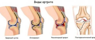
Kinds
The following types of illness are distinguished:
- Acquired (formed in the process of life) and congenital (genetically determined condition).
- Natural (anatomical fusion within normal limits, at the base of the skull, pelvic skeleton), artificial (created during surgery, mainly to eliminate or prevent defects), post-traumatic (fusion of bone structures as a result of traumatic exposure).
- Pathological (pathology of the connective tissue located between the bones can occur as a result of osteomyelitis, tuberculosis, typhoid fever, osteochondrosis) and physiological (the natural form into which bones come as the body grows).
According to the localization criterion, synostosis of the forearm, tarsus, skull, radioulnar, brachioradial, Klippel-Feil (a known anomaly of the spine), cervical vertebrae, sagittal, metopic, lambdoid or coronal suture, ribs, vertebral bodies, pelvic bone, radioulnar, articular processes, ulnar joint, hand, coronary, chest, etc.
Causes
It is mainly a consequence of diseases that directly affect the cartilaginous connective tissue and pathologies between the tibia bones. It can also be a trauma to the periosteum, a post-traumatic reaction after dislocations and fractures of bones and cartilage.
Features of the pathology
Synostosis is a condition in which fusion of bone tissue occurs. Natural manifestations are considered part of the norm, for example, when the pelvic bones fuse.
But there are also conditions of unnatural origin.
An important factor in diagnosis is the differentiation of synostoses from ankylosis or fusion after a bone fracture.
As the disease progresses, there is a continuous process of bone tissue reunification. As a result, areas of fusion are formed, which can either be asymptomatic or threaten the patient’s life. The disease was given an ICD code – Q74.0.
Clinical picture[edit | edit code]
Tarsal synostosis does not manifest itself until early adolescence. Pain during synostosis is often localized at the level of the sinus tarsus or in the projection of the medial edge of the subtalar joint. Usually the pain is dull, aching, and worsens with exercise. Lameness often occurs after exercise. In many cases, synostosis is not accompanied by pain and sometimes remains undiagnosed. There is evidence of familial forms of the disease.
Tarsal synostosis is associated with conditions such as fibular underdevelopment, syndactyly, Ahler's syndrome, Niefergelt's disease, and carpal synostosis. Synostosis is often discovered during surgery for congenital clubfoot, and in this case the subtalar joint is also ankylosed.
Main features[edit | edit code]
- Susceptibility to ankle ligament injuries.
- Pain on exercise.
- Pain in the sinus area or in the ankle joint.
- Limited mobility or immobility of the subtalar joint.
- Pain with pronation and supination of the foot.
- Spasm of the peroneal muscles.
Radiation diagnostics[edit | edit code]
Some signs of tarsal synostosis can be detected on conventional radiographs in frontal, lateral and oblique projections, as well as on radiographs of the calcaneus in axial projection. The easiest way to identify calcaneonavicular synostosis, which is clearly identified on oblique films by the bone fusion between the anterior part of the calcaneus and the external horn of the navicular bone. The lateral image shows a characteristic elongation of the anterior part of the calcaneus - the “anteater trunk”. In the same projection, a C-shaped line is determined, running through the arch of the leg, and then down and back to the lower edge of the support of the talus. As a result of synostosis, as well as after surgical treatment of clubfoot, a coracoid deformity of the anterior process of the talus occurs, often limiting mobility in the subtalar and transverse tarsal joints.
An axial radiograph of the calcaneus may reveal changes in the medial articular surface of the subtalar joint.
CT is ideal to confirm the diagnosis and clarify the location and extent of synostosis. The location of adhesions (especially below the subtalar joint) is even better demonstrated by three-dimensional CT reconstruction.
Cartilaginous and fibrous adhesions cannot be seen on CT. Sometimes their presence can be assumed by the roughness of the articular surfaces, but MRI is necessary to confirm fibrous or cartilaginous adhesions.
Types of synostoses
The disease has many types, which may differ in the causes of manifestation, as well as in certain symptoms. Moreover, the mechanism of manifestation of the disease itself may have individual characteristics.
Examples of synostoses
Congenital
The congenital form refers to pathological conditions that are provoked by corresponding conditions, including aplasia or hypoplasia of connective tissue between individual bone units in the joints. Most often manifests itself in the ulnar-radial bones. A little less often, fusion may be observed in other areas.
This disease is described in medical reference books as syndactyly on the fingers and synostosis of the ribs. If the connection of the vertebrae appears, then we are usually talking about Klippel-Feil syndrome. If Entley-Bixler syndrome manifests itself, then fusion is observed in many areas.
Congenital synostosis of the atlas and axis on x-ray
Post-traumatic
The post-traumatic form develops after injury to the corresponding elements of the joint, which are located nearby.
Typically this condition is observed in the lower leg, vertebrae or forearm.
But in the radial-ulnar and tibial-fibular bones, a similar disease develops due to the convergence of fragments of these joints at the site of injury.
In the area of the spinal column, the disease manifests itself in this form after fractures and dislocations.
In the photo there is ulnar synostosis
Physiological
Physiological is considered natural, that is, normal for humans. It correctly connects bones as a person grows. Areas of fusion are formed in adolescence and/or young adulthood, and, as a rule, in the place of the presence of synochondroses - cartilaginous joints. Normally, it manifests itself in people as:
- Pelvic area;
- Sacral vertebrae;
- The bones of the skull at its base.
In some situations, such processes slow down or speed up. Then we are talking about pathology, for example, Kashin-Beck disease, hypergonadism, and so on. They lead to disruption of the development of the muscle-bone system.
Deformations due to improper development of the sagittal suture
Metopic
The presented form refers to trigonocephaly or asymptomatic craniostenosis. That is, the metopic suture fusion occurs ahead of schedule.
Normally, this occurs from about 8 months of age until 2 years of age.
In the first case, the condition is intrauterine (less often - in the first two months from birth), but the second option develops after birth at about 3-4 months. Both conditions lead to deformation of the bones in the skull.
Artificial
The artificial type of pathology manifests itself due to the creation of fusion sites during surgery. Typically used to eliminate extensive bone defects, eliminate false articular elements, and so on.
This type of intervention is often performed in the area between the tibia. It is noteworthy that after manipulation, the load then passes to the fibula. This leads to its compensatory thickening.
Bone fusion
Radioulnar
This type of pathology is a congenital defect that manifests itself due to impaired differentiation of the osteoarticular apparatus. It appears in the forearm and is characterized by fusion of the ulna and radius in the proximal region.
Also, during diagnosis, dislocation of the radial head is often diagnosed. As a result, the child’s hand is unable to rotate at the wrist, although extension/flexion at the elbow is maintained. Accordingly, such a situation can make the patient’s life somewhat difficult.
see also
- Syndesmology
| This is a draft article on anatomy. You can help the project by adding to it. |
| Trochlear joint: | Interphalangeal joints • Humeral-ulnar joint |
| Condylar joint: | Atlanto-occipital joint • Knee joint |
| Saddle joint: | Carpometacarpal joints |
| Flat joint: | Facet joints |
| Stiff joint (amphiarthrosis): | Sacroiliac joint |
| Rib cage: | Sternoclavicular joint • Acromioclavicular joint |
| Taz: | Sacroiliac joint • Articulus pubis |
Localization
If we talk about localization, then in general there are three types of disease. Depending on the location of such a defect, there will be own forecasts. This will also determine what type of therapy doctors decide to use.
Craniostenosis
Craniostenosis occurs in the cranial region. It is observed somewhat more often in boys, in 1 case out of 2000.
In parallel, various heart defects can be observed. Typically reflected in the following symptoms:
- Early closure of sutures in the skull;
- Changing the shape of the head;
- Intracranial hypertension;
- Reducing the volume of the skull.
Intelligence is in normal dynamics from the very beginning, but then a secondary developmental delay may appear.
This condition is usually caused by a disturbance in the processes of laying the bones of the skull inside the womb. That is, this condition is usually affected by congenital or intrauterine pathologies.
Important! It is worth noting that if the disease developed during the prenatal period, then it manifests itself outwardly more intensely, while with a disease that appears in the first months of life, the deformation of the skull is not so clearly visible.
Synostosis of the forearm bones
With this type of pathology, the bones of the forearm are involved. For this localization, the manifestation of radioulnar synostosis, which is a congenital condition, is typical. In general, it can manifest itself in literally any area of the forearm, but the proximal zone suffers more often than others.
Fusion is quite capable of varying within 1-12 cm. Synostosis is sometimes combined with syndesmosis. Gross deformations are usually absent. The hand and forearm show signs of some atrophy, but the olecranon process is hypertrophied.
Klipell-Feil disease
This disease is considered quite rare due to its localization. The manifestation of synostosis occurs in the area of the spinal column quite rarely. It is a hereditary pathology that is combined with other defects and diseases of internal organs.
Prevention
There are no methods for preventing congenital synostosis. But the likelihood of its occurrence can be reduced if the required conditions are provided for the normal course of pregnancy, preventing the influence of pathological factors on the body of the expectant mother and fetus.
The following recommendations will help prevent the formation of the acquired form of this disease:
- avoiding situations that are fraught with injury to the hip joint, and if they cannot be avoided, using personal protective equipment;
- prevention of diseases and pathological conditions, against the background of which adhesions in the hip joint can form, and if such diseases have already arisen, their timely detection and adequate treatment;
- avoiding inadequate loads on the hip joint;
- physical education - to strengthen the joint, thereby reducing the risk of injuries and pathological damage to the hip joint.
Causes
In children, this condition is usually congenital and can manifest itself during fetal development or shortly after birth in the first four months.
If we talk about general factors, these include:
- Illnesses during the prenatal period;
- Congenital pathologies and anomalies;
- Injuries;
- Hereditary predisposition;
- Specific and nonspecific joint infections;
- Osteomyelitis;
- Degenerative-dystrophic diseases;
- Operations performed.
Such conditions can become factors that trigger the pathological process. But what exactly becomes the trigger mechanism can only be indicated by a doctor.
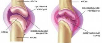
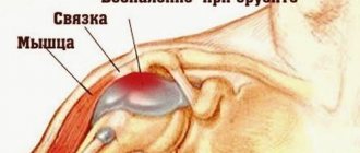
Continuous bone connections
Depending on the type of tissue located between the bones and connecting them, all synarthrosis is divided into four types:
- Syndesmoses
, syndesmoses – connections through connective tissue;
- Synchondroses
, synchondroses – connections through cartilaginous tissue;
- Synostosis
, synostoses – connections through bone tissue;
- Synsarcoses
, synsarcoses - connections through muscle tissue.
1. Syndesmoses
can be represented by ligaments, interosseous membranes, impaction, sutures (Fig. 2).
Ligaments
, ligamenta are relatively thick bundles of dense fibrous or elastic connective tissue. In most cases, ligaments are thrown across a joint from one bone to another.
Interosseous membranes
, membranae interosseae are flat connective tissue (formations) plates located between the bones. For example, interosseous membranes between the diaphyses of the bones of the forearm and lower leg.
IN
pounding
, gomphosis - represented by a thin layer of connective tissue between connecting structures. This is how the roots of the teeth are connected to the bone tissue of the dental alveoli. This layer of tissue is called periodontium, periodontum.
Sh
Fig. 2. Types of syndesmoses:
A – ligaments; B – hammering;
B – interosseous membranes; G – sutures between the bones of the skull.
You
, sutura - very thin layers of connective tissue located between the edges of bones, for example, between the edges of the skull bones. Depending on the shape of the edges of the connecting bones, three types of sutures are distinguished:
a) serrated seam
, sutura serrata, when the teeth of the edge of one bone intersect between the teeth of the edge of another, for example, a suture between the edges of the parietal bones;
b) scaly suture
, sutura squamosa, when the obliquely cut edges of the bones overlap each other, for example, the suture between the parietal and temporal bones;
c) flat seam
, sutura plana, when the edges of the connecting bones are smooth, for example, the sutures between the bones of the facial skull.
2. Synchondroses
may be represented by hyaline or fibrous cartilage tissue. According to the duration of existence, synchondrosis can be temporary or permanent (Fig. 3).
Temporary synchondrosis
exist only at a certain age, and then are replaced by bone tissue - synostoses, for example, synchondrosis between the epiphysis and diaphysis of tubular bones, between the sphenoid and occipital bones.
Permanent synchondrosis
exist throughout a person’s life, for example, between the pyramid of the temporal bone and the sphenoid bone, between the pyramid of the temporal bone and the occipital bone.
Fig.3. Types of synchondrosis
: A – temporary (the arrow indicates the connections of the diaphyses of tubular bones with the epiphyses); B – permanent (spheno-occipital synchondrosis).
3. Synostosis
- these are connections when connective or cartilaginous tissue is located between developing bones, and after the end of the osteogenesis process they are replaced by bone tissue. For example, temporary synchondroses in childhood between the bones of the pelvic girdle (pubic, iliac, ischial) are replaced by synostoses in an adult.
Fig.4. Synsarcoses:
A – shoulder blades with vertebrae, B – hyoid bone with lower jaw and sternum.1 – m. trapezius; 2 – m. levator scapulae; 3 – mm. rhomboidei major et minor; 4 – os hyoideum; 5 – m. mylohyoideus; 6 – m. omohyoideus; 7 – m. sternohyoideus.
4. Synsarcoses
- these are bone connections when muscle tissue is located between the connecting bones, for example, the connections of the scapula with the ribs and vertebrae, the hyoid bone with the lower jaw, sternum and scapula (Fig. 4).
All types of continuous joints, with the exception of synostoses, have one degree or another of mobility, which depends on the thickness of the layer of tissue connecting them. The larger the layer of connective, cartilage or muscle tissue, the more mobile these types of synarthrosis are. Phylogenetically continuous connections are more ancient with a limited range of movements.
Below is a diagram of the types of continuous bone connections.
Scheme 1
Symptoms
Symptoms will depend on the location. Manifestations of craniostenosis include:
- Violation of the shape of the head, obvious deformations, narrow face;
- Sleep disorders;
- Vomiting, nausea;
- Intracranial hypertension;
- Convulsions;
- Strabismus;
- Irritability, drowsiness, anxiety;
- Exophthalmos, strabismus;
- Headache;
- Mental retardation;
- Memory loss.
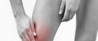
Synostosis of the bones in the forearm usually gives signs that are not so pronounced in the context of visual manifestations. Typically the main symptoms are:
- Rotation in the forearm is impossible;
- The area is fixed in the pronation zone;
- The hand and forearm appear atrophied;
- It is difficult for a person to carry out self-care and engage in certain types of work;
- A person is not able to accept small objects in the palm of his hand;
- Helical amplitude movements with the brush are impossible.
With Klippel-Feil disease, there are certain typical signs that can be explained both by the localization of the pathological focus and by the factors that led to such a disease:
- Short neck;
- Reduced hair growth at the back of the head;
- Limitation of range of motion in the upper part of the spinal column;
- Other developmental anomalies - from cleft palate to spina bifida.
Sometimes patients with this type of disease may have congenital anomalies that appear in the ribs, face, genitals, urinary tract, respiratory organs, head, and limbs.
Klippel-Feil syndrome
A rare congenital anomaly of the spine, accompanied by a decrease in the number or synostosis of the cervical vertebrae. Described by French doctors André Feil and Maurice Klippel in 1912. It is transmitted in an autosomal dominant manner. Characteristic signs are a short neck, limited mobility of the upper spine and decreased hair growth in the occipital region. Possible combination with other anomalies, including heart defects, cleft palate, spina bifida and scoliosis. In addition, in patients with Klippel-Feil syndrome, congenital anomalies of the respiratory system, ribs, kidneys, genitals, fingers, upper and lower extremities, face and head are sometimes detected.
To confirm the diagnosis, X-rays of the cervical and thoracic spine are performed. Treatment is often conservative; in case of a pronounced cosmetic defect, high-lying upper ribs are sometimes removed. To improve posture and prevent secondary deformities, patients are prescribed massage, physiotherapy and physical therapy. When radiculitis develops, analgesics are used. In case of severe compression of the nerve roots, surgical interventions are performed.
Treatment methods, what to do
The norm is the process of fusion of bones with the help of bone tissue, if they occur strictly within the specified time frame and do not give pathological deviations and complications. That is, if this process occurred earlier or later, then we are talking about a specific type of disease.
The same applies to those cases when synostosis develops in the wrong zone. In this case, they can be used depending on the type:
- Drug treatment to eliminate negative symptoms;
- Correction using splints and plaster casts;
- Correction using exercise therapy and massage;
- Maintaining and improving processes through physiotherapy;
- In the case of the forearm, occupational therapy will also become relevant, which will help maintain and develop hand motor skills.
If the condition is assessed as complex, there is a risk of complications, or correction of pronounced aesthetic defects is required, then the question of whether the operation is advisable is decided. In this case, the type of impact will depend on the specific goals.
Diagnostics
Despite the fact that synostoses have pronounced symptoms, hardware examinations will be required to establish a final diagnosis.
However, in any case, a primary diagnosis must be carried out, which is aimed at the clinician’s work with the patient. Among such measures it is worth highlighting:
- studying the medical history and life history of the patient - to identify the nature of the pathology;
- a thorough physical examination - aimed both at identifying the main defect and accompanying symptoms;
- a detailed survey of the patient - its necessity is for the doctor to receive complete information about the nature of the course of the disease.
Diagnosing synostosis does not imply laboratory tests of the patient’s blood, urine and feces, since such tests do not have diagnostic value.
Instrumental methods for establishing a final diagnosis involve:
- X-rays of the skull, ribs, all parts of the spine and the pelvic area - this procedure will help identify the presence of pathology between the ribs on the left or right, in the cervical region and coccyx;
- CT and MRI;
- Ultrasound.
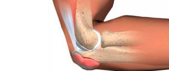
X-ray of the elbow joint
An orthopedist or traumatologist can perform such a diagnosis, but in addition, consultations with specialists from other fields of medicine, in particular cardiology, endocrinology, neurology and surgery, will be needed.
Forecast
Physiological synostosis is normal. That is why it is never considered as a pathology. This does not apply to the artificial form, which is obtained through surgery. On the other hand, other types can be considered as deviations in the ODA state.
In case of synostosis, surgical separation of joints is a rather controversial method, which often ends in failure and the separated areas grow back together, which leads to disability. Therefore, doctors, in the absence of a threat to life or serious aesthetic defects, try to use conservative methods whenever possible.
Types of bone connections and possible pathologies:
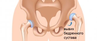
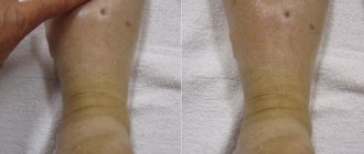
Treatment of hip synostosis
If synostosis of the hip joint was detected in children under 3 years of age, then this pathology is treated conservatively - the bone bridges have not yet acquired typical bone hardness, so you can try to destroy them using conservative methods. Appointed:
- staged corrective plaster casts;
- physiotherapeutic procedures;
- massage of the affected hip joint and lower limb on the affected side;
- individually selected exercise therapy complex.
Plaster casts are replaced every two weeks; this treatment may take 6-10 months.
Bone growths are removed surgically starting from the age of 6 years.
note
Surgical removal of adhesions for synostosis of the hip joint can be performed either open or arthroscopically.
In adults, conservative therapy for synostosis of the hip joint is ineffective, so surgery is performed to remove the bone bridges. After surgery the following are prescribed:
- antibacterial drugs for the prevention of postoperative complications;
- painkillers;
- physiotherapeutic methods;
- exercise therapy;
- massage.
How many ribs does a person have - a life-or-death question
Today, even from school, every child knows for sure that people have 12 pairs of ribs in their chest (occasionally - 13), that is, 24 or 26 pieces of ribs, and this figure does not depend on gender, that is, men and women have the same number of ribs.
But this was not always the case.
Thanks to biblical legends and church prohibitions that existed in ancient times regarding such a branch of medicine as pathological anatomy, it was believed for quite a long time that a man has one pair of ribs more than a woman. And from this extra pair, the Creator, they say, created Eve.
Despite the threat of being burned at the stake for heresy, some courageous aesculapians of antiquity, in order to learn how to heal correctly, which is impossible without an anatomical atlas, performed autopsies at their own peril and risk. The more autopsies were performed, the more convinced the doctors of those years were that the number of ribs in men and women, as well as their anatomical structure, are exactly the same, although the female skeleton is more fragile and the woman’s chest is less voluminous.
To get an answer to such a childish question that seems ridiculous today, many ancient doctors paid with their lives...
Anatomical structure of the chest
So, what do we know today about the chest:
- It consists in most cases of 12 pairs of ribs, symmetrically located on both sides of the skeleton (seven pairs on each side).
- Some individuals have an additional, extra 13th pair of ribs, which, in memory of the biblical tradition, is called “Adam’s” ribs. Any person (both man and woman) can also have this extra one pair, that is, “Adam’s” ribs are not some kind of male privilege or a sign of some kind of chosenness.
- Each adult rib consists of bony flat arched plates approximately 5 mm thick, ending in front with cartilage, and behind with a neck and head covered with cartilage, which enters the costovertebral articulation.
- In addition to the costovertebral joint, each rib is attached to the vertebra using the costotransverse joint, which connects the costal tubercle with the transverse process of the vertebra.
- In the anterior region, seven pairs of ribs, with the help of cartilage, form an elastic connection with the sternum, which consists of the manubrium, body and xiphoid process. These seven pairs are called true edges.
- The first pair of ribs are attached to the manubrium of the sternum by means of synchondrosis (an elastic cartilaginous connection), and the next six pairs are attached by means of flat costosternal joints (symphyses).
- The next five (in rare cases six) pairs are not attached to the sternum, so they are called free. Each of the rib pairs, starting from the 8th, forms a soft connective tissue syndesmosis (fusion) with the above pair. The last (12th or 13th) pair is attached only to the muscles.
- A child's rib differs from an adult's in that it consists almost entirely of cartilage, so the child's chest is very fragile and vulnerable.
- With age, the process of ossification of the rib is completed, and cartilage is preserved only at the ends of the ribs connected to the sternum.
- Each rib is covered with thin, hard hyaline cartilage and contains spongy bone tissue inside.
- The sternum consists of the outer periosteum, under which lies the red bone marrow.