Blood vessels of the human body (diagram)
Blood vessels
- elastic tubular formations in the body of animals and humans, through which the force of a rhythmically contracting heart or a pulsating vessel carries out the movement of blood throughout the body: to organs and tissues through arteries, arterioles, capillaries, and from them to the heart - through venules and veins.
Classification of blood vessels
Among the vessels of the circulatory system, arteries
,
veins
and vessels of the
microcirculatory
;
the latter carry out the relationship between arteries and veins and include, in turn, arterioles
,
capillaries
,
venules
and
arteriole-venular anastomoses
[1]. Vessels of different types differ not only in their diameter, but also in tissue composition and functional features [2].
- Arteries are vessels through which blood moves away from the heart. Arteries have thick walls that contain muscle fibers as well as collagen and elastic fibers. They are very elastic and can contract or expand, depending on the amount of blood pumped by the heart. The blood flowing through the arteries is saturated with oxygen (the exception is the pulmonary artery, through which venous blood flows)[3][4].
- Arterioles are small arteries (less than 300 microns in diameter), immediately preceding capillaries in the blood flow. Smooth muscle fibers predominate in their vascular wall, thanks to which arterioles can change the size of their lumen and, thus, resistance. The smallest arterioles - precapillary arterioles
, or
precapillaries
- retain only single smooth muscle cells in their walls [5] [6]. - Capillaries are tiny blood vessels that are so thin that substances can easily pass through their walls. The diameter of their lumen ranges from 3 to 11 microns, and the total number in the human body is about 40 billion. Through the wall of the capillaries (no longer containing smooth muscle cells), nutrients and oxygen are released from the blood into the cells and carbon dioxide and other waste products are transferred from cells into the blood[7][8].
- Venules are small blood vessels that provide in a large circle the outflow of oxygen-depleted blood saturated with waste products from the capillaries into the veins. postcapillary venules
adjacent to the capillaries (
postcapillaries
) with a diameter of 8 to 30 microns and collecting venules with a diameter of 30-50 microns, flowing into the veins [9]. - Veins are vessels that carry blood to the heart. As the veins enlarge, their number becomes less and less, and in the end only two remain - the superior and inferior vena cava, which flow into the right atrium. The walls of veins are less thick than the walls of arteries and contain correspondingly fewer muscle fibers and elastic elements [10] [11].
- Arteriolo-venular anastomoses are vessels that provide direct blood flow from the arteriole to the venule, bypassing the capillary bed. They contain in their walls a well-defined layer of smooth muscle cells that regulate such flow [12] [13].
Characteristic
A vein is an elastic blood vessel that transports blood from various areas of the body to the heart. The venous system has low pressure and needs muscle contraction to move the blood upward. Venous vessels are divided into superficial and deep.
One of the biggest differences between the two is the amount of blood carried. The deep ones are responsible for transporting most of the blood, and the superficial ones, although important, carry a much smaller amount.
The organs of the upper extremities include:
- shoulder girdle;
- free upper limb, consisting of the shoulder, elbow, forearm and hand.
The lower limbs include:
- lower limb belt;
- the free lower limb, which includes the thigh, knee, and foot.
The superficial veins of the upper extremity and lower extremity are located between two layers of tissue called superficial fascia or subcutaneous tissue, and are visible just under the skin.
They receive blood from the venules and then combine into the venous system. Valves play a great role in the movement of blood through the vessels, which occurs due to hydrostatic pressure in various parts of the circulatory system.
They are folds of the inner lining of the vessel.
Their role is to pass blood towards the heart and block its reverse flow, or prevent blood reflux, which they do with the help of 2 leaflets located at the beginning and end of the valve.
Some valves have a 3-leaf system. The vessels of the lower extremities have the largest number of valves. They play a special role at the junction of deep and superficial veins.
Venous vessels also collect blood from the numerous tributaries that flow into them.
| Veins of the lower limb | |||
| Great (long) saphenous vein | Source | Inflow | Stock |
| Medial marginal vein of the foot. |
| Femoral vein. | |
| Small (short) saphenous vein | Popliteal vein. |
| External iliac vein. |
| Veins of the upper limb
| |||
| Forearm |
| ||
| Brush |
| ||
| Hand |
| ||
| Shoulder |
| ||
Superficial veins are not connected to an artery, unlike deep veins, which are located next to the artery of the same name.
The structure of blood vessels (using the example of the aorta)
Structure of the aorta: 1. elastic membrane (external membrane or Tunica externa, 2. muscular membrane (Tunica media), 3. internal membrane (Tunica intima)
This example describes the structure of an arterial vessel. The structure of other types of vessels may differ from those described below. For more details, see .related articles.
The aorta is lined from the inside with endothelium, which, together with the underlying layer of loose connective tissue (subendothelium), forms the inner membrane (lat. tunica intima). The middle shell consists of a large number of elastic fenestrated membranes. It also contains a small number of smooth myocytes. On top of the middle shell lies loose fibrous connective tissue with a high content of elastic and collagen fibers (lat. tunica adventitia).
Functions
The main function of the venous vessels is to deliver deoxygenated blood to the heart. By the time the blood has passed through the capillaries and entered the venules, the pressure initially exerted on it by the contractions of the heart has decreased.
Veins resist much lower pressure from the blood that flows through them. Their walls are much thinner, and their lumen is correspondingly larger in diameter, which allows more blood to flow with less vascular resistance.
In addition, many veins in the body, especially the limbs, contain valves that help the blood flow in one direction to the heart. This is very important because blood flow in the extremities becomes sluggish due to lower pressure and gravity.
The venous system works smoothly, collecting blood that has given oxygen to the skin and superficial tissues, and then fills it with deep veins that run parallel to the muscles and bones.
Physiologically, the superficial veins are not as important as the deep veins, since they carry less blood, but they perform the function of cooling the body. When the body becomes too hot, blood is redirected from the deep to the superficial to facilitate the transfer of heat to the environment.
In addition to their primary function of returning blood to the heart, veins can be considered blood reservoirs since the systemic veins contain approximately 64% of the volume of blood held in them at any one time.
Their ability to hold this amount is due to their high capacity, that is, the ability to easily expand to store large volumes even at low pressure. The large lumens and relatively thin walls of venous vessels make them much more distensible than arteries, thus they are called capacitance vessels.
Notes
- Sapin and Bilic, vol. 2, 2009, p. 338—340, 344.
- Histology, cytology and embryology, 2004, p. 386-387.
- Sapin and Bilic, vol. 2, 2009, p. 338, 340-343.
- Histology, cytology and embryology, 2004, p. 386, 391.
- Sapin and Bilic, vol. 2, 2009, p. 340, 344.
- Histology, cytology and embryology, 2004, p. 394.
- Sapin and Bilic, vol. 2, 2009, p. 344-347.
- Histology, cytology and embryology, 2004, p. 399-400.
- Sapin and Bilic, vol. 2, 2009, p. 345.
- Sapin and Bilic, vol. 2, 2009, p. 338, 354.
- Histology, cytology and embryology, 2004, p. 402-403.
- Sapin and Bilic, vol. 2, 2009, p. 347.
- Histology, cytology and embryology, 2004, p. 400.
Anatomy and structure
The superficial veins of the upper limb are grouped together with the deep ones. Both groups connect to each other and return blood from all parts of the upper limb to other veins in the chest, which eventually enter the heart.
In the upper extremities, the veins run from their connections (anastomoses) in the fingers, through the forearm, armpit and up to the heart.
The vascular supply to this area occurs through the subclavian artery. It first passes into the axillary artery, then the axillary passes into the brachial artery to supply the shoulder. The brachial splits distally into the radial and ulnar arteries to supply the forearm and hand.
Oxygen-depleted blood drains through the cephalic, basal and brachial veins, which then enter the subclavian veins and ultimately into the heart. Extracellular fluid is cleared by the lymphatic system. The right upper limb feeds the right lymphatic duct, and the left upper limb drains into the thoracic duct.
There are 2 types of superficial veins of the upper limb: cephalic and basal, both originating from the venous networks of the dorsum of the hand.
- The hand is filled with blood (drained) by superficial and deep veins. In this area they are associated with the superficial and deep palmar (arterial) arches. The digital and metacarpal veins are located in the back of the hand; they connect with each other to form networks. Blood from the digital veins drains into the 3 dorsal metacarpal veins, which form the dorsal venous network. The superficial network combines to give rise to two extensive superficial veins, the cephalic and basal. A relatively small amount of blood is drained from the arm into the deep veins of the forearm. Most of the blood from the palm passes through the venous network in the back. All superficial veins of the arm drain into the cephalic and basal veins.
- The forearm is drained by numerous deep veins, and the cephalic and basal veins are the main superficial veins of the forearm.
- Like the forearm, the arm is drained by the brachial veins and all its branches. The superficial basal and cephalic veins pass upward through the subcutaneous tissue and drain the superficial areas of the arm. The basal ones perforate the deep fascia in the middle of the arm, while the head of the vessels passes through the groove between the deltoid and pectoral muscles and penetrates the clavicular fascia to enter the axillary vein.
- Axillary vein. The vein, located in the space between the shoulder and the side of the chest, bounded anteriorly and posteriorly by the axillary folds, passes up the medial side of the axillary artery and leaves the axilla, passing through its apex in front of the third part of the subclavian artery. On the upper surface of the first rib, in front of the anterior scalene muscle, the axillary vein continues as the subclavian vein.
There are also 2 main types of veins in the lower limb:
- superficial;
- deep, which accompany the main arteries, their branches, and are usually paired with arteries.
The superficial ones include:
- long saphenous vein;
- small superficial.
The source of the great saphenous vein is the venous arch of the foot and the dorsal vein of the big toe. It ascends the medial side of the leg, passing anteriorly to the medial aspect of the ankle and posteriorly to the medial condyle at the knee.
The great saphenous drains into the femoral vein, which ends in the inguinal ligament, becoming the external iliac vein. Moving up the leg, the vessel receives inflows from other small superficial veins.
There are six such tributaries:
- Posteromedial femoral vein - drains the superficial femoral region.
- The anterior cutaneous vein of the thigh is a continuation of the anterior veins in the distal thigh and crosses the femoral triangle before opening into the long saphenous vein.
- The superficial epigastric vein and the superficial peripheral iliac vein both drain the lower abdominal wall and open into the long saphenous vein.
- Superficial external pudendal vein - drains part of the scrotum or labia, opens into the long saphenous vein.
- The deep external pudendal vein also joins the long saphenous vein.
The long saphenous vein, distal to the knee, communicates with and receives blood from the small saphenous, anterior and posterior tibial veins. It has 10-20 venous valves.
The small vein is formed from the dorsal venous arch of the foot and the dorsal vein of the little toe. It runs along the back of the leg, moving towards the lateral malleolus, along the lateral border of the calcaneal tendon. It moves between the two heads of the gastrocnemius muscle and flows into the popliteal vein 3-7.5 cm above the knee joint. Has from 7 to 13 valves. On its way it receives many tributaries.
Treatment methods
Initially, care should be taken to possibly reduce the lumen of the veins so that the valves begin to close again. For this purpose, compression therapy is used in the form of bandaging the legs with elastic bandages in the morning after waking up. Additionally, venotonics are prescribed to strengthen the venous walls.
Treatment for varicose veins involves the injection of a sclerosing agent, which destroys the endothelial wall of the vein. The drug Ethoxysklerol, created on the basis of the active substance lauromacrogol, is often used. The medication is administered once a day, 2 ml per 1 kg of weight.
After treatment and to prevent further development of varicose veins, compression stockings are recommended.
Medications
The veins of the upper, and especially the lower extremity, their walls, are strengthened with the help of drugs.
- Warfarin is a sclerosing agent. The drug increases the formation of blood clots and scar tissue in superficial and other types of veins, which helps reduce the dilation of the veins. Used to treat small, uncomplicated spider veins and varicose veins on the legs. Not suitable for treating extensions with a diameter of less than 3 mm.
- Polidocanol is a sclerosing agent indicated for the treatment of uncomplicated spider veins (varicose veins less than 1 mm in diameter) and uncomplicated reticular veins (varicose veins with a diameter of 1 to 3 mm) in the lower extremity, small uncomplicated spider veins
- Heparin is an injectable drug used to prevent and treat venous, arterial, and pulmonary blood clots. The medicine belongs to anticoagulants (blood thinners), has the ability to reduce blood clotting. Heparin is often prescribed to people with atrial fibrillation, deep vein thrombosis, pulmonary embolism, and certain bleeding disorders. Dosage is based on weight, medical condition, and response to treatment. The drug is sometimes given 1 to 6 times a day or sometimes as a slow, continuous drip.
- Ticlopidine is an inhibitor that inhibits the ability of platelets to clump together and form blood clots. The drug is prescribed at a dose of 250 mg twice daily, and platelet inhibition begins within 24–48 hours.
Traditional methods
The superficial veins of the upper limb, their walls, can be strengthened using simple rules:
- Do physical exercise regularly to keep blood moving actively through the veins.
- Try to maintain a healthy weight, which reduces the risk of high blood pressure. High blood pressure can weaken the walls of blood vessels over time.
- Avoid long periods of standing or sitting.
- Do not cross your legs while sitting, change the position of your legs regularly.
- When working sedentarily, stand up and stretch as often as possible. Even while sitting, you can flex your ankles to stimulate blood flow.
Before taking medications, you should try wearing compression stockings all day. They compress the legs in a stable manner, helping the veins and muscles move blood more actively.
Using herbs in combination with a healthy lifestyle can play an important role in maintaining optimal blood flow to the heart and brain.
Horse chestnut extract helps reduce leg pain, heaviness and itching in people with chronic venous insufficiency, which is the main cause of varicose veins. It can be purchased at a pharmacy as a finished dosage form.
The combination of grape seed oil, jojoba oil and lemon is one of the most effective home remedies. According to a study, grape seed extract helps manage the symptoms of chronic venous disease. Lemon juice is enriched with vitamin C and acts as a blood purifier.
To prepare the mixture you will need:
- 3 - 4 tbsp. l. jojoba oils.
- 3-4 tbsp. l. grape oil.
- 8 -10 drops of lemon.
Mix all ingredients well and lubricate the affected area. After this, your feet should be massaged. Apply 2 times a day.
Herbal mixtures based on venotonics will help strengthen the walls of blood vessels and maintain their integrity.
- Blueberry extract improves blood flow in the body. Thanks to its antioxidant power, the plant protects the vessel wall from oxidative damage and also strengthens collagen and elastin, which allows blood vessels to function properly and maintain integrity. The anthocyanins contained in the berry have also been found to help relax the walls of blood vessels and improve blood flow.
- Collection of pine bark, periwinkle. They contain many different bioflavonoids. These compounds are able to protect vascular walls from inflammation, oxidative stress, and also increase blood flow.
Other methods
If there is no positive response to drug therapy or compression stockings, or the condition worsens, the doctor may suggest other treatment methods:
- Radiofrequency ablation is one of the minimally invasive methods of endovenous obliteration, in which high-precision currents cause thermal injury to the inner lining of the damaged vein. The heat generated causes the collagen fibers in the vein wall to shorten, resulting in complete closure of the treated vein. Currently, the operation is carried out using a new generation bipolar boat.
- Endovenous laser varicose vein surgery is a procedure that uses laser light to reduce the dilation of the veins. Under the influence of the operation, the veins are sealed, which makes it possible for the blood to seek other paths to flow.
- Sclerotherapy is effective for varicose veins and spider vein removal. During surgery, a solution is injected directly into the vein, which causes scarring of the vein, causing the blood to be redirected to healthier destinations. The compressed vein is reabsorbed into local tissue and eventually disappears. After surgery, the treated veins usually disappear within a few weeks, although sometimes it may take a month or more to see full results. In some cases, multiple sclerotherapy treatments may be required.
- Endoscopic surgery . The operation is performed when ulcers appear on the legs when other methods have failed. The operation is carried out using video cameras. With its help, varicose veins are identified, closed and removed through small incisions.
Medical procedures are performed to either remove varicose veins or close them. Removal or closure usually does not cause problems with blood flow because the blood begins to move to other veins.
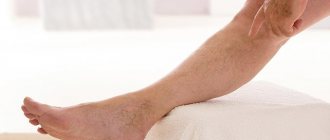
The superficial veins of the upper and lower extremities are not immediately affected. Diseases associated with deterioration of their functioning sometimes take decades to develop. At the beginning, a person notices slightly dilated veins of the lower limb, and subsequently swelling, pain in the calf muscles, and discomfort during sleep occur.
In severe cases, if clear fluid from dilated blood vessels leaks into the tissue, it can cut off circulation to the skin, causing an itchy rash or even a painful ulcer.
A dangerous complication is thrombophlebitis, when inflammation of the venous wall joins varicose veins and a blood clot forms. A blood clot can break loose and travel with the bloodstream, causing death.
Good blood flow throughout the body, and not just in the superficial and deep veins of the lower and upper extremities, and the brain, is extremely important if a person wants to stay young and healthy longer.
Why veins appear - reasons
Often problems with veins in the legs are hereditary.
In this case, it is necessary to begin taking preventive measures as early as possible. But more often the appearance of this disease is provoked by concomitant diseases, hormonal changes, and unhealthy habits. An incorrect diet and excessive consumption of unhealthy, fatty, high-calorie foods cause an increase in the level of bad cholesterol. And the love for fast carbohydrates provokes surges in blood glucose levels.
Other causes of varicose veins in women include:
- hormonal changes associated with teenage and menopausal changes, pregnancy;
- work that requires prolonged sitting or standing, constant stress on the legs;
- lack of physical activity, passive lifestyle, excess weight.
Prolonged immobility in any position provokes stagnation of blood circulation. To avoid this problem, you need to periodically warm up and change your body position.
Important! Beautiful, but tight and uncomfortable underwear, narrow high-heeled shoes can also lead to the formation of varicose veins.
Features of the structure of arteries
We indicated that arteries are much stronger vessels than veins. The structure of any artery is three-layered. The first layer is endothelial cells. It is called internal. The middle layer consists of smooth muscle fibers and elastic tissue. This is the most important difference between arteries and each other. Depending on the predominance of specific fibers, the arteries themselves differ. Large ones have a large amount of collagen and elastin. Small ones (arterioles) are almost 90% composed of muscle elements. The outer layer is connective tissue.
Features of the arterial system in men
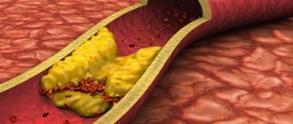
The most important difference between the male arterial system and the female one is the presence of testicular vessels. It is noted that cardiovascular diseases are more common among the male half of the world's population. Moreover, the greatest damage is caused to the circulatory system by cholesterol, which contributes to the development of atherosclerosis. This phenomenon also causes the development of myocardial infarction.
Features of the arterial system in women
Thanks to the presence of special hormones, women's blood circulation is protected from the effects of cholesterol. But at some point, estrogen stops being produced, which creates a risk of developing hypertension. A difficult situation arises during pregnancy, as this leads to an increase in the volume of circulating blood.
Where is the reason?
To say that varicose veins of the lower extremities is a ubiquitous phenomenon is to say nothing. But there are people who live to a ripe old age and have no idea about varicose veins. Why? Maybe initially they knew some secret or spent their lives on the couch with their feet up?
In order for the structural structure of the wall of a venous vessel to be disrupted, it doesn’t take much, just some congenital characteristics of an individual or conditions created during life can cause the onset of a pathological process:
- Hereditary predisposition, that is, a person receives from his parents, NOT a disease, but the structure of blood vessels, predisposing to varicose veins and disease;
- Female gender in connection with natural purpose.
- Loss of elasticity and tone of the vascular wall occurs as a result of:
- Professional characteristics (work involving long periods of standing in an upright position);
- Excessive load during pregnancy, obesity and sports;
- Hormonal background, where the leading place belongs to hormonal changes during childbearing, as well as during menopause;
- Constant violation of nutrition and food preferences for products that affect the vascular wall;
- Metabolic diseases;
- Liver diseases (hepatitis and cirrhosis);
- Arterial hypertension;
- Smoking;
- Venous-arterial fistulas, which can cause dilation of the veins in the legs even in adolescents.
Vein
Vienna is a city with a rich history, the capital of a once mighty empire. Here you can find magnificent sights, historical buildings and cultural monuments. But Vienna is especially famous for its museums, theaters and art galleries.
Hofburg
The Hofburg is one of the symbols of Vienna's imperial past. A huge luxurious palace, which was the winter residence of the Habsburgs. In the Middle Ages there was a castle here, from which a small chapel has survived. The Hofburg was expanded into a magnificent residence when Vienna became the capital of Austria-Hungary. Currently, you can find almost any architectural style here - from Gothic to Art Nouveau. And in its halls there is a national library, a treasury, a museum of musical instruments, weapons and ethnography and the famous “Spanish riding school”.
The huge palace square - Heldenplatz (Heroes' Square) is also impressive. Equestrian statues of Archduke Charles, who won the Battle of Aspern against Napoleon's troops (1809), and Prince Eugene of Savoy, who defeated the Turks, testify to Austria's glorious past. Heroes' Square is not just a huge square in the center of the Austrian capital, it is one of the symbols of glorious history.
Cathedral of St. Stefan
Cathedral of St. Stephen's (Stephandom) is one of Vienna's most famous landmarks and one of the most significant masterpieces of European Gothic architecture. The construction of the first church dates back to 1147. For a long time, Stephandom was the tallest building in Europe - 137 meters. The old church was rebuilt in the Gothic style by order of Duke Rudolf IV. In 1359 he laid the cornerstone of the nave, and in 1433 the South Tower was completed.
Cathedral of St. Stefan's is especially distinguished by its large beautiful roof and tall slender tower (136.7 meters). Interestingly, the number of medieval towers of this height in the world can easily be counted on one hand. Inside the cathedral there are many art treasures, such as: the tomb of Prince Eugene of Savoy, one of the greatest commanders of Europe (1754), the Wiener Neustadt altar, the pulpit of Anton Pilgram (1514-15), the tomb of Emperor Frederick III by Niklas Gerhart (1467-1513) ) and a Gothic altar.
Cathedral of St. Stephen's is the central link of Viennese legends and stories. On the left side of the main entrance is a strange indentation that was used to measure the size of a loaf of bread if a customer was unhappy with its size. The unfinished northern spire is attributed to a young architect who was in love with the daughter of the builder of the southern spire. Although the most likely reasons are financial problems due to the constant threat of a Turkish siege and Gothic going out of fashion. And dozens more such romantic, funny and mystical stories.
Church of St. Carla
Church of St. Karla is a Baroque masterpiece and the largest Baroque church north of the Alps. It was built in 1715 by the famous Austrian architect Johann Fischer von Erlach. It was erected in honor of the vow of Emperor Charles VI in gratitude for deliverance from a severe plague epidemic and was dedicated to Saint Charles Borromeo.
The church is located on one of the central squares of Vienna - Karlsplatz. The square in front of the cathedral was rebuilt in the 1970s by one of the most important sculptors of the 20th century, Henry Moore.
The unusually wide pediment of the Church of St. Carla consists of several contrasting elements that surprisingly complement the unique and harmonious overall image. Two bells with an allegorical depiction of the life of Saint Borromeo are reminiscent of the Italian Renaissance. They form the main portal, reminiscent of a Greek temple. The oval nave of the church has a beautiful dome (72 m high), impressively decorated inside.
Anchor (anchor) clock
The anchor clock is located on Vienna's oldest square, the Hoher Markt, and represents the Art Nouveau style. The clock was built in the early 20th century by von Matz and forms a bridge between two parts of the Anker insurance company building. Over the course of 12 hours, twelve historical figures move along this unique “bridge.” Among them are the medieval lyricist Walter von der Vogelweid, Empress Maria Theresa and Prince Eugene of Savoy. Every day at exactly noon the clock strikes a small show accompanied by music from different eras.
Gazebo
Belvedere is one of the most beautiful palace complexes in the Austrian capital. Consists of two magnificent palaces in the middle of a magnificent park. The Belvedere was built for Prince Eugene of Savoy by the famous Baroque master von Hildebrandt. Interestingly, the palace complex was originally located outside the city walls, but today it is part of the third district of Vienna, which is located near the historical center. The architecture and design of the Belvedere halls is made in the Rococo style. Currently, both palaces house museums with Austrian paintings from the 18th to 20th centuries. The park is a collection of more than 4,000 plants from the alpine ecosystem. It is especially beautiful in spring and summer.
Ring Boulevard
Ring Boulevard is the main avenue of Vienna. It is a 4 km long boulevard ring that encircles the center of the Austrian capital. The Ring Boulevard was founded in 1857 by Franz Joseph I. It was built on the site of old fortress walls and fortifications. Walking here you can look at a large number of monumental historical buildings of various architectural styles.
National Theater
The National Theater (Burgtheater) is located in the first district of Vienna opposite the town hall on the famous ring boulevard. This is a monumental building from the late 19th century, the façade of which is faced with white marble. It is one of the most famous theaters in Europe.
Vienna Opera
The Vienna Opera is located in the very center of Vienna in the southern part of Kärntnerstrasse. This is one of the most famous opera houses in the world. The opera building was built in the second half of the 19th century in the Italian Renaissance style.
Prater
The Prater is Vienna's most popular fun fair. Its most significant part is the huge Ferris wheel. It was erected at the end of the 19th century in honor of the anniversary of the reign of Emperor Franz Joseph. The height of the Ferris wheel is 60 meters.
Blutgasse
Blutgasse is an area of narrow winding streets, medieval and baroque buildings. This area is located near the Cathedral of St. Stefan
Maria am Gestad
Maria am Gestad is one of the oldest buildings in the capital of Austria, a 14th-century Catholic church in Gothic style. Located in the northern part of the historical center.
Scottish monastery
The Scottish Priory is a Benedictine monastery founded in the 12th century. Located in the central part of Vienna on Frejung Square.
Plague Column
The Plague Column is a monumental Baroque sculpture built in marble at the end of the 17th century. It is interesting that the column is dedicated to the Holy Trinity, and not to the Virgin Mary.
Church of St. Augustine
Church of St. Augustine - court church of the Habsburgs. It was founded in the 14th century by Augustinian monks. This Gothic religious building is located on Josefplatz square in the center of Vienna.
Church of St. Petra
Church of St. Petra is an early 18th century Roman Catholic Baroque church on the Graben Street. The church was built on the site of an old medieval religious building by Gabriel Montana under Emperor Leopold I.
Mariahilfer
Mariahilfer is a 17th century Roman Catholic Baroque church located in the sixth district of Vienna.
Vienna City Hall
The Vienna City Hall is a grand neo-Gothic building from the late 19th century, located in the historical center of Vienna. Built according to the design of Friedrich Schmidt.
Parliament
The Parliament is a monumental building from the late 19th century, faced with marble. It is the seat of the national parliament.
Special types of varicose veins
Female varicose veins
Of course, varicose veins are most common in the veins of the lower extremities, but dilation of the pelvic veins is also a common phenomenon and one can imagine what the organs there can reach, because the eye cannot see what is happening in the veins of the internal organs. The female body “succeeds” especially in this regard; women even have a separate group of “their own” varicose veins.
The physiological characteristics of representatives of the fair half of humanity predispose them to this disease more, since nature has endowed them with the function of childbirth and provided them with special organs where blood circulation is very intense, requiring a large number of veins and arteries. When a woman prepares to become a mother, her body must restructure itself and ensure normal pregnancy. Of course, the load on the body is colossal and stagnation occurs both in the pelvis and in the lower extremities.
Varicose veins during pregnancy will become the starting point for the development of the disease in the future, where the expansion of the veins of the small pelvis, as well as the legs, may take a “worthy” place and over time will begin to pose a threat not only to health, but also to human life.
The trouble is that you can’t just see the dilation of the pelvic veins; this requires special equipment, because the venous vessels of the pelvis are not as accessible as on the legs. However, they also form nodes, similar to the veins of the legs, they can contain blood clots that can break off and “set free to float”, unfortunately, not for long... Varicose veins of the small pelvis can cause internal bleeding. Surgeries on the pelvic organs, especially in gynecology, require the mandatory use of compression stockings (elastic stockings and tights) to prevent life-threatening complications associated with blood clot rupture.
Read more about varicose veins of the pelvis and uterus at the link.
Male varicose veins
Varicose veins occur in men to a lesser extent than in women, but in the case of damage to the legs, they manifest themselves similarly. Therefore, everything that is said above and below is fully applicable to the stronger sex.
It is worth adding that men can “boast” of a special male varicose veins - an enlargement of the veins of the spermatic cord (varicocele). Fortunately, this type of male varicose veins can be easily operated on and is not particularly life-threatening, but it is still better to do without it.
Reticular (cosmetic) varicose veins
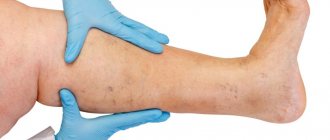
It happens that a network of small blood vessels suddenly begins to appear through the skin of the leg or thigh. Particularly impressionable people can be reassured immediately - this is the so-called reticular varicose veins, or varicose veins of the superficial veins. It is benign and, as it were, not entirely real. Progression and serious complications are not inherent in it; in general, apart from a small cosmetic defect, it does not threaten anything. Therefore, reticular varicose veins of the lower extremities are also called cosmetic.
But those who are not particularly impressionable should remember why varicose veins on the legs are dangerous and begin to fight them in the early stages, which are simply ignored and missed by the patient himself. However, having learned about the disease, future patients of the vascular surgeon still try to do without surgery in the future, are interested in traditional medicine recipes and often quite successfully cope with the progression of the disease at home.