A brain cyst is a rather dangerous diagnosis for a person, after which it is necessary to strictly follow all the instructions and recommendations of the attending physician. If the disease is detected in the early stages and the patient follows all instructions, then in most cases it is possible to prevent unwanted complications. A cystic tumor can be located anywhere in the skull: the development of pathology and treatment features largely depend on this.
General information about pathology
A cyst is a fluid-filled cavity located in the substance of the brain or its membranes. When the pathology is small in size, it has a subclinical course and is diagnosed accidentally during a neuroimaging examination of the head. Since the intracranial space has limited dimensions, with a significant increase in the volume of the formation, intracranial hypertension develops.
The size of the cavity with liquid is largely determined by the compensatory capabilities of the formation and its localization. Due to the pliability of the skull bones in children at an early age, the cyst may not manifest itself for a long time.
The formation can be found in people of different ages, both in infants and in elderly patients. Even if the fluid cavity is congenital, it can make itself felt only by the age of 30-50.
According to generally accepted practice, treatment is prescribed only in the case of a pronounced clinical picture and when complications develop. If the cavity with fluid is frozen or slowly progressing and its volumes are insignificant, a wait-and-see approach is chosen, which involves regular monitoring of the patient.
Classification
Classification by location:
- Cerebral (intracerebral). It is formed in areas of dead brain tissue in the internal structures of the brain.
- Arachnoid. It is formed as a result of the accumulation of cerebrospinal fluid in places of adhesions formed as a result of inflammatory processes and in places of their congenital duplication. The advantage is localized in the meninges.
The following types of brain cysts are distinguished separately:
- dermoid;
- colloidal;
- choroid plexus;
- pineal gland.
According to its genesis, a cavity with fluid can be congenital or acquired. In turn, congenital is divided into colloid and dermoid, and due to its formation into post-infectious, post-traumatic, post-stroke and echinococcal.
Pineal gland cyst: treatment, risks, symptoms, causes
The structure of the brain is represented by multiple neurons. Cells store and transmit information through electrical and chemical impulses. Their connection ensures uninterrupted control over the entire body. Some pathologies can disrupt this balance, causing mental health problems. Where is the pineal cyst located, and what is its threat to the body?
They disrupt control over the entire body, causing a disruption in mental health.
Peculiarities
A benign tumor develops in the pineal region—the epiphysis. This area is located in the deep layers of the brain; the full range of its functions is not fully defined. When the pineal gland is damaged by a cyst, the outflow of hormones responsible for the cycle of sleep and wakefulness and puberty changes.
The outflow of hormones responsible for the cycle of sleep and wakefulness and puberty changes.
A cavity with liquid contents is rarely identified during a routine visit to a therapist. This usually occurs during a comprehensive examination for the presence of other pathologies.
Sometimes a cyst in the pineal region of the brain forms before birth. The patient can live with it all his life, sometimes experiencing minor symptoms.
Causes of the disease
- Blockage of the channel through which endocrine secretions are removed. The outflow of melatonin is disrupted, which leads to its constant accumulation. This process often occurs as a result of various injuries. The flow is slow.
- Helminthiasis is the cause of a parasitic cyst in the epiphysis of the brain.
The larva of echinococcus enters the tissue through the circulatory system. The parasite is enveloped in a protective capsule. Products and toxins from its development constantly accumulate in it. - Hemorrhages can be one of the reasons for the appearance of a cyst in the pineal gland.
Active blood supply to the pineal gland for unknown reasons leads to the formation of a capsule. Hemorrhages lead to the formation of a capsule. - A microcyst of the epiphysis may be congenital. It is formed due to defects during embryonic development, infections during pregnancy and lack of oxygen.
Signs
A pineal cyst in the brain manifests itself in rare cases. As the capsule grows, symptoms similar to other pathologies that are not associated with neuropsychic disorders are observed.
The main symptom of a pineal cyst is recurrent or constant headaches.
As the pineal organ tumor grows, other symptoms appear, but they may not be intense:
Double vision, blurred images as with cataracts.
- visual disturbances (diplopia or double vision, blurred images as with cataracts);
- change in coordination of movements (unclear gait, staggering);
- nausea or vomiting due to severe headaches;
- dropsy (formed after compression of the pineal gland duct and slowing down the circulation of cerebrospinal fluid).
The number and intensity of manifestations depend on the size of the cystic formation of the pineal gland. The larger the cavity in diameter, the more it compresses the surrounding tissue. One of the complications of an epiphysis cyst is blocking the movement of the spinal substance, which leads to persistent disorders in the body.
Echinococcal lesions of the pineal gland pose a great danger. The parasite can grow into a large capsule in just a few years. It is with a parasitic tumor that mental disorders clearly manifest themselves - depression, mental retardation, delusional consciousness and epileptic seizures.
Diagnosis of pineal gland neoplasm
Cystic transformation of the pineal gland can only be detected by extensive instrumental examination. Early diagnosis practically does not yield results due to weak manifestations.
At later stages of development of a cyst in the pineal gland of the brain, a neurological examination reveals a violation of the motor function of the eye. The optic discs become swollen.
MRI determines the size and localization of the pathology.
When cerebrospinal fluid is taken through a puncture from the spine, the level of protein mass and the number of cells - lymphocytes and monocytes - are determined. The study of cerebrospinal fluid allows us to determine other disorders, for example, hidden inflammatory processes and chronic pathologies.
Magnetic resonance imaging is considered the most informative diagnostic method. Layer-by-layer images are displayed on the monitor and photographs. MRI of the brain clearly determines the size, location and level of spread of the cystic process of the pineal gland. An analogue of MRI is computed tomography.
Treatment
For microcysts of the pineal gland, treatment is abandoned if it does not progress and there are no symptoms. Usually such a tumor is discovered by chance. To control the formation, the patient is prescribed a scheduled MRI once every 12-24 months.
Drug therapy
For microcysts in the epiphysis of the brain that have pronounced manifestations, symptomatic treatment is prescribed. Pain in the head helps to eliminate diuretics, which remove a significant part of the fluid from the cystic cavity of the epiphysis. If the tumor has reached a large size and affects the electrical impulses of the brain, the patient requires antiepileptic drugs.
Diuretics help relieve pain.
Sedative medications will help reduce psycho-emotional stress. For small echinococcal cysts of the pineal gland, antiparasitic treatment is carried out. The use of medications is necessary for patients who are contraindicated for surgery. In other cases, radical treatment is used.
Cyst removal
Before preparing a patient for surgery, the doctor tells him why such a cyst is dangerous and what consequences may occur after surgical treatment. Sometimes, before intervention, several years of constant monitoring of the cavity in the pineal gland take place. And only for serious indications that threaten the health and life of the patient, one of the radical treatment methods is used.
The fluid is removed using an endoscope.
- Shunting. Thanks to the implantation of an artificial vessel into the pineal organ, the outflow of fluid is restored to the patient. After the shunt is installed, various side effects may occur, but without increasing intracranial pressure.
- Endoscopy. A relatively gentle surgical method. The fluid from the pineal cavity is removed using an endoscope, after which its walls collapse. The outflow of secretory substances through the drainage is resumed.
- Trepanation of the skull. One of the most difficult brain surgeries, associated with a high level of injuries and complications. Surgical intervention is advisable for persistent brain disorders, progression of the cyst or growth of its walls into malignant tissue. Successful surgery depends on the type of education and experience of the neurosurgeon. In 70% of cases, neurological disorders are observed after treatment.
Consequences of the disease
Epiphysis cysts are not dangerous if they are of normal size. A threat to humans is the growth of a tumor, which provokes hydrocephalus and epilepsy. As well as formations that exceed 1 cm in diameter and capsules with parasitic contents.
A tumor size of 2 cm carries a great danger of neurological abnormalities.
Children with congenital complicated cysts are sometimes diagnosed with autism, but a direct relationship between the pathologies is not observed. Most often, the cause is more complex defects.
Preventive actions
Prevention of epiphysis cysts includes routine examinations of the brain. It is necessary to promptly pay attention to changes in the child’s behavior and complaints of headaches. The main indicator of a brain disorder is neurological disorders. It is necessary to undergo an MRI to accurately establish the diagnosis and begin early therapy.
A brain cyst is not a death sentence. But if the patient is already aware of his diagnosis, he will need regular thorough examinations and consultations with a neurologist.
Source: https://kistateka.ru/golova/shishkovidnaya-zheleza
Etiology and pathogenesis
The causes of a congenital cyst are unfavorable factors that occur during the development of the fetus. These are:
- fetal hypoxia during delivery;
- fetoplacental insufficiency;
- taking a specific group of medications by a pregnant woman;
- Rh conflict between mother and child;
- intrauterine infections.
Factors that provoke the development of a congenital cavity with fluid are drug, alcohol or nicotine addiction of the mother. In this case, the child’s development takes place under conditions of intrauterine intoxication, which negatively affects the brain structures. The causes of the cavity can also be chronic decompensated diseases of the expectant mother.
An acquired cavity with fluid in the head develops as a result of:
- inflammatory diseases (encephalitis, brain abscess, arachnoiditis, meningitis);
- traumatic brain injuries;
- injuries of newborns received during childbirth;
- cerebrovascular accidents (subarachnoid hemorrhage, ischemic stroke, hemorrhagic stroke).
Depending on the etiology, the following types of liquid cavities are distinguished:
- A cyst of iatrogenic origin forms as a complication after brain surgery.
- Parasitic - develops with paragonimiasis, cerebral form of taeniasis and echinococcosis.
A cavity with fluid can also replace cerebral tissue during degenerative and dystrophic processes in the head.
If a cyst is present, there are a number of factors that can trigger its growth. These include obstruction of venous outflow from the skull, strokes and other vascular disorders, as well as head injuries, hydrocephalus, and neuroinfections.
Pineal gland cyst: causes, symptoms, treatment
The discovery of such a rare disease as a pineal gland cyst initially drives patients into a stupor. This is a very rare and little-studied condition, which in itself does not harm the body, but can compress the surrounding membranes of the brain and lead to the development of various pathologies.
Therefore, patients who learn about their diagnosis begin to be overwhelmed by many questions: what kind of disease is this, why it could have arisen and whether there is a chance to get rid of it forever. All this is discussed in the article below.
What is this
A pineal gland cyst of the brain is a benign neoplasm that is located in the pineal region of the pineal gland. Located deep in the brain, between both hemispheres and behind the thalamus. Depending on the reasons that caused it, it may take the form of a capsule filled with viscous contents, or a compaction that is surrounded by inflamed vessels.
The disease is extremely rare and is always benign. Detection of a pineal gland cyst occurs accidentally during an MRI or other brain examinations. The pathology does not manifest itself for a long time, and a person may not be aware of its existence.
The gland itself is responsible for the following processes in the body:
- Melatonin production and regulation of sleep-wake cycles.
- Early puberty and human sexual desire.
- Preventing and slowing the growth of cancer cells.
- Immune system support.
Pathological changes in it occur extremely rarely and are little studied. Currently, experts distinguish between hemorrhage, cysts and neoplasms in this organ.
The pineal gland grows actively in young children; during adolescence, this process gradually slows down. With the onset of middle age, iron does not grow, mineral deposits begin to accumulate in it.
Causes
It is not known for certain why a pineal gland cyst can form. Experts agree that the causes of the tumor may be as follows. Firstly, difficulties with the outflow of melatonin due to blockage of the gland's excretory channels. Occurs as a result of previous inflammatory diseases of the brain, injuries, and unsuccessful surgical interventions.
If the patient has suffered a stroke, the risk of developing a tumor increases. Since the pineal gland is actively supplied with blood, this can lead to the development of hemorrhage. As a result, a pineal gland cyst appears.
Infection of the body with echinococcosis. Parasites enter the brain through the bloodstream and provoke the development of inflammation. In this case, the epiphysis cyst begins to rapidly increase in size, and the patient feels unwell.
It can appear in babies due to intrauterine developmental defects and birth injuries, as a consequence of neurological infectious diseases.
All these conditions lead to disruption of the outflow of cerebrospinal fluid, so the tumor gradually grows and begins to compress the surrounding tissues.
Symptoms
As a rule, the signs of a pineal gland cyst, given its small size, are practically not expressed. The patient may not be bothered by headaches or visual disturbances, and he learns about the presence of pathology quite by accident. The situation will be different if the pineal cyst is caused by a parasitic infestation. In this case, the symptoms increase rapidly:
- The patient begins to suffer from constant headaches of unknown etiology, while the blood pressure level is normal. Often this condition is accompanied by nausea and vomiting.
- It becomes difficult for the patient to rotate the eyeballs, especially if it is necessary to look up.
- Deterioration of vision, decreased acuity and double vision are noted.
- Constant drowsiness, consciousness becomes confused.
- Coordination problems may occur, and patients' gait becomes unsteady.
- Mental abnormalities and depressive states are observed, and the patient may periodically experience epileptic seizures.
- Women. Those who are trying to get pregnant report difficulties conceiving.
If at least several of the above symptoms occur, you should seek qualified help as soon as possible, since these conditions can lead to irreversible consequences.
Brain cyst
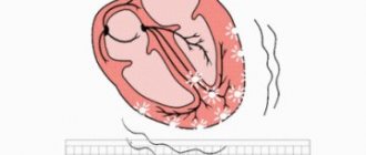
The growth of the formation at the initial stage is in most cases accompanied by symptoms of intracranial hypertension. Patients constantly complain of nausea, which has nothing to do with food, deterioration in general health and decreased performance, constant cephalalgia and pressure on the eyeballs.
In some cases, the main symptoms include a constant feeling of pulsation in the head, sleep disturbance, mild hearing loss, dizziness, motor dysfunction, fainting and tremors of the limbs. Possible visual impairment, namely double vision, visual hallucinations, deterioration of visual acuity. With high intracranial hypertension, the patient is worried about constant vomiting.
There are cases when the first signs indicating a cavity with fluid are the first occurrence of epileptic paroxysm. Subsequently, epileptic seizures recur. Paroxysms can take the form of focal Jacksonian epilepsy or absence seizures and be of a primary generalized nature.
Compared to general cerebral manifestations, focal symptoms are observed in fewer cases. These may be sensory disorders, monoparesis and hemiparesis, brainstem symptoms. The latter include dysarthria, eye movement disorders, and swallowing disorders.
One of the complications of formation is cyst rupture. In this case, hemorrhage due to rupture of the vessel, compression of the brain, the formation of an epileptogenic focus and occlusive hydrocephalus are possible.
In the congenital form of the cyst, episyndromes and intracranial hypertension are recorded at an early age. Education in the brain can cause the child to develop mental retardation and mental development disorders.
Clinical signs.
Clinical manifestations usually occur in early childhood. In adults, symptoms appear much less frequently. They depend on the location. Often a cyst in the brain in an adult is asymptomatic, is an accidental finding during examination and does not require treatment.
Typical clinical manifestations of a brain cyst in an adult:
- General cerebral symptoms: headache, nausea, vomiting, drowsiness.
- Epileptic seizures.
- Protrusion of the skull bones.
- Focal symptoms: monoparesis, hemiparesis, sensitivity disorders of the mono- and hemitype, speech disorders in the form of sensory, motor or mixed sensorimotor aphasia, loss of visual fields, paresis of cranial nerves.
- Sudden deterioration, which is accompanied by depression of consciousness up to coma:
- Due to hemorrhage into the cyst;
- Due to cyst rupture.
Types of brain cysts and their symptoms
Arachnoid - occurs in almost 4% of the population. A cavity with fluid can be congenital or acquired. In the latter case, it develops in response to traumatic brain injury. The formation is localized on the surface of the brain in its membranes. The cavity is filled with cerebrospinal fluid.
In most cases, an arachnoid cyst does not make itself felt for quite a long time and is discovered by chance. Severe symptoms appear only if a large amount of fluid accumulates in the cavity. In this case, cerebrospinal fluid is produced by the cells lining the cavity.
With a sharp increase in the volume of the cavity with liquid, it can rupture and, as a result, die.
Colloid cysts are recorded in 15-20% of all cases of formations inside the ventricles of the brain. Most often it is localized above the foramen of Monroe in the anterior region of the 3rd ventricle. Less common in the area of the transparent cerebral septum in the 4th ventricle.
The liquid filling the cavity of the colloid cyst has high viscosity. Patients experience symptoms of hydrocephalus and, with a certain position of the head, a paroxysmal increase in cephalgia is noted.
In rare cases, memory loss, behavioral disorders and weakness in the limbs occur.
Pineal cyst of the pineal gland - according to statistics, 10% of patients have formations of this type that are small in size and do not make themselves felt. Cystic formations are localized in the epiphysis of the brain, in most cases they are no more than 1 cm in size. Otherwise, symptoms appear. When it grows, it can block the entrance to the “plumbing” of the brain and block the circulation of cerebrospinal fluid, causing occlusive hydrocephalus.
Epidermoid or dermoid is a formation that is an anomaly of intrauterine development. In this case, the cells of the baby's future skin and its appendages remain inside the brain. Accordingly, in addition to liquid, elements of the ectoderm are present, namely sebaceous glands and hair follicles.
After the birth of a child, such a cyst quickly increases in size. The only possible treatment is surgical removal of the formation.
Choroid plexus cyst - forms regardless of the person’s age. In this case, the space between the plexus vessels is filled with cerebrospinal fluid. Symptoms are rarely present, sometimes accompanied by epileptic seizures and symptoms of intracranial hypertension.
With a congenital choroid plexus cyst, the formation is diagnosed at the 20th week of intrauterine development using ultrasound. By the 28th week, such formations resolve.
Causes of pathology
In the pineal region of the brain, cysts form for a variety of reasons, however, the main factors that influence this process should be described in more detail:
- The channel responsible for excretion becomes clogged. This phenomenon leads to the fact that the output of melatonin produced by the gland is inhibited, and this becomes the result of the accumulation of a large amount of fluid;
- a pathology such as echinococcosis, which develops quite quickly, can act as a catalyst for the formation of parasitic cysts. As a result of this process, the formation in the pineal gland is filled with all the substances that the parasite produces in abundance, which can lead to an increase in size. And this already poses a serious danger, since it has a direct effect on the activity of the brain.
Diagnostic measures
A neurologist may suspect the presence of an intracranial formation of significant size based on the patient’s neurological status and clinical symptoms. In this case, the patient is sent for examination to an ophthalmologist and otolaryngologist to check vision and hearing. Specialists perform ophthalmoscopy, audiometry, perimetry and visometry. With severe hydrocephalus, ophthalmoscopy reveals congested optic discs.
By referring the patient for echo-encephalography, it is possible to diagnose increased intracranial pressure. If the patient experiences epileptic paroxysms, he is additionally sent for electroencephalography.
It is extremely important to differentiate a cavity with fluid from a tumor, abscess and hematoma. It is not possible to do this on the basis of collected clinical data alone. Therefore, to make a clear diagnosis, neurologists use neuroimaging diagnostic methods.
By performing an ultrasound examination, it is possible to diagnose certain types of congenital cysts even at the stage of intrauterine development of the fetus. After birth, until the baby's large fontanelle closes, neurosonography is performed to make the correct diagnosis. In adulthood, to visualize a brain cyst, the patient is sent to a magnetic resonance or computed tomography scan of the head.
MRI and CT are performed with contrast in order to differentiate a cyst from a tumor. A cavity with liquid is not able to accumulate a contrast agent, unlike a tumor.
After diagnosis, constant monitoring of the patient with a cystic formation is important. During regular examinations, the doctor monitors the volume of the cyst over time.
If the cyst is a consequence of a stroke, additional vascular examinations are carried out: ultrasound, MRI and CT of vessels, duplex scanning.
Diagnostics
If a scalp cyst is suspected in an adult or child, the doctor will refer the patient for examination. The main method for diagnosing such a tumor is magnetic resonance imaging (MRI). However, MRI alone is not enough; this procedure must be repeated periodically to monitor the dynamics of the development of the pathology. In newborns, this disease is diagnosed mainly by ultrasound examination of the head. It is important to notice in time if the formation begins to grow. If your child starts to get sick every week, this could be a sign of a cyst. The most common symptoms of a cyst in the head are:
- Noticeable deterioration in hearing and vision in a short time;
- Nausea;
- Sleep disturbance;
- Temperature;
- Partial paralysis of the limbs;
- Development of mental disorders;
- Constant noise in the ears and “ripples” before the eyes;
- Feeling of pulsation inside the head;
- Cramps.
Other symptoms depend on the location of the formation. For example, if a cyst appears in the frontal part of the head, in the area of the nasal sinuses, the patient will have a stuffy nose all the time, and he will always feel discomfort, as if diving under water. Sometimes the disease does not appear for quite a long time. In newborn children, a cyst can result in deformation of the fontanel.
General principles of therapy
Drug therapy for cystic formations has practically no results. The only possible treatment is surgical removal of the fluid cavity. But, most forms are small in size and remain in a “dormant” state for many years. In this case, no methods of therapy are used; a wait-and-see regimen is chosen and the patient is regularly examined.
Formations that are accompanied by symptoms of hydrocephalus, complicated by bleeding and rupture, compressing the brain and rapidly increasing in size must be removed. Only a neurosurgeon decides which method of surgical treatment to use.
If the patient has a disorder of consciousness (coma or stupor), he is urgently referred to external ventricular drainage. This method allows you to reduce compression of the brain by the cyst and intracranial pressure. If the cyst ruptures or hemorrhages, surgery is performed. The patient undergoes craniotomy and the formation is excised.
In the absence of complications and disturbances of consciousness, the operation is performed planned and endoscopically. The advantage is the patient’s quick recovery period and low trauma. With endoscopic access, a milling hole is made in the skull, through which fluid is sucked out from the cavity. In order to avoid subsequent accumulation of fluid, several holes are made in the cavity (connected to the cerebrospinal fluid space) or cystoperiotoneal shunting is performed (a special shunt is installed).
The postoperative period involves rehabilitation therapy, including exercise therapy, reflexology and massage. The patient is prescribed drugs to improve blood supply to the brain, absorbents and decongestants.
Diagnosis of a cyst in the brain in an adult.
Computed tomography or magnetic resonance imaging are mandatory research methods.
Additional diagnostic methods:
- Cisternography and ventriculography are contrast studies of the cerebrospinal fluid pathways. They are rarely required, for example, when examining median suprasellar cysts and when affecting the posterior fossa for the purpose of differential diagnosis with Dandy-Walker anomaly.
- Examination of the fundus by an ophthalmologist for hypertension syndrome.
- Electroencephalography (EEG) if there was an epileptic seizure.
Forecasts
In most cases, a frozen cavity with liquid of insignificant size does not bother the patient and does not cause any symptoms. In other cases, with adequate and timely treatment, the outcome is favorable.
In rare cases, patients after surgical removal of a cyst experience a residual moderate-to-severe liquor-hypertensive symptom. If a focal neurological deficit develops, it persists after treatment.
With the removal of the cyst, epileptic paroxysms disappear, but often appear later. This is justified by changes in the operated area of the head, in particular the formed adhesions. Secondary epilepsy is practically uncontrollable by anticonvulsant therapy.
Treatment of post-traumatic cyst
Small brushes are amenable to medication. The natural processes of resorption of dying gray matter cells are restored, and cavities disappear. Epileptic manifestations are relieved and intracranial pressure is normalized.
When drug therapy does not produce the desired effect, surgical excision of space-occupying formations is provided. Especially when there is a tendency for their growth, this is accompanied by vascular hemorrhages.
During treatment and in the postoperative period, for speedy rehabilitation, patients should adhere to a healthy diet and moderate physical activity. A prerequisite is the cessation of drinking alcohol and smoking, which disrupt metabolic processes in the body.
Be healthy !
Prevention
An acquired cyst most often becomes a consequence of developing inflammatory, vascular, infectious and post-traumatic processes. Therefore, it is absolutely obvious that only correct and timely treatment of any pathologies, including resorption and neuroprotective therapy, can serve as a preventive measure for the development of formations in the brain.
The only measure to prevent congenital cysts is to protect the woman and fetus from exposure to provoking factors. Equally important are correct management of pregnancy and delivery.