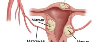
Myomatous nodes. Types of myomatous nodes
Myomatous node is a benign neoplasm that affects the female genital organs. A myomatous node is formed from cells of smooth muscle fibers and elements of connective tissue. In some patients, fibroids are represented by a single tumor, however, most often the pathology is characterized by the proliferation of several myomatous nodes.
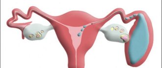
Myomatous nodes are located within the uterus and differ in:
- sizes;
- quantity;
- location.
Myomatous nodes are of four types:
- First. The presence of a single or several intramural or subserous nodes is observed. The size of each of them should not exceed three centimeters.
- Intramural myomatous nodes are neoplasms. Located in the middle muscle layer. With this type of pathology, the uterus enlarges several times, the menstrual cycle is disrupted or completely absent, nagging pain and a feeling of fullness in the lower part of the abdominal cavity are observed.
- Subserous myomatous nodes form on the outer walls of the uterus and often affect the external pelvic cavity. In this case, the menstrual cycle is not disrupted, however, subserous fibroids often become an obstacle to conception and gestation of the embryo.
- Second. The pathology is characterized by the development of one or more intramural and subserous nodes. The size of tumors should not exceed six centimeters.
- Third. Neoplasms of this type have similar characteristics to the second type, differing only in size. Nodes of the third type are more than six centimeters.
- Fourth. The growth of a submucosal neoplasm, the size of which is difficult to predict. The tumor develops under the layer of the uterine endometrium, gradually enveloping the entire uterine cavity. Patients complain of too long and heavy menstruation. The submucosal node grows into the lumen of the uterus and becomes the root cause of the impossibility of conceiving and carrying an embryo.
Pain and discomfort indicate the rapid formation of myomatous nodes from the tumor.
Necrosis of myomatous node
Necrosis of the myomatous node tends to manifest itself in the following types of fibroids:
- subserous (progresses on the outside of the uterus, heading into the pelvic cavity);
- submucous (grows outside the uterine cavity);
- intramural (grows and is retained in the middle muscular layer of the uterus).
Characteristic signs of pathology are observed in the affected areas:
- swelling;
- aseptic inflammation;
- hemorrhages;
- deformation.
Based on morphological characteristics, several types of necrosis of the myomatous node are distinguished:
- Coagulation. The areas of the node subject to necrosis shrink, resembling cavernous cavities in shape, in which fragments of dying tissue accumulate.
- Weeping. Necrotizing tissue softens and forms pathological cystic capsules.
- Hemorrhagic infarction (red necrosis of the myomatous node). The affected tissues acquire a soft consistency, taking on a reddish-brown color. Dilatation of the veins of the node and thrombosis of its vessels are also observed. At risk are women during gestation and after childbirth.
- Aseptic. Necrotizing tissues are accompanied by infections of hematogenous or lymphogenous origin. The most common pathogens are E. coli, staphylococci and streptococci. This type of necrosis of the myomatous node creates a high risk of developing peritonitis and sepsis.
Causes
In fibroids, as in other tissues of the body, metabolism constantly occurs, ensuring its development and increase in volume. Metabolism requires normal blood supply, so the fibroids are approached by blood vessels that bring oxygen and nutrients. With partial disruption of normal blood flow, ischemia develops in them, with complete disruption - necrosis.
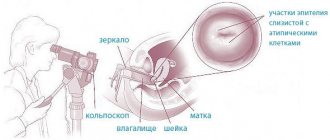
Read more Colposcopy of the cervix staining with iodine
Factors that lead to stopping blood circulation:
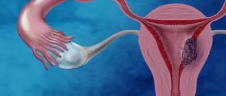
- Torsion of the leg - the neoplasm grows, continuing to stay on the leg in which the nerves and blood vessels are located. Any intense impact provokes a change in the configuration of the node, squeezing the area on which it rests and thanks to which it is nourished. This situation develops very quickly, and in a short period of time, necrosis of the nodal structure occurs;
Leg torsion
- Compression of blood vessels by growing fibroids . In this case, cell death occurs more slowly;
- Violation of the rheological properties of blood - the formation of blood clots, deterioration of the vascular wall provoke the formation of an obstacle to blood flow. This factor is closely related to diseases of the circulatory system and infectious diseases;
- Pregnancy - along with the uterus, fibroids also grow at this time. The growing fetus puts pressure on the surrounding tissues, including the tumor. In addition, increased blood flow to it leads to a decrease in microcirculation in the benign formation.
Note! A woman diagnosed with a benign nodule should be under the supervision of a doctor. Depending on her well-being, the extent and location of the pathological muscle growth in the uterus, the doctor will prescribe either conservative or surgical treatment. In this way, complications are prevented.
There are several morphologically different types of myomatous node necrosis.
- Wet - characterized by the formation of cysts containing liquid contents. These cavities are formed as a result of the gradual decomposition of dead fragments. The eating disorder occurs smoothly and slowly, so the liquid component enters the tissues;
- Dry - a disorder of cell nutrition leads to the development of cavernous cavities consisting of necrotic parts. Ischemia contributed to the drying out of some areas of the neoplasm, their wrinkling and filling with dead cells;
- Hemorrhagic or red - occurs most often with an intramural form of a tumor located inside the uterine wall. Its appearance can be provoked by dilation of veins extending from a pathological accumulation of muscle fibers. Against the background of various coagulopathies, the risk of developing thrombosis increases. Similar phenomena are mainly observed in girls who are pregnant or in women in labor. When examining the myomatous node, red-brown contents are determined.
There is also another type - aseptic, which does not belong to the histological classification, but has a pathogenetic basis. When necrosis forms, blood and lymphatic vessels are drawn into the pathological process. There is a danger of infection spreading through lymphogenous and hematogenous routes. Pathogens represented by streptococcus, staphylococcus or E. coli, entering other parts of the body, cause sepsis or inflammation of the peritoneum. This type of necrosis is the most dangerous, since the pathology quickly covers the entire body.
Causes of necrosis of myomatous node
The myomatous node, like any organ of the internal system that is not a pathology, needs a stable blood supply and trophism, since it is represented by uterine cells. Necrosis of the myomatous node is considered a complication of uterine fibroids. The most dangerous consequence of necrosis is the occurrence of irreversible processes in the tissues of the uterus and the further death of tissues not involved in the tumor process.
Necrotization of myomatous node tissue occurs for the following reasons:
- bending or twisting of the leg;
- administration of uterine contraction stimulants;
- significant physical activity;
- sudden movements;
- abortion;
- carrying an embryo;
- increased vascular tone;
- inflammatory diseases of the pelvic organs;
- obesity;
- sudden weight loss;
- prolonged constipation;
- exposure to stressful situations.
After delivery or instrumental abortion, disruption of the blood supply to fibroids is provoked by a rapid contraction of the uterine myometrium, which is caused by stimulant drugs. The muscles, actively contracting, compress the node, disrupting its trophism and causing the death of viable cells.
If the blood supply to the myomatous node is unstable, its degeneration, swelling, hemorrhage, ischemia, and necrosis are observed. At the first stage of necrosis, the process of cell death of the myomatous node is aseptic in nature, unrelated to an infectious lesion. However, very quickly an infection is connected to the outbreak, entering through the blood and lymph. Since the process occurs in the pelvic area, the pathogenic bacterium E. coli poses a high danger.
Infection of a myomatous node after its necrosis is extremely dangerous. When timely and adequate treatment is not provided, the infection spreads in the abdominal cavity and rises through the bloodstream to the upper organs and tissues.
Symptoms and complications of myomatous node necrosis
With the relatively painless development of uterine fibroids, in most cases, necrosis of the myomatous node has a fairly clear clinical picture. The main manifestation of this pathology will be the symptom of “acute abdomen”.
Women with necrosis of the myomatous node describe the following symptoms:
- cutting or dull pain in the lower abdomen;
- nausea;
- vomiting;
- increase in body temperature to 380 C and above;
- feeling of obstruction when passing gases;
- painful urination;
- frequent false urge to urinate;
- a feeling of incomplete emptying of the bladder and bowels.
Palpation of the abdominal cavity with necrosis of the myomatous node causes tension and the appearance of nagging pain in the suprapubic region and around it. Unfortunately, pathology can only progress and provoke dangerous conditions in neighboring tissues and organs, as well as transmit infection through the blood and lymph flow to the entire body. Symptoms also develop progressively. The patient experiences:
- tension and pain in the anterior abdominal wall;
- chills;
- frequent attacks of nausea;
- vomiting;
- increased gas formation;
- cramping type pain;
- dryness of the oral mucosa;
- persistently high body temperature;
- constipation;
- diarrhea;
- rapid heartbeat;
- coating of the tongue with a whitish coating;
- unnatural pallor of the skin;
- dizziness;
- loss of performance.
Pain from the abdominal cavity radiates to the lumbar region, acquiring a systematic aching character and the ability to suddenly appear and disappear. The intensity of painful manifestations during necrosis of the myomatous node directly depends on the nature of the occurrence:
- blood supply disorders;
- thrombophlebitis;
- squeezing by enlarging fibroids;
- twisting of the leg.
A myomatous node that has “survived” necrosis and has not received proper timely treatment can provoke the following dangerous complications:
- sepsis;
- peritonitis;
- adhesions in the abdominal cavity;
- constant pain in the pelvic area;
- infertility;
- increased risk of ectopic conception;
- dysfunction of the pelvic organs.
Unfortunately, all of the above complications can lead to death.
Features of pathology during pregnancy
It was previously indicated that one of the factors provoking necrosis of the myomatous node is gestation. Indeed, the uterus, which increases in size, adapts to the normal development of the child. Myoma grows along with it. But the fact is that blood flows to a greater extent to the fetus, while the surrounding tissues experience a slight deficiency of nutrients and oxygen. The tumor vessels, which have reduced their functionality, gradually regress, and for this reason it suffers from ischemia. Long-term lack of trophism leads to necrosis.
On the other hand, compression of surrounding organs by the enlarging uterus plays a role. The arteries and veins of the nodular formation are pinched and the blood flow in them slows down, even to a complete stop.
Based on the above, we can conclude that if a pregnant woman has a benign tumor, the doctor should carefully monitor her condition. In the absence of contraindications, resection of the pathological neoplasm is recommended in order to prevent complications that could eventually lead to fetal death, miscarriage, premature birth and death of the woman.
Note! Pregnancy contributes to the progression of many cancers in the female body, as hormonal levels change and the body is rebuilt. A vicious circle is formed, because in many cases, oncology also has an extremely negative effect on pregnancy.
Methods for diagnosing necrosis of myomatous node
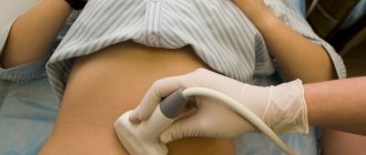
During a vaginal examination, an examination by a gynecologist visually determines the enlargement of the uterus in size and some soreness in its walls. Palpation of the lower abdominal cavity reveals myomatous formations. A myomatous node that has undergone necrosis reveals itself as severe pain when pressed.
The gynecologist evaluates his patient’s complaints, clarifies the presence of uterine fibroids in the history, and also prescribes a general examination with the following tests and studies:
- general blood test;
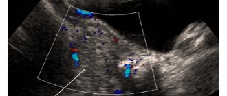
- ultrasound examination of the pelvic organs;
- Dopplerography;
- laparoscopy;
- differential diagnosis of internal diseases.
Ultrasound with Doppler can determine not only the morphological characteristics of the tumor, but also the specifics of its trophism and blood supply. Thus, the doctor is able to make a diagnosis as accurately as possible. Ultrasound signs of necrosis of a myomatous node are the following:
- rounded cavities;
- presence of cysts;
- tissue heterogeneity;
- disruption of blood supply in the body of the node and neighboring tissues;
- proliferation of the anterior and posterior sections of the uterus;
- deformation of the fibroid contour.
Laparoscopy reveals the most complete picture of the state of the pathology site. Treatment can also be carried out together with this laparoscope. The device detects dark red or bluish areas with white speckles and hemorrhages. The inflamed node is covered with a dull peritoneal film with signs of an acute inflammatory process.
In differential diagnosis, specialists try to identify or exclude the following pathologies that can provoke necrosis of the myomatous node:
- ovarian apoplexy with pronounced pain syndrome;
- ectopic pregnancy;
- chronic appendicitis;
- inflammatory processes in the appendages and uterus;
- tubo-ovarian abscess;
- pyosalpinx;
- piovar.
In addition to the gynecologist, the consultation is also carried out by a surgeon.
conclusions
If you have a disease such as a benign uterine tumor, you should be regularly examined by a doctor. It is advisable to do ultrasound diagnostics every time in order to notice the formation of a stalk in this type of tumor in time. This will allow you to remove the fibroid and avoid torsion of the myomatous node.
If the operation is postponed for any reason, then heavy lifting, sports, and pregnancy should be avoided so as not to disturb the position of the tumor and prevent twisting of the leg.
Video: why are uterine fibroids dangerous? Subserous, nodular, interstitial fibroids
Why are uterine fibroids dangerous? Subserous, nodular and interstitial.
Large uterine fibroids (large myomatous node)
Large uterine fibroids (large myomatous node)
Video: uterine fibroids. Surgery
Uterine fibroids. Surgery.