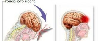
A brain tumor is a neoplasm of nervous or other tissue that arises as a result of abnormally rapid cell division that is not sufficiently controlled by the immune system.
Such structures may be malignant or benign. Strictly speaking, it is not always cancer. Since only neoplasias from epithelial tissue have this name.
Cerebral neoplasms have their own classification according to ICD-10, and are located in the classifier under the code C71: malignant brain tumors.
Structures that are not prone to rapid growth, benign neoplasia, are defined as D33. Various postfixes are possible that clarify the etiology of the disorder or pathological process.
Tumors almost always need to be treated, because otherwise complications cannot be avoided. Including severe neurological deficits.
An oncologist and a neurosurgeon work with patients of this profile. As a rule, both, in tandem.
Causes of tumors
In children, the main cause of tumors is a disruption of the structure of genes responsible for the proper formation of the nervous system, or the appearance of one or more pathological oncogenes, which are responsible for controlling the life cycle of cells, in the structure of normal DNA. Such anomalies can be of congenital origin, or they can also appear in an immature brain (a child is born with a not yet fully formed, “ready” nervous system).
Congenital changes occur in the following genes:
- NF1 or NF2. This causes Recklinghausen syndrome, which in ½ cases is complicated by the development of pilocytic astrocytoma;
- ARS. Its mutation leads to Turcot's syndrome, and it leads to medulloblastoma and glioblastoma - malignant tumors;
- RTSN, whose change leads to Gorlin's disease, and it is complicated by neuromas;
- P53, associated with Li-Fraumeni syndrome, which is characterized by the appearance of various sarcomas - malignant non-epithelial tumors, including in the brain;
- some other genes.
The main changes affect the following protein molecules:
- hemoglobin is a protein that carries oxygen to cells;
- cyclins – proteins that activate cyclin-dependent protein kinases;
- cyclin-dependent protein kinases are intracellular enzymes that regulate the cell life cycle from birth to death;
- E2F are proteins responsible for cell cycle control and the functioning of those proteins that are supposed to suppress tumors. They must also ensure that DNA viruses do not alter human DNA;
- growth factors - proteins that give a signal to grow a particular tissue;
- proteins that “translate” the language of the incoming signal into a language understandable to the cell organelles.
It has been proven that those cells that are actively dividing are the first to undergo changes. And children have much more of these than adults. Therefore, a brain tumor can be activated even in a newborn baby. And if a cell accumulates many changes in its own genome, it is impossible to guess at what speed it will divide and what descendants it will have. Thus, benign tumors (for example, glioma - the most common formation of the brain) can, due to uncontrolled mutations of their constituent cellular structures, degenerate into malignant ones (glioma - into glioblastoma).
Triggering factors for brain tumors
When there is a predisposition in the brain to the appearance of a tumor, or there is a decrease in the rate of damage recovery, then the appearance of a tumor can be provoked (and in adults, initially caused) by:
- ionizing radiation;
- electromagnetic waves (including from abundant communication);
- infrared radiation;
- exposure to vinyl chloride gas, which is needed when making things from plastic;
- pesticides;
- GMOs in food;
- human papillomatosis viruses types 16 and 18 (they can be diagnosed by blood PCR, and their treatment consists of supporting the immune system at a good level, which is helped not only by medications, but also by hardening, and plant phytoncides, and vegetables in the diet).
Tumor risk factors
The following are more likely to “get” a formation in the cranial cavity:
- men;
- persons under 8 years of age or 65-79 years of age;
- liquidators of the Chernobyl Nuclear Power Plant;
- those who constantly carry a mobile phone near their head or talk on it (even through a hands-free device);
- working in toxic production where there is contact with mercury, petroleum products, lead, arsenic, pesticides;
- if an organ transplant was performed;
- HIV-infected;
- receiving chemotherapy for a tumor of any location.
That is, being aware of the risk factors, if you think that you or your child have enough of them, you can talk to a neurologist and get a referral from him for magnetic resonance imaging (MRI) or positron emission tomography (PET) of the brain.
How to cure brain cancer
This disease belongs to one large group of oncological diseases, but it differs greatly from other organ tumors. The reason for this is the spread of cancer cells in the nervous system. The share of brain cancer is one and a half percent of all cancers.
There are 5 stages of development of tumors in the brain, but the last, fifth, means death. Therefore, it is believed that the last stage is still the fourth. In this case, a person has practically no chance of a full recovery, but doctors are able to prolong life in at least 20 cases out of 100 . The difficulty of treatment lies in the fact that metastases gradually develop. You can understand what the patient’s chances of a full recovery are only by tracking the first signs.
Of course, even stage 4 brain cancer is being treated, but even stage 3 is sometimes considered inoperable. The therapy carried out in this case is not aimed at treatment, because it is practically impossible, but at alleviating the patient’s condition. It is because of unbearable pain in advanced stages that strong drugs are prescribed, which calm the nerves and reduce pain.
Classification of tumors
Based on their origin, brain tumors can be:
- Primary: which develop from those structures that are in the cranial cavity, be it bones, white or gray matter of the brain, vessels feeding all these structures, nerves emerging from the brain, meninges.
- Secondary: they are made up of modified cells of any other organ. These are metastases.
Depending on the cellular and molecular (one type of cell may contain different receptor molecules) structure, many types of brain tumors are distinguished. Here are the main, most common ones:
- Developing from brain tissue - neurons and epithelium. These are benign ependymoma, glioma, astrocytoma.
- Originating from the meninges: meningiomas.
- Neuromas growing from cranial nerves.
- Whose origin is pituitary cells. This is a pituitary adenoma.
- Dysembryogenetic tumors that arise in the prenatal period, when normal tissue differentiation is disrupted. In this case, a ball of hair, tooth buds, or other tissues unsuitable for this location can be found in the brain.
- Metastases from organs outside the cranial cavity. They enter the brain through the bloodstream, and less often through lymph.
There is also a classification that takes into account the differentiation of tumor cells. Here it must be said that the more differentiated the tumor is (that is, the cells are more similar to normal ones), the slower it grows and metastasizes.
The classification of primary tumors suggests that they are divided into 2 large groups: gliomas and non-gliomas.
Gliomas
This is the general name for tumors that arise from cells surrounding the nervous tissue - the basis of the brain. They provide neurons with a “microclimate” and conditions for normal functioning. Gliomas account for 4/5 of all malignant brain tumors.
There are 4 classes of gliomas. Classes 1 and 2 are the least malignant, slow-growing. The third class is considered already malignant; it grows moderately quickly. Class 4 is the most malignant of all primary tumors, known as glioblastoma.
They are divided into the following types:
Astrocytomas
This species accounts for 60% of all primary brain formations. They consist of astrocytes - cells that delimit, nourish and ensure the growth of neurons. They make up a barrier that separates brain cells from the blood.
Oligodendrogliomas
The origin of these tumors is from oligodendrocyte cells, which also protect neurons. This is a rare type of neoplasm. They are represented by moderately differentiated and moderately malignant tumors; found in young and middle-aged people
Ependymomas
Ependyma are the cells that line the walls of the ventricles of the brain. It is they who, exchanging components with the blood, synthesize cerebrospinal fluid, which washes the spinal cord and brain.
Ependymal tumors come in 4 classes:
- Highly differentiated: myxopapillary ependymomas and subependymomas. They grow slowly and do not metastasize.
- Moderately differentiated: ependymomas. They grow faster and do not metastasize.
- Anaplastic ependymomas. They grow quite quickly and can metastasize.
Mixed gliomas
Such tumors contain a mixture of different cells of different differentiation. They almost always contain mutated astrocytes and oligodendrocytes.
Non-gliomas
This is the second main type of malignant brain tumors. It also consists of several types of different tumors.
Pituitary adenomas
Such tumors are most often benign; more common in women. The early stage of these neoplasms is characterized by symptoms of endocrine disorders associated with an increase (less often, a decrease) in the production of one or more hormones. Thus, when a large amount of growth hormone is synthesized, acromegaly develops in adults - excessive growth of individual parts of the body; in children - gigantism. If the production of ACTH increases, obesity develops, slower wound healing, acne, and increased hair growth in hormonally active areas.
Lymphomas of the central nervous system
In this case, malignant cells form in the lymphatic vessels, which are located in the cranial cavity. The causes of such a tumor are not fully known, but it is clear that they develop in immunodeficiency states and after transplant operations. Learn more about the symptoms, diagnosis and treatment of lymphoma.
Meningiomas
This is the name given to neoplasms originating from modified cells of the meninges. Meningiomas are:
- benign (grade 1);
- atypical (class 2), when mutated cells are visible in the structure;
- anaplastic (grade 3): there is a tendency to metastasize.
Signs in women
The first signs of a brain tumor in women are similar to those observed in men. Therefore, there is no need to talk about specific signs of the development of tumors in women.
Signs often repeat those of other pathological conditions, for example, headache, tinnitus, decreased vision, nausea and others.
However, symptoms that last a long time (several weeks) and are persistent, increasing in nature, indicate the occurrence of tumor processes.
Therefore, it is important to seek medical help as early as possible. This will allow diagnosis and effective treatment methods to be applied.
Symptoms of brain tumors
Disturbances in the normal structure of the cell periodically occur in each organ during the process of its renewal (when cells divide), but normally such abnormal cells are quickly recognized and destroyed by the immune system “prepared” for such events. The “problem” of the brain is that it is surrounded by a special cellular barrier that prevents the immune system (it acts as a “policeman”) from “inspecting” all the cells of this organ. Therefore, until:
- the tumor will not compress neighboring tissues;
- or will not spread the products of its vital activity into the blood,
no symptoms will appear. Some pituitary tumors are even detected only posthumously, since their signs are so insignificant that they do not attract attention. An MRI of the brain with intravenous contrast, which could detect them, is not included in the list of required examinations.
Early symptoms of any brain tumor are as follows:
- Headaches due to brain tumor. It appears at night or in the morning (this is due to the fact that the membranes swell during the night, since at this time the cerebrospinal fluid is less reabsorbed). The pain has a bursting or pulsating character, intensifies when turning the head, coughing, tensing the abs, but disappears after a while after taking a vertical position (when cerebrospinal fluid and blood flow better from the cerebral vessels). The syndrome is not eliminated by taking painkillers. Over time, the headache becomes constant.
- Nausea and vomiting not related to food. They accompany a headache, but, occurring at its height, do not alleviate the person’s condition. The state of the vomit depends on how long ago the person ate: if this happened recently, then there will still be undigested food, if long ago there will be an admixture of bile. This does not mean that poisoning has occurred with these products.
- Other symptoms in the early stages are:
- memory impairment;
- worse at analyzing information;
- I have trouble concentrating;
- changing the perception of what is happening.
In some cases, the first symptoms may be convulsions - twitching of the limbs or stretching of the entire body, while the person loses consciousness, and in some cases, stops breathing for a certain period of time.
Subsequent symptoms of a brain tumor may include:
- General cerebral. This:
- Depression of consciousness. Initially, a person - against the background of a severe headache - becomes more and more drowsy until he begins to sleep for days without waking up to eat (at the same time, awakening when a person does not understand where he is and who surrounds him can occur in order to go to the toilet).
- Headache. It has a constant character, it is stronger in the morning, when taking a diuretic, the pain decreases slightly (list of diuretics).
- Dizziness.
- Photophobia.
- Depending on the location of the tumor:
- If it is located in the motor cortex, paresis (movements are still possible) or paralysis (complete immobilization) may develop. Usually paralyzes only one half of the body.
- Hallucinations. If the tumor is in the temporal lobe, the hallucinations will be auditory. When it is located in the visual occipital cortex, the hallucinations will be visual. When the mass affects the anterior portions of the frontal lobe, olfactory hallucinosis may occur.
- Hearing impairment up to deafness.
- Impaired speech recognition or production.
- Visual impairment: loss of vision - complete or partial; double vision; distortion of the shape or size of objects.
- Impaired recognition of objects.
- Inability to understand written text.
- Nystagmus (“running pupil”): a person wants to look in one direction, but his eyes “run around”.
- The difference is in the diameter of the pupils and their reaction to light (one can react, the other cannot).
- Asymmetry of the face or its individual parts.
- Inability to write text.
- Coordination problems: staggering when walking or standing, missing objects.
- Autonomic disorders: dizziness, causeless sweating, feeling hot or cold, fainting due to low blood pressure.
- Impaired intellect and emotions. A person becomes aggressive, does not get along well with others, thinks worse, and finds it more difficult to coordinate his activities.
- Impaired pain, temperature, vibration sensitivity in certain areas of the body.
- Hormonal disorders. They occur with tumors of the pituitary gland or pineal gland.
All these symptoms are similar to a stroke. The difference is that they do not develop immediately, but there is a certain stage.
The stages of a brain tumor are as follows:
- The tumor is located superficially. The cells that make it up are non-aggressive, they are only engaged in maintaining their vital functions, that is, they practically do not spread in depth and breadth. Detection of a tumor at this stage is difficult.
- The growth and mutation of cells progress, they penetrate into deeper layers, soldering adjacent structures to each other, affecting blood and lymphatic vessels.
- At this stage, symptoms appear: headache, dizziness, emaciation, increased body temperature. Loss of coordination, visual disturbances, nausea and vomiting may develop, after which it does not get better (unlike poisoning).
- At this stage, the tumor grows into all the meninges, which is why it can no longer be removed, and also metastasizes to other organs: lungs, liver, abdominal organs, causing symptoms of their damage. Consciousness is impaired, epileptic seizures and hallucinations appear. The headache is so significant that fighting it takes up all my thoughts and time.
Symptoms that should prompt a visit to a neurologist:
- headache appeared for the first time after 50 years;
- headache appeared before age 6 years;
- headache + nausea + vomiting;
- vomiting occurs early in the morning, but there is no headache;
- changes in behavior;
- Fatigue sets in quickly;
- focal symptoms appeared: facial asymmetry, paresis or paralysis.
Signs in men
The first signs of a brain tumor in men depend on the size of the tumor, its type and location. For example, benign neoplasms develop slowly, without symptoms, and proceed for several years in a latent form with periodic exacerbations of the clinical picture.
Malignant neoplasms develop quickly and acutely, imitating vascular diseases (stroke, viral infectious meningoencephalitis, etc.).
Symptoms in men include the following:
- headache is a general cerebral symptom, the nature of which depends on the histostructure of the tumor and its location;
- vomiting is a general cerebral symptom that is observed extremely often and is sudden, gushing, and reflexive in nature. Often occurs on an empty stomach, regardless of meal time;
- dizziness - occurs with a sharp increase in cerebrospinal fluid and intracranial pressure, accompanied by decreased hearing, tinnitus, vomiting and headache;
- mental disorders - appear gradually with impaired lymph and blood circulation, cerebral edema, increased intracranial pressure, hypoxia, changes in the cellular structures of the cortex of both hemispheres. Men often experience mental disorders such as decreased attention, increased criticism of themselves and others, weakened memory, and deafness.
Diagnostics
If a brain tumor is suspected, only a neurologist can prescribe an examination. First, he will examine the patient, check his reflexes and vestibular functions. Then he will refer you for examination to related specialists: an ophthalmologist (he will examine the fundus of the eye), an ENT doctor who will evaluate your hearing and sense of smell. Electroencephalography will be performed to determine the focus of convulsive readiness and its degree of activity. Simultaneously with assessing the damage, it is necessary to make a diagnosis by identifying the location and volume of the tumor. The following methods will help with this:
- Magnetic resonance imaging is a method that can be used by persons without metal parts in the body (pacemaker, joint replacements, fragments of explosive devices).
- CT scan. It is not as effective as MRI in diagnosing a brain tumor, but can be performed if the first method is not possible.
- Positron emission tomography helps to clarify the size of the tumor.
- Magnetic resonance angiography is a method that allows you to examine the vessels feeding the tumor. This requires the introduction of a contrast agent into the bloodstream, which will also color the capillaries of the neoplasm.
All these methods can only “give” an idea about the histological structure of the tumor, but accurately determining it in order to draw up a treatment plan for a brain tumor and prognosis is only possible with the help of a biopsy. It is performed after constructing a 3D model of the brain and the tumor in it in order to insert the probe strictly into the pathological area (stereotactic biopsy).
In parallel with the diagnosis, other research methods are performed to determine the stage of the tumor: instrumental diagnostics of those cavitary organs into which the brain tumor could metastasize is performed.
Signs of early stage brain cancer
The main symptom is periodic headaches. Painful sensations in the head can arise for a great variety of reasons, therefore, they usually do not cause any suspicion. The person thinks it’s just overwork, the weather, stress, and takes a routine pill that relieves pain.
On the other hand, there is no point in doing an MRI after every attack of pain in the head. It is important to monitor your well-being and “catch” serious manifestations of possible pathology . If attacks occur very often, their intensity and duration increase, and medications do not bring a noticeable effect, skipping a trip to the doctor can be expensive.
What other symptoms to if you suspect a brain tumor:
- Nausea and vomiting in the morning
- Dizziness or the impression of objects spinning around you
- Problems with spatial orientation and coordination
- Epilepsy
- Hearing problems, temporary hearing loss
- Visual disturbances: floaters and fog
Manifestations of a tumor in the brain in the first stages
Summarize. Headache is considered the first manifestation of a tumor in the brain. It is characterized by increasing duration and strength, accompanied by attacks of nausea and vomiting. The pain may occur in the occipital region, frontal region, temples, including only on one side. If the tumor affects the cerebellum, then symptoms of disruption of its functioning occur: dizziness, loss of orientation and coordination.
Initial phase in adults
In adults, both men and women, already in the early stages of the appearance of head tumors, a decrease in body weight is possible, as the processes of normal metabolism are destroyed. If atypical tissues have already entered the blood, then general weakness, high temperature, and changes in the skin are noted.
The group at greatest risk is the white male population over 65 years of age . Factors that provoke the disease include:
- Prolonged exposure to magnetic or radiation fields, usually in connection with professional activities
- Previous exposure of the head to radiation
- Presence of diseases that reduce immunity, such as HIV
- Previous exposure to chemotherapy
First signs and manifestations in children
The development of brain gliomas in children is quite common. At a more mature age, the likelihood of occurrence decreases and increases only in old age. The signs of brain cancer in children are generally exactly the same - headaches, vomiting - as in adults, but there are some features associated with the growth and adaptation of the child’s body to the outside world:
- Scoliosis appears with back pain
- Slanty eyes
- Growth slowdown
- Incorrect gait, lack of precise coordination in space
- Muscle cramps
- Chronic vision problems
Treatment of tumors
The main treatment for the pathology is surgery to remove the tumor. It is possible only if there are boundaries between the neoplasm and intact tissues. If the tumor has grown into the meninges, it cannot be performed. But if it compresses an important part of the brain, sometimes an emergency operation is performed, during which not the entire tumor is removed, but only part of it.
Before the operation, preparation is carried out: drugs are administered (these are the osmotic diuretic Mannitol and the hormonal drugs Dexamethasone or Prednisolone) that reduce cerebral edema. Anticonvulsant and painkillers are also prescribed.
To reduce the volume of the tumor and more clearly delimit it from healthy areas, radiation therapy can be performed in the preoperative period. In this case, the radiation source can be located either remotely or introduced into the brain (this also requires a three-dimensional model and equipment capable of implanting a capsule with a radioactive substance at given coordinates - a stereotactic technique).
If the tumor blocks the free flow of cerebrospinal fluid, or impedes the movement of blood through the vessels, then as a preoperative preparation, bypass surgery can be performed - installation of a system of flexible tubes that will act as artificial liquor-carrying pathways. This operation is performed under MRI guidance.
Direct removal of a brain tumor can be performed:
- scalpel;
- laser: it will evaporate mutated cells using high temperature;
- ultrasound: the tumor is broken into small pieces by high-frequency sound, after which each of them is removed from the cranial cavity using negative pressure suction. Possible only on confirmed benign tumors;
- radioknife: in addition to vaporizing the tumor tissue, which immediately stops tissue bleeding, nearby brain areas are irradiated with gamma rays.
After removal of the tumor, if indicated, external radiation therapy (that is, the radiation source is located outside the body) can be performed. It is especially necessary if the formation was not completely removed or there are metastases.
Radiation therapy begins 2-3 weeks after surgery. 10-30 sessions are carried out, 0.8-3 Gy each. Such treatment, due to its severe tolerability, requires medicinal support: antiemetics, painkillers, hypnotics. Can be combined with chemotherapy.
The goal of both chemotherapy and radiation treatment is to stop the vital activity of tumor cells (in this case, the effect also occurs on healthy tissue) that could remain after the operation.
Alternative to surgery
If the tumor cannot be removed using the above methods, doctors try to maximize the person’s quality of life using one or a combination of several of the methods below.
Radiotherapy
This second name is radiation therapy. If it is impossible to remove a brain tumor, stereotactic irradiation of the tumor and its metastases is performed, when gamma rays are directed at pathological areas from several points. Such an intervention is planned using a three-dimensional model of the brain of a particular patient, and in order for the rays to receive a clear direction, the head is fixed in a special plexiglass mask.
Radiation therapy can also be carried out as brachytherapy, when the radiation source is placed - again using stereotaxis - directly into the lesion. A combination of external beam radiation and brachytherapy is possible.
If the brain tumor is secondary, remote irradiation of the entire head is required, but with lower doses than in the above case. The hair then falls out, but grows back a few weeks after the end of radiation treatment.
Chemotherapy
This method involves the introduction into the body (most often intravenously) of drugs that will have the most selective effect on tumor cells (they differ from the cells of a healthy body). To do this, it is important to carefully verify the tumor by determining its immunohistotype (identify specific proteins that are found only in tumor tissue).
The course of chemotherapy is 1-3 weeks. The intervals between drug administration are 1-3 days. Additional drug support is required to facilitate the tolerability of chemotherapy drugs and accelerate the recovery of normal tissue cells after them (the bone marrow, the most actively dividing structure that promotes the renewal of human blood, is almost always affected during chemotherapy).
Side effects of chemotherapy: vomiting, headache, hair loss, anemia, increased bleeding, weakness.
Targeted therapy
This is a subtype of chemotherapy that was recently invented. In this case, not drugs that suppress division are introduced into the body, but those that will block only reactions associated with the growth of tumor cells, which is why the toxicity will be much lower.
The following drugs are used for targeted therapy of brain tumors:
- selectively blocking the growth of blood vessels that feed the tumor;
- selectively inhibiting proteins that command the growth of tumor cells;
- inhibitors of the enzyme tyrosine kinase, which in tumor cells should regulate signal transmission, their division and programmed death.
Combined use of radiation and chemotherapy
If you combine external irradiation with gamma rays and administer drugs that inhibit the division of tumor cells, the prognosis significantly improves. Thus, when treating low-grade tumors with combination therapy, the three-year survival rate increases from 54% to 73%.
Cryosurgery
This is the name of the method when the tumor focus is exposed to extremely low temperatures, while the surrounding tissue is not injured. This type of therapy can be used as an independent treatment - for inoperable tumors that grow into neighboring tissues; it is also performed during surgery for a clearer vision of the tumor boundaries.
You can freeze tumor tissue using a cryoapplicator, which is applied to the pathological focus from above. A cryoprobe inserted deep into the lesion can also be used.
Indications for cryosurgery are:
- the tumor is located deep in important areas of the brain;
- multiple tumors (metastases), located deep;
- traditional surgical methods cannot be used;
- after the operation, tumor fragments remained “glued” to the membranes of the brain;
- tumors are located in the pituitary gland;
- a neoplasm was discovered in an elderly person.
Treatment
Therapy for adults (both men and women) is selected by the doctor individually for each patient and depends on the degree of tumor development, its size and location, as well as the patient’s age, concomitant diseases and the general condition of the patient.
- The surgical method is considered the most effective method of treatment. The tumor is either completely or palliatively, that is, partially removed from the skull in order to reduce pressure. However, surgery is not always indicated. Contraindications are the late stage of tumor development, its difficult localization, the patient’s serious condition;
- Radiation treatment is used as a complement to surgery or as an independent method. Represents ionizing remote irradiation;
- Chemotherapy is considered a powerful treatment. A toxin that is specially selected to combat a specific tumor is injected into the patient’s body in courses. In most cases, the poison has a lethal effect on the tumor. However, the body as a whole is also exposed to chemicals, which is accompanied by the death of healthy cells of the gastrointestinal tract and bone marrow - the main organ of immunity and hematopoiesis. As a result, this treatment method is accompanied by many side effects - nausea, vomiting, exhaustion, lethargy, hair loss, itching and pigmentation of the skin, loss of appetite, drowsiness, impaired kidney function and signs of menopause in women;
- Symptomatic therapy is used to overcome not the cause itself, but the unpleasant manifestations that accompany the disease. They use sedatives, antiemetics, glucocorticosteroids, and narcotic analgesics.
What are the consequences of a brain tumor?
The following are the main consequences of brain tumors:
- Convulsions – twitching of the limbs or their stretching with a sharp straightening of the entire body, accompanied by loss of consciousness. In this case, foam may appear at the mouth, the tongue may be bitten, and convulsions are also often accompanied by stopping breathing for a while. This condition is treated with anticonvulsants.
- Disruption of the normal flow of cerebrospinal fluid, which causes hydrocephalus. Symptoms of this complication are severe headache, vomiting, convulsions, drowsiness, nausea, blurred vision, and dizziness. This complication is treated with shunt surgery, when tubes are installed into the cranial cavity through which the cerebrospinal fluid will drain. Read more about the symptoms of hydrocephalus.
- Depression is a depressed mood when there is no desire to engage in any activity, speech is slow, and reactions are inhibited. In such cases, psychotherapeutic treatment methods are used, and antidepressants are also prescribed (list of antidepressants sold without prescription).
Description
The brain is the heart of any personality, that amazing organ that guides everything and makes a person an individual. This is why being affected by a tumor causes such fear. The starting point for the occurrence of a neoplasm is a genetic failure in the developmental system. A group of causative factors puts a spoke in the wheels of normal cell formation. As a result, the body is replenished with atypical, uncharacteristic strangers, who, on top of everything else, are immortal. They begin to divide endlessly, creating an army of their own incorrect clones.
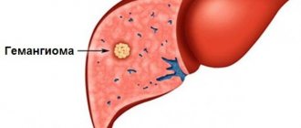
Regardless of the location of the lesion, all brain tumors are divided into benign and malignant. The former are the preferred variant of a dangerous disease and develop at an early stage. This group includes meningioma (20%), hemangioblastoma (1%), schwannoma (9%), pituitary gland (15%). New growths grow quite slowly, without squeezing or growing into neighboring organs. They do not spread metastases. Their cells are not so atypical and differ slightly from normal ones. You can determine what tissue they came from. However, in a protracted course of the disease, they can turn into an oncological form.
A malignant tumor is very dangerous. Sarcomas, gliomas, meningiomas, neuromas grow extremely quickly, literally before our eyes, sweeping away everything in their path. They compress organs, grow, destroy, and spread metastases. The malignant neoplasm is so atypical and uncontrolled that the origin of these cells cannot be determined. Even after treatment, a recurrent course of the disease is possible. Most brain tumors, unfortunately, are malignant.
Currently, there is no exact reason that leads to the appearance of neoplasms. But there is an established specific list of factors that predispose the genetic system to cellular failure:
- Hereditary burden - if there were relatives in the family with such a disease, then the likelihood of the occurrence of pathology increases;
- Ionizing radiation is a precipitating factor for many developmental defects;
- Toxic effects on the body of harmful chemicals.
Clinic
The symptom complex of the disease consists of gradually increasing general cerebral and focal signs. The progression of the disease is determined by the growth of the tumor. In this regard, depending on the degree of prevalence of the primary space-occupying lesion, 4 stages are distinguished.
The first symptoms are limited to general cerebral manifestations. These include nonspecific symptoms, indicating general ill-being of the brain, which often remains without due attention. However, it is advisable to diagnose a space-occupying lesion already at this stage. During this period, based on a person’s complaints, the following can be identified:
- headache, often of a bursting, painful nature, occurring in the morning, intensifying when changing the position of the head. It is the earliest and most obligate symptom of the disease;
- nausea and vomiting;
- dizziness;
- general fatigue;
- lability of emotional mood;
- mental disorders;
- disturbances of consciousness.
The early stages of the disease are represented by general cerebral symptoms. The attachment and dynamic change of focal signs occurs at later stages and indicates tumor growth.
It is the area of the brain affected by the tumor that determines the clinical picture of the disease. Focal manifestations of the tumor can be represented by symptoms of irritation and symptoms of prolapse. Symptoms of irritation include local or secondary generalized epileptic paroxysms. They appear as:
- somatomotor seizures - convulsive twitching of individual parts of the body;
- somatosensory seizures - abnormal sensations on the skin in the form of tingling, burning, crawling, and so on;
- hallucinations (auditory, visual, olfactory or gustatory);
- vegetative symptoms – pain in the epigastrium, chest, changes in skin color, increased sweating;
- mental symptoms - sudden transient speech impairment (aphasic seizures), feelings of euphoria, distorted perception of reality.
These variants of paroxysms can develop into secondary generalization with the development of major tonic-clonic seizures.
Symptoms of prolapse include the development of motor and sensory disorders, extrapyramidal disorders, and pathology of higher cortical functions (gnosis, praxis and phasis). With hemispheric neoplasms, paresis and paralysis of the limbs may occur with the development of anesthesia, loss of visual fields, and ataxia. These clinical signs occur on the side opposite to the lesion. Cerebellar tumors cause static and dynamic ataxia on the side corresponding to the localization of the space-occupying lesion. Tumors of the trunk lead to damage not only to the motor, sensory and cerebellar tracts, but also to the cranial nerves. This pathology is manifested by oculomotor disorders, facial asymmetry, difficulty swallowing, blurred speech, and possibly the addition of respiratory disorders.